Download presentation
Presentation is loading. Please wait.

1
RESPIRATORY PHYSIOLOGY (HUMAN PHYSIOLOGY I) Dr. Waheeb Alharbi
 Dr. Waheeb Alharbi")
2
References (1) Physiological basis of medical practice. By; John B. West (2) Concise Human Physiology By; M. Y. Sukkar, H. A. El-Munshid & M. S. m. Ardawi (3) Human physiology By;Guyton By;Guyton
 Concise Human Physiology By; M. Y. Sukkar, H. A. El-Munshid & M. S. m. Ardawi (3) Human physiology By;Guyton By;Guyton.")
3
Lecture 1 -Ventilation -Gas transport -Tissue respiration -Functional anatomy of the respiratory system -Basic mechanism of V E -Lung volume and capacities -Dead space -Alveolar V E -VD and uneven V E

4
Resp is the use of O 2 by the living cell for oxidation of nutrients. This result in production of CO 2. It can be divided into 4 main events; 1) pulmonary VE 2) gas diffusion 3) gas transport 4) regulation of resp VE is the movement of air between the environment and the alveoli. It can be spontaneous or artificial. Air is a mixture of gases. According to Dalton’s Law, the total pres of a mixture of gases is the sum of the pres of the individual gases (P total = P 1 + P 2 + P 3 ……).I.e. partial pressure. V E = fr X V T
 pulmonary VE 2) gas diffusion 3) gas transport 4) regulation of resp VE is the movement of air between the environment and the alveoli. It can be spontaneous or artificial. Air is a mixture of gases. According to Dalton’s Law, the total pres of a mixture of gases is the sum of the pres of the individual gases (P total = P 1 + P 2 + P 3 ……).I.e. partial pressure. V E = fr X V T.")
5
Gas transport Most gases transported in the blood in 2 forms; 1- Dissolved in the plasma 2- Combine with Hb Under normal circumstances, more than 98% of the O 2 in a given vol of blood is transported in RBCs, bound to Hb.

6
Tissue resp It means getting energy out of glucose. The most efficient form of resp is aerobic (require O 2 ) and anaerobic resp (does not require O 2 ). Aerobic resp: It is the normal process by which food substances are broken down and oxidized to provide energy. Glucose + O 2 → CO 2 + H 2 O + energy released Glucose + O 2 → CO 2 + H 2 O + energy released Anaerobic resp: It means that energy can be derived from food substances without the simultaneous utilization of O 2. Glucose → lactic acid + much less energy released Glucose → lactic acid + much less energy released
 and anaerobic resp (does not require O 2 ). Aerobic resp: It is the normal process by which food substances are broken down and oxidized to provide energy. Glucose + O 2 → CO 2 + H 2 O + energy released Glucose + O 2 → CO 2 + H 2 O + energy released Anaerobic resp: It means that energy can be derived from food substances without the simultaneous utilization of O 2. Glucose → lactic acid + much less energy released Glucose → lactic acid + much less energy released.")
7
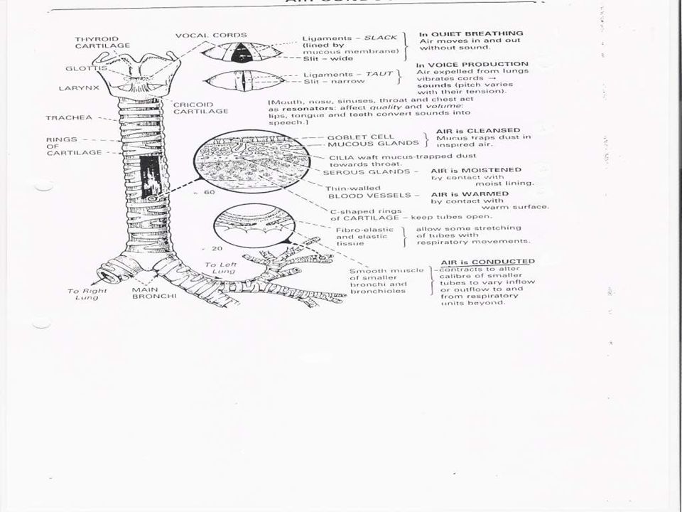
Functional anatomy of the resp system Anatomy of the resp system is composed of; 1) the resp air ways 2) the lungs 3) the resp muscles 4) the neural centers The main function of the lungs is to provide continuous gas exchange between inspired air and blood in the pulmonary circulation, supplying O 2 and removing CO 2, which is then cleared from the lungs by subsequent expir. The functional structure of the lung can be divided into; 1- The Conducting zone, and 1- The Conducting zone, and 2- The respiratory zone. 2- The respiratory zone. The Conducting zone (air flow): Air comes into the nose and the mouth through the pharynx, larynx and then through the trachea. The respiratory zone (gas diffusion): It begins when alveoli start to appear in the walls of the bronchioles.
: Air comes into the nose and the mouth through the pharynx, larynx and then through the trachea. The respiratory zone (gas diffusion): It begins when alveoli start to appear in the walls of the bronchioles..")
13
Basic mechanism of vent Breathing consists of 2 phases; inspiration (active process) and expiration (passive process). During inspir: The diaphragm and intercostals muscles contract. The diaphragm moves downwards increasing the vol of the thoracic cavity, and the intercostals muscles pulls the ribs up expanding the rib cage and further ↑ this vol. During expir: The diaphragm and intercostals muscles relax. This returns the thoracic cavity to its original vol, ↑ the air pressure in the lungs, and forcing the air out.

15
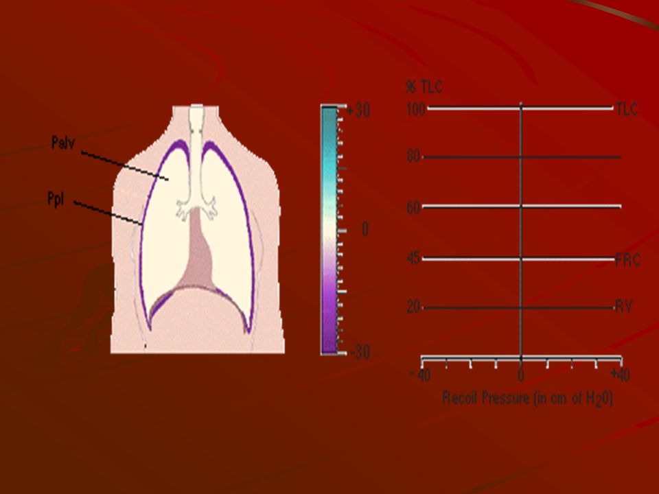
Pleural pressure: It is the pres in the narrow space between the lung pleura and chest wall pleura. Alveolar pressure: It is the pres inside the lung alv.

18
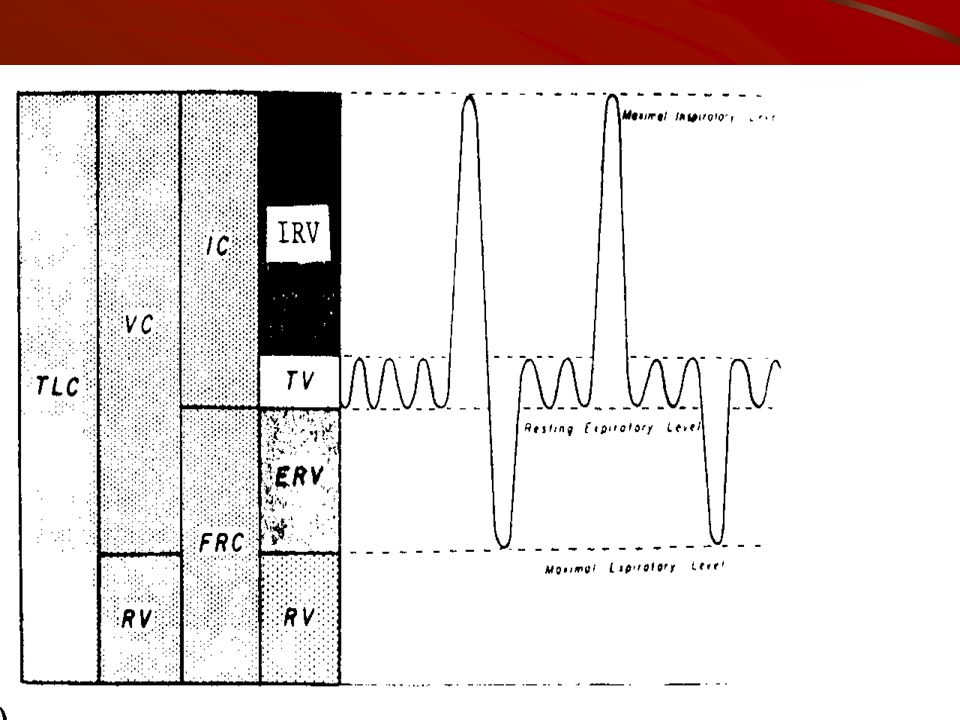
Lung volumes and capacities Capacity is the sum of 2 or more vols. Lung vol and capacity can be measured by a spirometer. It also can be measured by vitalograph, gas dilution and body plethysmography. Lung vol includes; 1) Tidal volume (V T ): It is the vol of air expired and inspired in each breath (500 ml). 2) Inspiratory reserve volume (IRV): It is the max vol of additional air that can be inspired from the end of a normal insp (3100 ml). 3) Expiratory reserve volume (ERV): It is the max vol of additional air that can be expired from the end of a normal exp (1200 ml). 4) Residual volume (RV): It is the vol of air that remains in the lung after maximal exp (1200 ml). Lung capacities include; 1) Inspiratory capacity (IC): V T + IRV. 2) Functional residual capacity (FRC): ERV + RV. 3) Vital capacity (VC): IC + ERV. 4) Total lung capacity (TLC): IC + FRC.
 Tidal volume (V T ): It is the vol of air expired and inspired in each breath (500 ml). 2) Inspiratory reserve volume (IRV): It is the max vol of additional air that can be inspired from the end of a normal insp (3100 ml). 3) Expiratory reserve volume (ERV): It is the max vol of additional air that can be expired from the end of a normal exp (1200 ml). 4) Residual volume (RV): It is the vol of air that remains in the lung after maximal exp (1200 ml). Lung capacities include; 1) Inspiratory capacity (IC): V T + IRV. 2) Functional residual capacity (FRC): ERV + RV. 3) Vital capacity (VC): IC + ERV. 4) Total lung capacity (TLC): IC + FRC..")
19
Spirometer

21
Normal values of lung vol and capacities in both male & female Parameter Volume (liter) Volume (liter) MaleFemale VT0.50.5 IRV3.31.9 ERV1.00.7 RV1.21.1 IC3.82.4 FRC2.21.8 VC4.83.1 TLC6.04.2
 Volume (liter) MaleFemale VT IRV ERV RV IC FRC VC TLC6.04.2")
22
LUNG CAPACITIES AND RESP DISEASES A) Restrictive Disease. Resp disease which make it more difficult to get air in to the lungs. They “restrict” inspiration. Includes fibrosis, sarcoidosis, muscular diseases, and chestwall deformities. B) Obstructive Disease. Resp disease which make it more difficult to get air out of the lungs. Includes emphysema, chronic bronchitis, asthma. C) A summary of lung capacity changes during disease such as follow; –Restrictive Disease: ↓ VC; ↓ TLC, ↓ RV, ↓ FRC. –Obstructive Disease: ↓ VC; ↑ TLC, ↑ RV, ↑ FRC.
 Obstructive Disease. Resp disease which make it more difficult to get air out of the lungs. Includes emphysema, chronic bronchitis, asthma. C) A summary of lung capacity changes during disease such as follow; –Restrictive Disease: ↓ VC; ↓ TLC, ↓ RV, ↓ FRC. –Obstructive Disease: ↓ VC; ↑ TLC, ↑ RV, ↑ FRC..")
24
Anatomical and physiological V D V D is defined as the vol of inspired air that does not participate in GE. The normal VD in a young adult man is about 150 milliliters. This ↑ slightly with age. There are two types of V D anatomical and physiological. (1) Anatomic V D is the vol of an inspired breath which has not mixed with the gas in the alv. It is anatomical because it measures the anatomical vol of the conducting airways leading up to the alv. It can be measured from the vol of expired gas leaving the mouth and nose before the 'front' of alveolar gas containing CO 2 arrives at the lips. (1) Anatomic V D is the vol of an inspired breath which has not mixed with the gas in the alv. It is anatomical because it measures the anatomical vol of the conducting airways leading up to the alv. It can be measured from the vol of expired gas leaving the mouth and nose before the 'front' of alveolar gas containing CO 2 arrives at the lips. (2) Physiological V D is the vol of an inspired breath which has not taken part in GE. It is physiological because it assesses one of the functions of the lungs (GE). It can be estimated using the Bohr equation, which is derived from the fact that the vol of gas expired equals the vol from the V D plus the vol from the alv. (2) Physiological V D is the vol of an inspired breath which has not taken part in GE. It is physiological because it assesses one of the functions of the lungs (GE). It can be estimated using the Bohr equation, which is derived from the fact that the vol of gas expired equals the vol from the V D plus the vol from the alv.
 Anatomic V D is the vol of an inspired breath which has not mixed with the gas in the alv. It is anatomical because it measures the anatomical vol of the conducting airways leading up to the alv. It can be measured from the vol of expired gas leaving the mouth and nose before the front of alveolar gas containing CO 2 arrives at the lips. (1) Anatomic V D is the vol of an inspired breath which has not mixed with the gas in the alv. It is anatomical because it measures the anatomical vol of the conducting airways leading up to the alv. It can be measured from the vol of expired gas leaving the mouth and nose before the front of alveolar gas containing CO 2 arrives at the lips. (2) Physiological V D is the vol of an inspired breath which has not taken part in GE. It is physiological because it assesses one of the functions of the lungs (GE). It can be estimated using the Bohr equation, which is derived from the fact that the vol of gas expired equals the vol from the V D plus the vol from the alv. (2) Physiological V D is the vol of an inspired breath which has not taken part in GE. It is physiological because it assesses one of the functions of the lungs (GE). It can be estimated using the Bohr equation, which is derived from the fact that the vol of gas expired equals the vol from the V D plus the vol from the alv..")
25
In a normal person, the anatomic and physiologic V D are nearly equal because all alv are functional in the normal lung, but in a person with partially function or nonfunctional alv in some parts of the lungs, the physiologic V D may be as much as 10 times the vol of anatomic V D.

26
VAVAVAVA V A is the total vol of new air entering the alv and adjacent GE area each minute. It is equal to the resp frequency times the amount of new air that enters these area with each breath; V A = fr X (V T - V D ) What is the V A in a normal person? V A = …. X (…. - ….) = ……… ml/min Because of the V D, rapid, shallow resp produces much less V A than slow, deep resp at the same minute vol (see table).
 What is the V A in a normal person. V A = …. X (…. - ….) = ……… ml/min Because of the V D, rapid, shallow resp produces much less V A than slow, deep resp at the same minute vol (see table)..")
27
Table: Effects of variations in respiratory rate & depth on V A. Respiratory rate 30 b/min 10 b/min VTVTVTVT 200 ml 600 ml Minute vol 6000 ml VAVAVAVA ….(…. – ….) = 1500 ml ….(…. – ….) = 4500ml
 = 1500 ml ….(…. – ….) = 4500ml.")
28
V D and uneven V E In the upright subject the bases of the lungs are found to be better ventilated than the apices. This can be demonstrated by breathing radioactive xenon. The uneven V E is due to the effect of gravity. Similarly, a subject in the supine position will have better V E of the posterior parts of the lungs than the anterior parts. Uneven V E can significantly affect gas exchange in the lungs. V E is preferentially distributed to the more dependent portions of the lungs because, as a result of the weight of the lungs, the intrapleural pres is lower (i.e. less negative). A clinical correlate of the effect of gravity on V E is that arterial oxygenation is improved in unilateral lung diseases when patients lie on their sides so that the good lung is in the dependent position.
. A clinical correlate of the effect of gravity on V E is that arterial oxygenation is improved in unilateral lung diseases when patients lie on their sides so that the good lung is in the dependent position..")
Similar presentations


 Role of surfactant Lung volumes and capacities Anatomical and physiological VD Alveolar space and.>")


