Download presentation
Presentation is loading. Please wait.

1
History of Fluorescent Proteins
1960s : Curiosity about what made the jellyfish Aequorea victoria glow Green protein was purified from jellyfish by Osamu Shimomura in Japan. Its utility as a tool for molecular biologists was not realized until 1992 when Douglas Prasher reported the cloning and nucleotide sequence of wt-GFP in Gene. - The funding for this project had run out, and Prasher sent cDNA samples to several labs. 1994 : Expression of the coding sequence of fluorescent GFP in heterologous cells of E. Coli and C. elegans by the lab of Martin Chalfie : publication in Science. Although this wt-GFP was fluorescent, it had several drawbacks, including dual peaked excitation spectra, poor photo-stability, and poor folding at 37°C.

2
1996 : Crystal structure of a GFP
Providing vital background on chromophore formation and neighboring residue interactions. Researchers have modified these residues using protein engineering (site directed and random mutagenesis) Generation of a wide variety of GFP derivatives emitting different colors ; CFP, YFP, CFP by Roger Y. Tsien group ex) Single point mutation (S65T) reported in Nature (1995) - This mutation dramatically improved the spectral characteristics of GFP, resulting in increased fluorescence, photostability, and a shift of the major excitation peak to 488 nm, with the peak emission kept at 509 nm. Applications in many areas including cell biology, drug discovery, diagnostics, genetics, etc. 2008 : Martin Chalfie, Osamu Shimomura and Roger Y. Tsien shared the Nobel Prize in Chemistry for their discovery and development of the fluorescent proteins.
 Generation of a wide variety of GFP derivatives emitting different colors ; CFP, YFP, CFP by Roger Y. Tsien group. ex) Single point mutation (S65T) reported in Nature (1995) - This mutation dramatically improved the spectral characteristics of GFP, resulting in. increased fluorescence, photostability, and a shift of the major excitation peak to 488 nm, with the peak emission kept at 509 nm. Applications in many areas including cell biology, drug discovery, diagnostics, genetics, etc : Martin Chalfie, Osamu Shimomura and Roger Y. Tsien shared the Nobel Prize in Chemistry for their discovery and development of the fluorescent proteins.")
3
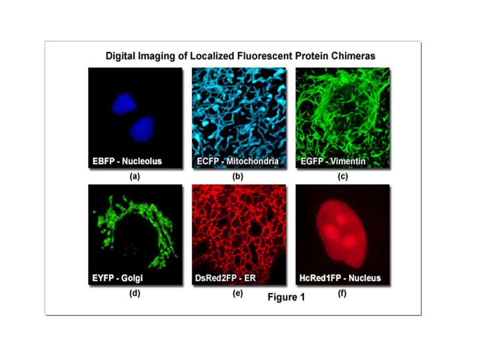
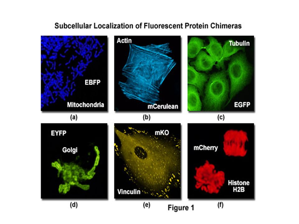
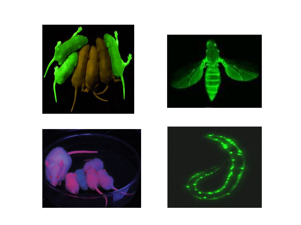
Fluorescent proteins Revolutionized medical and biological sciences by providing a way to monitor how individual genes are regulated and expressed within a living cell ; Localization and tracing of a target protein in the cells Widespread use by their expression in other organisms as a reporter usually fused to N- or C terminus of proteins by gene manipulation Key internal residues are modified during maturation to form the p-hydroxybenzylideneimidazolinon chromophore, located in the central helix and surrounded by 11 ß-strands (ß-can structure) GFP variants : BFP, CFP, YFP Red fluorescent protein from coral reef : tetrameric, slow maturation - Monomeric RFP by protein engineering Quantum yield : 0.17 (BFP) ~ 0.79 (GFP)
 GFP variants : BFP, CFP, YFP. Red fluorescent protein from coral reef : tetrameric, slow maturation. - Monomeric RFP by protein engineering. Quantum yield : 0.17 (BFP) ~ 0.79 (GFP)")
4
GFP (Green Fluorescent Protein)
Jellyfish Aequorea victoria A tightly packed -can (11 -sheets) enclosing an -helix containing the chromophore 238 amino acids Chromophore Cyclic tripeptide derived from Ser(65)-Tyr(66)-Gly(67) Wt-GFP absorbs UV and blue light (395nm and 470nm) and emits green light (maximally at 509nm)
 enclosing an -helix containing the chromophore. 238 amino acids. Chromophore. Cyclic tripeptide derived from Ser(65)-Tyr(66)-Gly(67) Wt-GFP absorbs UV and blue light (395nm and 470nm) and emits green light (maximally at 509nm)")
5
Chromophore formation in GFP

6
GFP and chromophore Covalently bonded chromophore : 4-(p-hydroxybenzylidene)imidazolidin-5-one (HBI). HBI is nonfluorescent in the absence of the properly folded GFP scaffold and exists mainly in the unionized phenol form in wt-GFP. Maturation (post-translational modification) : Inward-facing side chains of the barrel induce specific cyclization reactions in the tripeptide Ser65–Tyr66–Gly67 that induce ionization of HBI to the phenolate form and chromophore formation. The hydrogen-bonding network and electron-stacking interactions with these side chains influence the color, intensity and photo-stability of GFP and its numerous derivatives
 : Inward-facing side chains of the barrel induce. specific cyclization reactions in the tripeptide Ser65–Tyr66–Gly67 that induce ionization of HBI to. the phenolate form and chromophore formation. The hydrogen-bonding network and electron-stacking interactions with these side chains influence. the color, intensity and photo-stability of GFP and its numerous derivatives.")
7
Diverse Fluorescent Proteins by Protein Engineering
wtGFP : Ser(65)-Tyr(66)-Gly(67)
-Tyr(66)-Gly(67)")
8
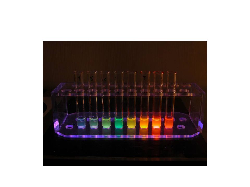
Fluorescence emission by diverse fluorescent Proteins
The diversity of genetic mutations is illustrated by this San Diego beach scene drawn with living bacteria expressing 8 different colors of fluorescent proteins.

10
Absorption and emission spectra
a) Normalized absorption and b) Fluorescence profiles of representative fluorescent proteins: cyan fluorescent protein (cyan), GFP, Zs Green, yellow fluorescent protein (YFP), and three variants of red fluorescent protein (DS Red2, AS Red2, HC Red). From Clontech.
 Normalized absorption and. b) Fluorescence profiles of representative fluorescent proteins: cyan fluorescent protein (cyan), GFP, Zs Green, yellow fluorescent protein (YFP), and three variants of red fluorescent protein (DS Red2, AS Red2, HC Red). From Clontech.")
Similar presentations
 introduced into them Used to increase survival rates Introducing.>")
 from jellyfish : Revolutionized medical and biological science by providing a way to monitor how.>")


![Some structures Dansyl chloride 1,5-I-AEDANS Fluorescein isothiocyante ANS Ethidium bromide 5-[2-[(2-iodoacetyl)amino]ethylamino] naphthalene-1-sulfonic.](/15/4639132/big_thumb.jpg "Some structures Dansyl chloride 1,5-I-AEDANS Fluorescein isothiocyante ANS Ethidium bromide 5-[2-[(2-iodoacetyl)amino]ethylamino] naphthalene-1-sulfonic.>")


 from jellyfish : Revolutionized medical and biological science by providng a way to monitor how individual.>")


