Download presentation
Presentation is loading. Please wait.

1
صدق الله العظيم الاسراء اية 58

2
By Dr. Abdel Aziz M. Hussein Assist. Prof. of Physiology Member of American Society of Physiology SYNAPSE

3
Def: Functional connection between 2 neurons Or Areas of contact between neurons in the C.N.S.

4
2)Types of Synapses: A. Chemical Most common Slow and fatigue One direction only B. Electrical Extremely rare Fast and resist fatigue Occurs in both directions

8
Physiological anatomy: Presynaptic small round or oval knobs→ terminal buttons or synaptic knobs Contains vesicles and mitochondria Postsynaptic Show a thickening directly opposite the synaptic knob (active zone) Contains receptors for neurotransmitters Synaptic cleft
 Contains receptors for neurotransmitters Synaptic cleft")
10
Types: Axodendritic Between axon and dendrites Most common and less excitable Axosomatic Between axon and soma Axoaxonic Between axon and axon Less common and most excitable

13
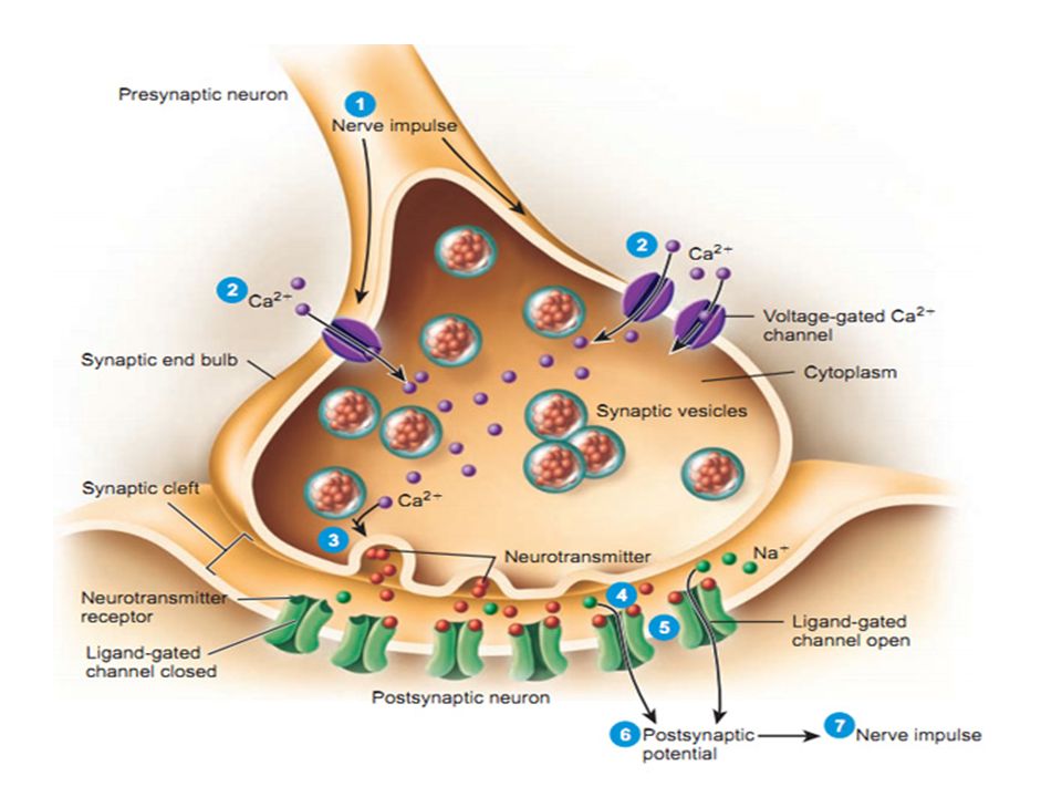
1) Release of neurotransmitter into synaptic cleft. 2) Action of the neurotransmitter on postsynaptic membrane. 3) Termination of synaptic transmission. It involves 3 steps:
 Action of the neurotransmitter on postsynaptic membrane. 3) Termination of synaptic transmission. It involves 3 steps:.")
16
It involves the following steps: During rest, both the pre. and postsynaptic membrane have R.M.P is about -70 mV. Stim. of presynaptic neuron → generation of AP → AP reaches the synaptic knob→ transient opening of the VGCa 2+ channels Ca 2+ influx → Ca 2+ causes the vesicles to fuse with the knob membrane at active zones vesicles release the transmitter by exocytosis in cleft

17
The number of vesicles ruptured depends upon the concentration of Ca 2+ within the active zone of the knob. The empty vesicles then separated from the knob membrane and mobilized back and fuse with the smooth E.R.

20
Post-synaptic receptors are 2 types; Ligand-gated ion channels When neurotransmitter bind to these receptors → produces structural changes →↑ the channels permeability to ions 2 types; cation and anion channels G-protein coupled receptors These receptors bind to G-protein (attached to GTP and GDP) G-protein consists of 3 components; α; β, and γ
 G-protein consists of 3 components; α; β, and γ")
22
Cation Channels Anion Channels Its transmitter excitatory transmitterinhibitory transmitter StructureTheir inner walls are lined with -ve charges →allow the passage of Na, K and Ca and not allow Cl They have small bore, so not allow the passage of Na, K, and Ca because their hydrated forms have large sizes ActionWhen these channels open Na influx, K efflux occur, but the total influx of Na is greater than K efflux because the driving force for Na influx is greater than K efflux (about 7.5 times) →depolarization of the postsynaptic membrane Opening of these channels as Cl channels→ ↑ influx of Cl → causing hyperpolarization of the membrane→ inhibits the postsynaptic neuron.
 →depolarization of the postsynaptic membrane Opening of these channels as Cl channels→ ↑ influx of Cl → causing hyperpolarization of the membrane→ inhibits the postsynaptic neuron.")
23
23 Biophysics, Abdelaziz Hussein + + - - + + - - + + + + + + + + + + - - + + + + + + + + - - + + __________ __________

25
25 Biophysics, Abdelaziz Hussein - - - - - - + + - - - - - - + + - - + + - - - - + + - - + + - -

28
Binding of transmitter to its receptor→ G- protein is activated (by replacement of its GDP with GTP) → separates the α component from the G-protein. The separated active α component can perform; 1.Opening specific ion channels e.g. 2 nd - messenger gated K channels 2.Activation of particular enzymes→ catalyze the formation of the 2 nd messengers, such as cyclic AMP, cyclic GMP, IP3,diacylglycerol (DAG) 3.Regulation of gene transcription→ control synthesis and production of certain proteins within the neuron
 3.Regulation of gene transcription→ control synthesis and production of certain proteins within the neuron.")
31
Postsynaptic potentials (PSPs) are 2 major types: 1.EPSPs 2.IPSPs
 are 2 major types: 1.EPSPs 2.IPSPs")
32
1)Def. It is a state of partial depolarization which occurs in the postsynaptic membrane due to single presynaptic impulse

33
2)Mechanism: When the excitatory chemical transmitter bind to and open ligand-gated cation channels→ allow much Na influx than K efflux→ depolarization of postsynaptic membrane → brought the postsynaptic membrane potential close to the threshold for excitation → called EPSP.
Mechanism: When the excitatory chemical transmitter bind to and open ligand-gated cation channels→ allow much Na influx than K efflux→ depolarization of postsynaptic membrane → brought the postsynaptic membrane potential close to the threshold for excitation → called EPSP.")
35
3) Characters or properties : 1.Partial depolarization not reach firing level 2.Increases the excitability as it carries the membrane potential to FL 3.Localized i.e. not spread. 4.Short duration i.e. decays within 15 m.sec. 5.Its amplitude (very small) about 0.5 mv To produce action potential must be summated. The summation is of 2 types: spatial and temporal summation
 about 0.5 mv To produce action potential must be summated. The summation is of 2 types: spatial and temporal summation.")
37
Summation : A depolarization of at least 10 to 20 mV is required to reach the usual threshold, so EPSPs must be summated a) Spatial: By stimulation of several presynaptic fibers that converge on the post synaptic neuron at the same time. b) Temporal: By stimulation of a single presynaptic neuron repetitively (successively) within very short duration (less than 15 m.sec).
 Temporal: By stimulation of a single presynaptic neuron repetitively (successively) within very short duration (less than 15 m.sec)..")
39
When the summated EPSPs reach the firing level, This will generate an action potential at the initial segment of the axon (axon hillock).
.")
40
During rest: some Ca 2+ enters the synaptic knob release of few vesicles of neurotransmitter → weak depolarization of postsynaptic membrane → miniature EPSP

41
1)Def. It is a state of partial hyperpolarization which occurs in the postsynaptic membrane due to single presynaptic impulse

42
2)Mechanism: When the inhibitory chemical transmitter bind to and open ligand-gated anion channels→ allow much Cl influx → hyperpolarization of postsynaptic membrane → brought the postsynaptic membrane potential away from the threshold for excitation → called IPSP.
Mechanism: When the inhibitory chemical transmitter bind to and open ligand-gated anion channels→ allow much Cl influx → hyperpolarization of postsynaptic membrane → brought the postsynaptic membrane potential away from the threshold for excitation → called IPSP.")
44
3) Characters or properties : 1.Partial hyperpolarization not reach firing level 2.Decreases the excitability as it carries the membrane potential to FL 3.Localized i.e. not spread. 4.Short duration i.e. decays within 15 m.sec. 5.Its amplitude (very small) about 0.5 mv To produce action potential must be summated. The summation is of 2 types: spatial and temporal summation
 about 0.5 mv To produce action potential must be summated. The summation is of 2 types: spatial and temporal summation.")
46
During rest: some Ca 2+ enters the synaptic knob release of few vesicles of neurotransmitter → weak hyperpolarization of postsynaptic membrane → miniature IPSP

47
A Postsynaptic neuron may receive many presynaptic terminals from several hundreds of neurons, some of these terminals are excitatory and the others are inhibitory. So, both EPSP & IPSP are produced and the effect on the postsynaptic membrane depends upon the net ability of summated postsynaptic potential to drive the membrane either towards or away from threshold level

51
It occurs when the transmitter is removed from the synaptic cleft by one or more of the following ways: i) Active reuptake i) Active reuptake of the transmitter into the presynaptic terminal. ii) Enzymatic degeneration ii) Enzymatic degeneration e.g.: hydrolysis or oxidation….etc. iii) Diffusion iii) Diffusion to the interstitial fluid.
 Enzymatic degeneration ii) Enzymatic degeneration e.g.: hydrolysis or oxidation….etc. iii) Diffusion iii) Diffusion to the interstitial fluid..")
52
ANS, Abdelaziz Hussein 52 ENZYME NT HYDROLYSIS DIFFUSION AND URINE EXCRETIO N Neuronal Uptake

53
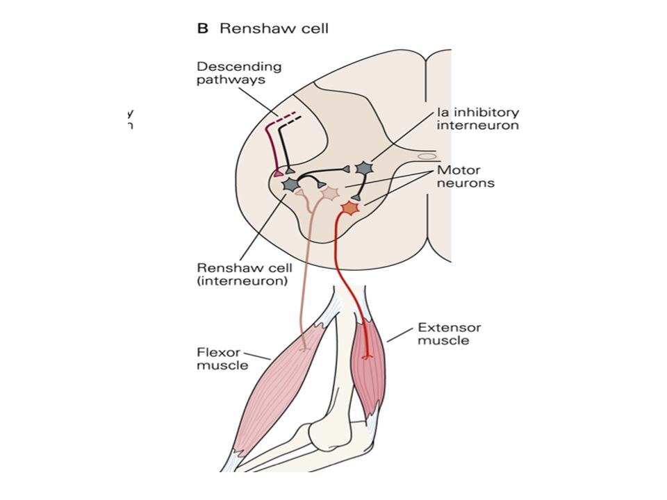
Types : 3 types Presynaptic Occurs when excitatory synapse is inhibited by inhibitory interneuron Postsynaptic Occurs when the pre-synaptic terminal release an inhibitory transmitter Recurrent Occurs by recurrent inhibitory interneuron circuits

54
C A B

55
Postsynaptic inhibition

56
Presynaptic inhibition Mediated by inhib. interneurons → end on excitatory pre-synaptic terminals (Axo-axonic Synapses) These inhibitory interneurons, thus can modify the rate of release of neurotransmitter
 These inhibitory interneurons, thus can modify the rate of release of neurotransmitter.")
57
Presynaptic inhibition Mechanism: GABA opens the Cl - channels in the excitatory presynatpic terminal increase Cl - influx leading to decrease in the amplitude of the action potentials arriving at the knob decrease Ca 2+ influx decrease the amount of neurotransmitter released decrease in post-synaptic response ( EPSP)
")
58
Presynaptic inhibition Significance: It occurs in many sensory and motor pathways in nervous system. It is involved in: 1.Process of lateral inhibition → strongly stimulated neurons inhibit the less stimulated surrounding neurons. 2.Pain control system→ plays a role in blocking the transmission of pain signals at the level of SGR cells.

59
Lateral Inhibition

60
Pain Control system

61
Recurrent inhibition

64
Synapse allow conduction of the impulses in one direction only; from the presynaptic to the postsynaptic neurons → Bell- Magendie law. This is because transmitter is present in the presynaptic neuron not the postsynaptic neuron

65
Def., It is the time that passes between arrival of an action potential to the synaptic knob and the occurrence of response in the postsynaptic neuron Time: 0.5 msec Cause: This represents the time required; 1.Release and diffusion of the neurotransmitter 2.Its binding with its postsynaptic receptors and generation and summation of EPSPs. Signficance: calculate number of synapses in reflex action

66
Def., Rapid and intense stimulation of synapse→ progressive decline in synaptic transmission and the synapse may in severe conditions, stop functioning

67
Ca ions...........Ach................. Rate of release Rate of reuptake

68
Cause: It results mainly from depletion of the neurotransmitter stores the in the synaptic knobs→ because in intensive stimulation the resynthesis and reuptake mechanisms that fill these stores are unable to provide all the demands for the transmitter Significance Protective mechanism against excessive neuronal mechanism e.g. in epileptic fit

69
Def., Rapid stimulation of presynaptic terminals for a period of time, the synapse becomes more responsive to subsequent stimulation than normal for a period of seconds or even minutes

70
Cause: Due to ↑ of the Ca ions concentration in the synaptic knob (due to weak Ca pump) → more and more vesicles to release their transmitter→ greater response of the postsynaptic neuron Significance: May have a role in the memory processes
 → more and more vesicles to release their transmitter→ greater response of the postsynaptic neuron Significance: May have a role in the memory processes")
71
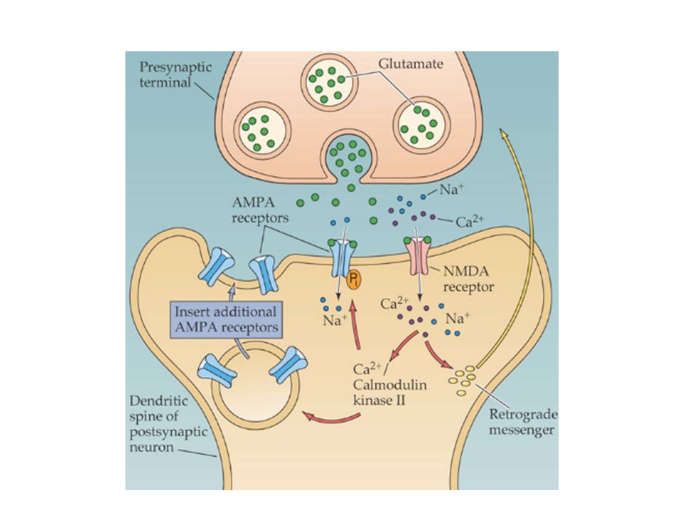
Def., Brief but repetitive, stimulation of presynaptic terminals results in long-lasting enhancement of synaptic transmission (lasts for several days). Cause: Caused mainly by an↑ of the intracellular Ca concentration in the postsynaptic neuron rather than the presynaptic knob produced by activation and opening of certain Ca channels in the postsynaptic membrane

74
Significance: Occurs in many parts of the CNS Has a special significance in the hippocampus which has an important role in learning and memory

75
-Normal function of the neuron is highly dependent upon an adequate O 2 supply - Marked hypoxia for a very short period, (a few seconds), causes loss of excitability of many neurons and stop of synaptic transmission. - When the blood supply to the brain is markedly reduced→ coma occurs within less than 7 seconds.

76
Alkalosis→ ↑es excitability of neurons→ at pH 7.8 to 8.0 severe convulsions occurs. Acidosis→ ↓es excitability of neurons→ at pH around 7.0 coma occurs. This latter condition is always seen in severe uremic or diabetic acidosis.

77
i)Caffeine and theophylline→↑ neuronal excitability by ↓ing the threshold for excitation of the postsynaptic neurons. ii)Strychnine →↑ neuronal excitability by interfering with the action of glycine (an inhibitory neurotransmitter) on the neurons. iii)Anesthetics and hypnotics →↑ threshold for excitation of the neurons→↓ synaptic transmission.
Strychnine →↑ neuronal excitability by interfering with the action of glycine (an inhibitory neurotransmitter) on the neurons. iii)Anesthetics and hypnotics →↑ threshold for excitation of the neurons→↓ synaptic transmission..")
79
Neurotransmitters of the C.N.S. classified into 2 groups: 1) Small- molecular weight neurotransmitters: are rapidly acting 2) Large- molecular weight neurotransmitters: are slowly acting
 Small- molecular weight neurotransmitters: are rapidly acting 2) Large- molecular weight neurotransmitters: are slowly acting.")
81
They are classified into 4groups: Class (I): Acetyl choline Class II): Biogenic Amines e.g. Noradernaline, adrenaline, Serotonin, Histamine & dopamine Class (III): Amino acids neurotransmitters e.g. Glutamate & Asparate (excitatory) GABA & Glycine (inhibitory) Class (IV): Nitric Oxide (NO)
: Amino acids neurotransmitters e.g. Glutamate & Asparate (excitatory) GABA & Glycine (inhibitory) Class (IV): Nitric Oxide (NO).")
82
They include : 1. Substance-P: neuropeptide for pain transmission. 2. Opioid peptides: e.g.: (Enkephalins & Endorphins). Neuropeptides prevent pain transmission. 3. Some neuropeptides coexist with Acetyl choline as V.I.P and with Noradrenalin as neuropeptide Y (N P Y).
. Neuropeptides prevent pain transmission. 3. Some neuropeptides coexist with Acetyl choline as V.I.P and with Noradrenalin as neuropeptide Y (N P Y)..")
83
Small M.W. neurotransmittersLarge M.W. neurotransmitters Rapidly actingSlowly acting Most of them are synthesized and stored at presynaptic terminals They are synthesized and stored in the soma of the presynaptic neuron and then transported along axons into the presynaptic terminals Synaptic vesicles contain neuropeptides co-exist with synaptic vesicles containing neurotransmitters and both are released simultaneously by the action potential that reaches the synaptic knob. Important for fast responses e.g.: 1.Transmission of sensory information to C.N.S. 2.Transmission of motor impulses from C.N.S to ms. Neuropeptides can prolong the actions of low M.W. transmitters at the post- synaptic neurons.

84
Small MW. Neurotransmitter

85
Large MW. Neurotransmitter

86
Small M.W. neurotransmittersLarge M.W. neurotransmitters Rapidly actingSlowly acting Most of them are synthesized and stored at presynaptic terminals They are synthesized and stored in the soma of the presynaptic neuron and then transported along axons into the presynaptic terminals Synaptic vesicles contain neuropeptides co-exist with synaptic vesicles containing neurotransmitters and both are released simultaneously by the action potential that reaches the synaptic knob. Important for fast responses e.g.: 1.Transmission of sensory information to C.N.S. 2.Transmission of motor impulses from C.N.S to ms. Neuropeptides can prolong the actions of low M.W. transmitters at the post- synaptic neurons.

87
THANKS

Similar presentations


 Nervous system functions Structure of a neuron Sensory, motor, inter- neurons Membrane potential Sodium.>")


