Download presentation
Presentation is loading. Please wait.

1
Eye anatomy

2
Conjunctiva

3
Conjunctiva Covers the inner surface of the eyelids and the anterior surface of the eye. Membrane which produces mucous that lubricates the eye and prevents dryness. Protects the eye.

4
Fibrous Tunic

5
Fibrous Tunic Cornea Functions: Transparent window for light entry
Refracts light Sclera Functions: Protects eye Shapes eye Anchors eye muscles

6
Vascular Tunic

7
Vascular Tunic Choroid Functions:
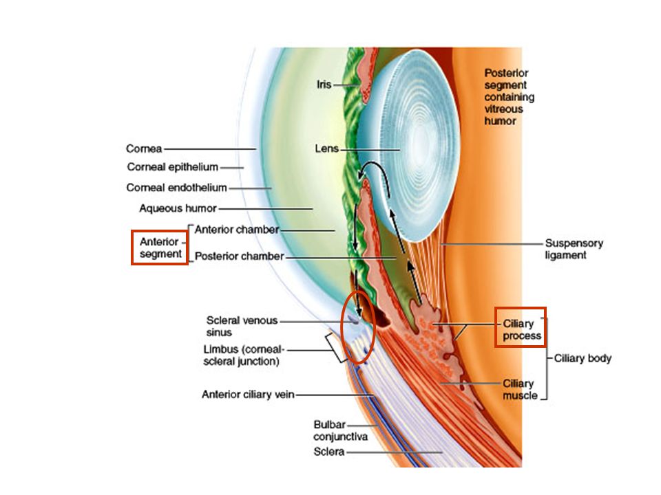
Provides nutrients to all eye tunics. Absorbs light preventing reflecting & scattering of light within the eye. Ciliary Body Functions: Ciliary processes secrete aqueous humor. Suspensory ligaments hold lens in place. Ciliary muscles pull on the ligaments to change the thickness of the lens. Iris Functions: Constricts or dilates to adjust the amount of light entering the eye.

8
Vascular Tunic Ciliary Muscles Ciliary Processes

10
Aqueous Humor Helps support the eye internally due to the intraocular pressure it produces inside the eye. Supplies nutrients & oxygen to the cornea, lens and portions of the retina. Carries away metabolic wastes from the cornea, lens and portions of the retina.

11
The iris constricts or dilates to adjust size of the pupil.
The pupil allows light to enter the posterior segment of the eye.

13
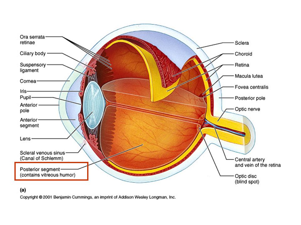
Vitreous Humor Transmits light within the posterior segment.
Supports the lens posteriorly. Holds the retina in place. Contributes to intraocular pressure.

14
Sensory Tunic

15
Retina Pigmented Layer Neural Layer Absorbs light
Carries out phagocytosis Stores Vitamin A Neural Layer Contains photoreceptors (rods and cones) for visual perception Contains bipolar cells & ganglion cells for visual impulse transmission
 for visual perception. Contains bipolar cells & ganglion cells for visual impulse transmission.")
16
Retina Fovea Centralis Contains only closely packed cones
Provides acute color vision in bright light Macula Lutea Contains more widely spaced cones Other areas of Retina Contain only rods Provide night, dim light & peripheral vision Shades of grey only Optic Disc Contains no receptors Blind spot

17
Retina Optic Disc

18
Photoreceptors

19
Cones Are located in macula lutea but are most highly concentrated in the fovea centralis. Are sensitive to bright light (daylight) situations in which light is very intense. Each cone synapses with a single bipolar cell which synapses with a single ganglion cell. The axons of ganglion cells form the optic nerve to conduct visual images to the brain. Provide acute (sharp) color images (vision).
 situations in which light is very intense. Each cone synapses with a single bipolar cell which synapses with a single ganglion cell. The axons of ganglion cells form the optic nerve to conduct visual images to the brain. Provide acute (sharp) color images (vision).")
20
Cones

21
Photoreceptors

22
Rods Most highly concentrated in the retina outside the macula lutea
Many rods synapse with a single bipolar cell Many bipolar cells may synapse with a single ganglion cell which carries stimuli to brain More sensitive & function only in dim light, night and peripheral vision Images are blurry and only in shades of gray

23
Visual Pigments Composed of two components
Retinal - light absorbing molecule (made from Vitamin A) Opsin (four types made from protein) Opsin combined with retinal = visual pigment OPSIN + RETINAL = Visual Pigment Depending on the type of opsin retinal is bound to, each of the four pigments will only absorb certain wavelengths of light.
 Opsin (four types made from protein) Opsin combined with retinal = visual pigment. OPSIN. + RETINAL. = Visual. Pigment. Depending on the type of opsin retinal is bound to, each of the four pigments will only absorb certain wavelengths of light.")
24
Visual Pigments: RODS Retinal + Opsin = Rhodopsin (visual purple)
Absorbs light throughout entire visible light spectrum (most sensitive to green) Functions only in dark, dim light & peripheral vision Light causes Retinal to change shape & separate from opsin causing nerve impulse Regenerate only in dark or dim light situations (Light) Impulse OPSIN RETINAL RHO DOPSIN
 Functions only in dark, dim light & peripheral vision. Light causes Retinal to change shape & separate from opsin causing nerve impulse. Regenerate only in dark or dim light situations. (Light) Impulse. OPSIN. RETINAL. RHO. DOPSIN.")
25
Visual Pigments: Cones
Retinal + Red, Green or Blue Opsin = Red, Green or Blue visual pigments Each Opsin absorbs light only in the area of the visible light spectrum it is sensitive to, ie, red cones, green cones & blue cones Function only in bright light (daylight) Provide sharp color images (Light) Impulse Red Opsin RETINAL Red Cone Impulse Green Opsin RETINAL Green Cone
 Provide sharp color images. (Light) Impulse. Red Opsin. RETINAL. Red. Cone. Impulse. Green Opsin. RETINAL. Green. Cone.")
26
Lens Refracts (bends) light
Focuses precise image on the retina (fovea) through accommodation (changing thickness)
 through accommodation (changing thickness)")
Similar presentations


 1. Cornea 2. Sclera Middle Tunic (pg. 470-474) 3. Choroid Coat 4. Ciliary Body 5. Lens & Accommodation 6. Aqueous.>")


 separated by the palpebral fissue Eyelashes Tarsal glands Lacrimal apparatus Vision Accessory structures.>")


