Download presentation
Presentation is loading. Please wait.

1
Eyes & Vision

3
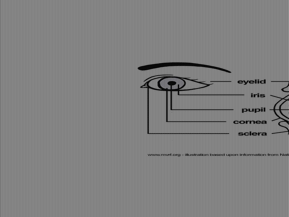
Outermost layer of the Eye Cornea – ‘window’ – bulges slightly outward, allows light to enter – only truly transparent portion. Absence of blood vessels, abundant pain receptors Sclera – ‘whites of eyes’ – helps with shape of eye. Attachment of extrinsic muscles

5
Middle layer of the Eye Choroid – contains blood vessels, and melanin. Melanin helps absorb light, reducing amount that reflects within eye, increasing visual sharpness Ciliary Bodies – ring of muscle tissue that holds lens in place, & functions in shaping lens for focusing Iris – pigmented area – regulates amount of light thru pupil via smooth muscle fibers

7
Innermost layer of the Eye Retina – contains about ¼ billion receptors [ 70% of all sensory receptors in body] primary cells of retina – Photoreceptors: rods & cones 125 million rods: 6 million cones [fovea] red, blue & green
![Innermost layer of the Eye Retina – contains about ¼ billion receptors [ 70% of all sensory receptors in body] primary cells of retina – Photoreceptors: rods & cones 125 million rods: 6 million cones [fovea] red, blue & green](http://images.slideplayer.com/16/5031243/slides/slide_7.jpg "Innermost layer of the Eye Retina – contains about ¼ billion receptors [ 70% of all sensory receptors in body] primary cells of retina – Photoreceptors: rods & cones 125 million rods: 6 million cones [fovea] red, blue & green")
8
Fovea The center of the macula; gives the sharpest vision. When we fixate or look directly at an object it is imaged on the fovea.

9
cells of retina Photoreceptors: contain retinal [ pigment] bound to a protein called opsin – 4 types of opsin, retinal will absorb a different color wavelength depending on the opsin its bound to Rods – rhodopsin Cones – red, blue, green – colors overlap and brain interprets color depending on strength of cone’s stimulation
![cells of retina Photoreceptors: contain retinal [ pigment] bound to a protein called opsin – 4 types of opsin, retinal will absorb a different color wavelength depending on the opsin its bound to Rods – rhodopsin Cones – red, blue, green – colors overlap and brain interprets color depending on strength of cone’s stimulation](http://images.slideplayer.com/16/5031243/slides/slide_9.jpg "cells of retina Photoreceptors: contain retinal [ pigment] bound to a protein called opsin – 4 types of opsin, retinal will absorb a different color wavelength depending on the opsin its bound to Rods – rhodopsin Cones – red, blue, green – colors overlap and brain interprets color depending on strength of cone’s stimulation")
10
cells of retina Bipolar cells – approx. 6 million – begin to process info received by photoreceptors Between bipolar cells and photoreceptors are horizontal cells & between bipolar cells and ganglion cells are amacrine cells – which relay or inhibit info laterally across retina and between cells Ganglion cells –axons form optic nerve

11
Optic Nerve [ CN II] The bundle of over one million nerve fibers that carries visual messages from the retina to the brain.
![Optic Nerve [ CN II] The bundle of over one million nerve fibers that carries visual messages from the retina to the brain.](http://images.slideplayer.com/16/5031243/slides/slide_11.jpg "Optic Nerve [ CN II] The bundle of over one million nerve fibers that carries visual messages from the retina to the brain.")
12
chambers Anterior chamber: between cornea & lens Contains aqueous humor –produced by capillaries of ciliary bodies, exits via Canal of Schlemm, replaced every 90min. creates pressure, maintains shape, nutrients/wastes. Posterior chamber: between lens & retina Contains vitreous humor – born with. Maintains shape of eye

14
Normal vision: Emmetropia Emmetropia (“normal” vision). What is referred to as “normal” vision, or emmetropia, happens when light rays focus precisely on the retina

17
Myopia

18
Hyperopia

19
Astigmatism (“ghost” vision). Astigmatic people see with double or “ghost” vision. Both far and near objects appear out of focus. This is because of the uneven diameter of the cornea. (Oblong-shaped, for instance.) For light rays to focus precisely on the retina, the cornea usually needs to be more evenly round.
 For light rays to focus precisely on the retina, the cornea usually needs to be more evenly round..")
20
presbyopia Presbyopia (reduced focus- adjusting ability). Presbyopic people have a reduced focus-adjusting ability. This results from a loss of elasticity in the eye's lens, often as part of the aging process.

21
"Isn't it creepy how they seem to follow you around the room?"Isn't it creepy how they seem to follow you around the room?"

Similar presentations


 1. Cornea 2. Sclera Middle Tunic (pg. 470-474) 3. Choroid Coat 4. Ciliary Body 5. Lens & Accommodation 6. Aqueous.>")

: the two.>")


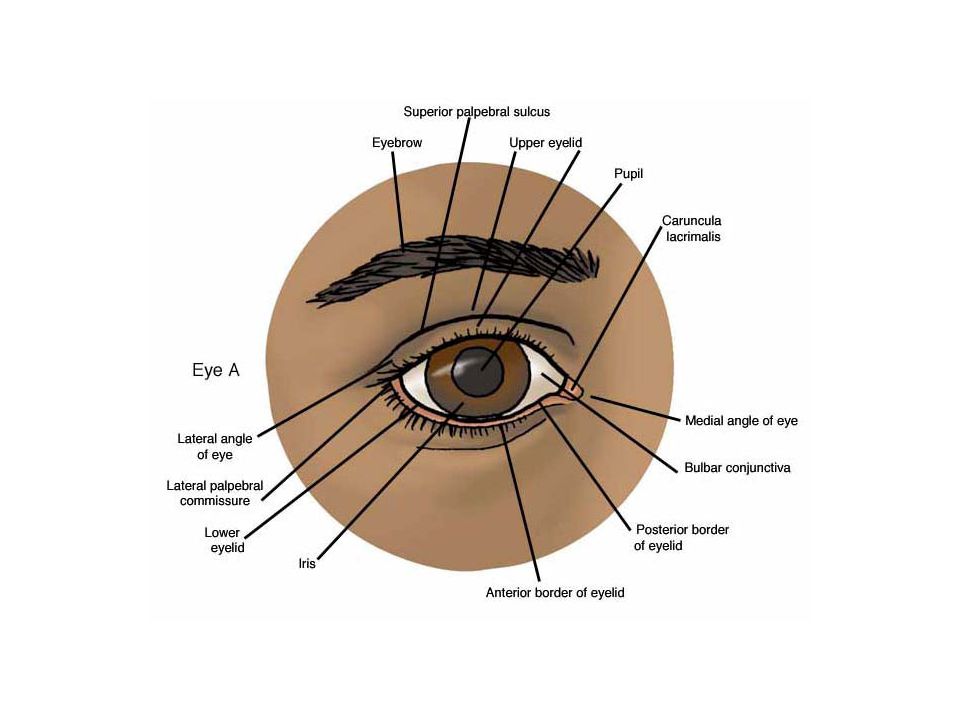
 separated by the palpebral fissue Eyelashes Tarsal glands Lacrimal apparatus Vision Accessory structures.>")


 Outer Layers of The Eye 3 layers surrounding inner fluid:3 layers surrounding inner fluid: –Outermost = Sclera.>")

