Download presentation
Presentation is loading. Please wait.

1
Forth Lecture: Development of Bone Dr. Wahda Kharofa

2
Objectives: To give information about: Bone development. What is intramembranous ossification? What is endochondral?

3
Development of bone (Ossification): The process of ossification in embryo start in the early embryonic stages and continuous several years after puberty (20-21) years of age. There are two types of ossification:

4
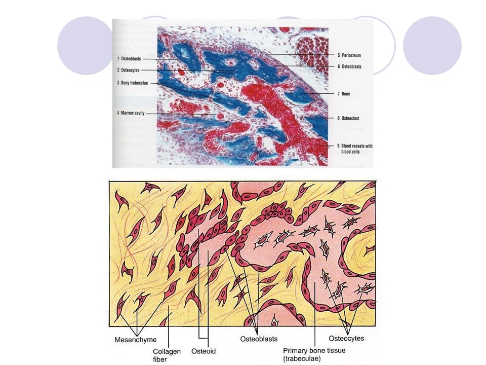
Intramembranous ossification: This type occurs in flat bones of face and skull and clavicle, it is responsible for the thickening of the flat bone. It is called “Intramembranous ossification” because takes place within membranes of connective tissue which is called “Mesenchymal connective tissue” which contains branched mesenchymal cell + collagen fibers + matrix.

5
The following steps occur during this development: 1- The starting point of ossification called “primary ossification” center in mesenchymal connective tissue which becomes highly vascular and groups of mesenchymal cells divided and enlarged in size and becomes polygonal in shape, some of them differentiated into osteogenic cells which gives rise into osteoblasts,then some of them begin to secret bony matrix, later the matrix becomes calcified by the activity of the osteoblasts,so the cells imprisoned in the matrix inside lacunae and canaliculi forming osteocytes, the remaining osteoblasts located at the periphery forming trabeculae.

6
2. Gradually, the trabeculae increases in size and separated by a space in which filled with mesenchymal tissue is specialized and changed into myloid tissue (bone marrow), so spongy bone is formed. 3. The outer part of mesenchymal connective tissue does not undergo ossification, so it forms the periosteum and the same thing happen in the lining form the endosteum. 4. After birth the outer part of spongy bone is change into compact bone by osteoblasts activity which add more successive layers of bone and osteocytes arrange themselves in lamellae surrounding blood vessels.
, so spongy bone is formed. 3. The outer part of mesenchymal connective tissue does not undergo ossification, so it forms the periosteum and the same thing happen in the lining form the endosteum. 4. After birth the outer part of spongy bone is change into compact bone by osteoblasts activity which add more successive layers of bone and osteocytes arrange themselves in lamellae surrounding blood vessels..")
8
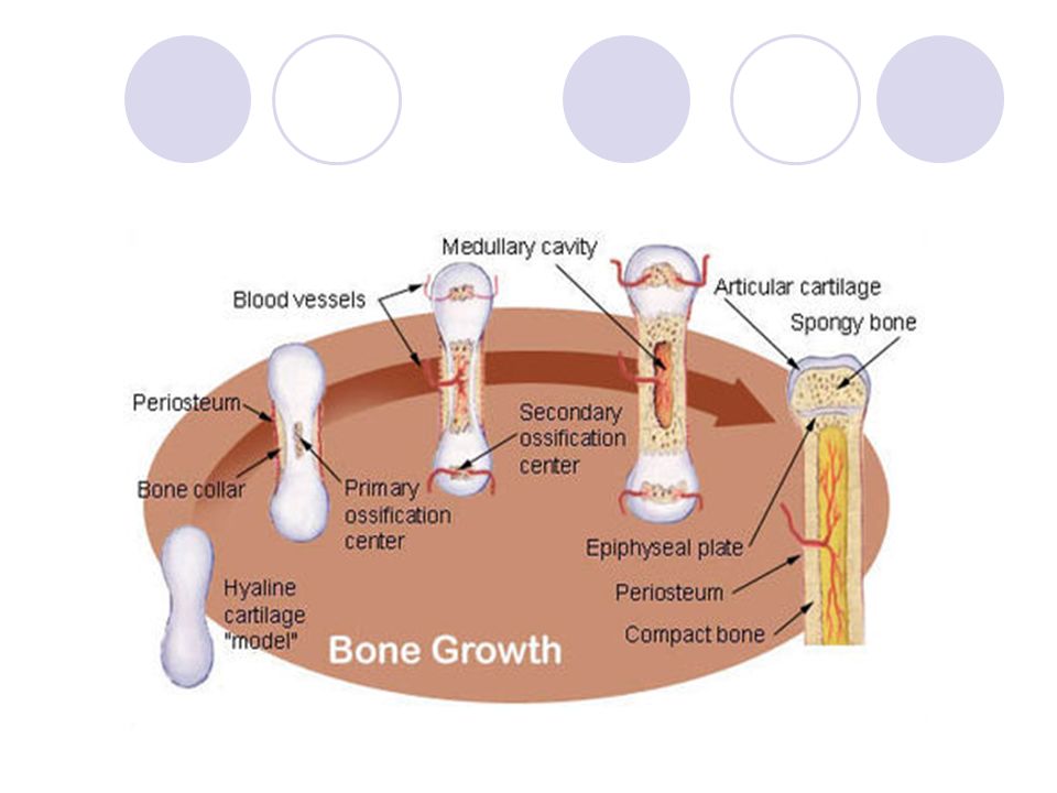
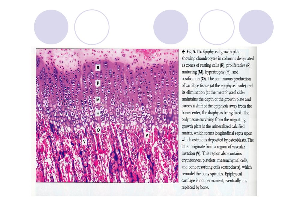
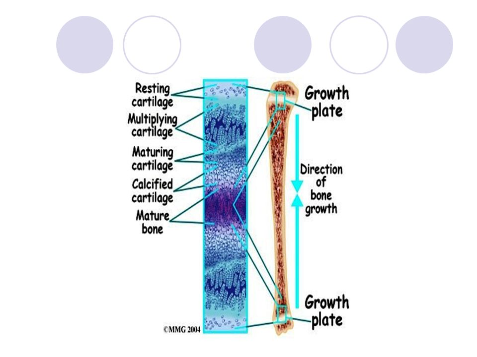
Intracartilaginous (Endochondral ossification): It takes place Intracartilaginous (Endochondral ossification): It takes place over a piece of hyaline cartilage whose shape is similar to a bone which it formed.This type of ossification responsible for the formation of short and long bones. In the shaft of the long the primary and secondary ossification centers occur. Endochondral bone formation divided into: 1. Reserve (Resting) zone: Chondrocytes here randomly arranged. 2.Proliferation (Multiplication) zone: This is an active zone showing numerous mitosises of chondrocytes divided and produce daughter cells which arrange themselves in columns parallel with the long axis of the cartilage. Each raw consists of number of cells which are crowded flattened and separated by little matrix. 3. Hypertrophy (Maturation) zone: The chondrocytes and the lacunae are enlarged the cytoplasm of the cells accumulated the cartilage matrix become reduced. 4. Calcification zone: The matrix in this zone is surrounding the enlarge lacunae. The perichonderium which surrounds the center of diaphysis becomes richly vascularized then the chondrogenic cells activated and changed into osteogenic cells, so the perichonderium called “Periosteum”. These osteoblasts start to deposit bony matrix around the middle portion of the shaft called “l bone collar” which is responsible for the thickening of the bone.
 zone: Chondrocytes here randomly arranged. 2.Proliferation (Multiplication) zone: This is an active zone showing numerous mitosises of chondrocytes divided and produce daughter cells which arrange themselves in columns parallel with the long axis of the cartilage. Each raw consists of number of cells which are crowded flattened and separated by little matrix. 3. Hypertrophy (Maturation) zone: The chondrocytes and the lacunae are enlarged the cytoplasm of the cells accumulated the cartilage matrix become reduced. 4. Calcification zone: The matrix in this zone is surrounding the enlarge lacunae. The perichonderium which surrounds the center of diaphysis becomes richly vascularized then the chondrogenic cells activated and changed into osteogenic cells, so the perichonderium called Periosteum . These osteoblasts start to deposit bony matrix around the middle portion of the shaft called l bone collar which is responsible for the thickening of the bone..")
9
5. Invasion zone: Osteoclasts in the bone collar, mesenchymal cells with blood vessels enter to the cavities (periosteal buds). These mesenchymal cells divided and some of them form a continuous layer over the calcified cartilage matrix. 6. Ossification zone: The mesenchymal cells which surrounding the calcified cartilage matrix into osteogenic cells which changed into osteoblasts. These osteoblasts start to secret bony matrix and become osteocytes, so the trabeculae formed having at the center. Calcified cartilage matrixes covered by a bony matrix, osteoclasts are active and here start to absorb calcified cartilage matrix which replaced by bone matrix the trabeculae are formed. The mesenchymal cells which lie between these trabeculae form the bone marrow cells, so spongy bone is formed.
. These mesenchymal cells divided and some of them form a continuous layer over the calcified cartilage matrix. 6. Ossification zone: The mesenchymal cells which surrounding the calcified cartilage matrix into osteogenic cells which changed into osteoblasts. These osteoblasts start to secret bony matrix and become osteocytes, so the trabeculae formed having at the center. Calcified cartilage matrixes covered by a bony matrix, osteoclasts are active and here start to absorb calcified cartilage matrix which replaced by bone matrix the trabeculae are formed. The mesenchymal cells which lie between these trabeculae form the bone marrow cells, so spongy bone is formed..")
10
7. Resorption zone: The marrow cavity increase in size owing to resorption of bone in the center of the diaphysis by the activity of osteoclasts.At about the time of birth the secondary ossification centers appear in each end of long bones (epiphysises).The cartilage in these centers passes through the same changes as happen in diaphysis..The epiphysis remain separated from diaphysis by a plate of cartilage called epiphyseal plate which cause longitudinal growth of the bone.The chondrocytes in this plate divided and gradually changed into bone in the same way of primary center and the development in this plate occur in both faces(epiphyseal and diphyseal) and no further longitudinal growth happen when growth of this plate stoped and is completely replaced by bone tissue at 20-21 years old. Most of spongy bone in the diaphysis and the outer part of epiphysis changed into compact bone by the activity of osteoblasts which add more successive layers of bone and the osteaocytes arrange themselves into lamellae surrounding blood vessel.
.The cartilage in these centers passes through the same changes as happen in diaphysis..The epiphysis remain separated from diaphysis by a plate of cartilage called epiphyseal plate which cause longitudinal growth of the bone.The chondrocytes in this plate divided and gradually changed into bone in the same way of primary center and the development in this plate occur in both faces(epiphyseal and diphyseal) and no further longitudinal growth happen when growth of this plate stoped and is completely replaced by bone tissue at years old. Most of spongy bone in the diaphysis and the outer part of epiphysis changed into compact bone by the activity of osteoblasts which add more successive layers of bone and the osteaocytes arrange themselves into lamellae surrounding blood vessel..")
Similar presentations