Download presentation
Presentation is loading. Please wait.

1
Arterial Blood Gas Interpretation
Dr. Kapila Hettiarachchi

2
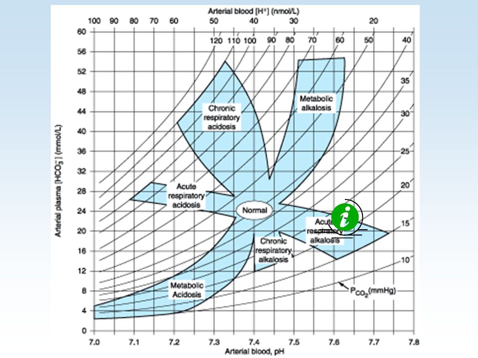
pH is inversely related to [H+]; a pH change of 1
pH is inversely related to [H+]; a pH change of 1.00 represents a 10-fold change in [H+] pH [H+] in nanomoles/L
![pH is inversely related to [H+]; a pH change of 1](http://slideplayer.com/slide/9409551/28/images/2/pH+is+inversely+related+to+%5BH%2B%5D%3B+a+pH+change+of+1.jpg "pH is inversely related to [H+]; a pH change of 1.00 represents a 10-fold change in [H+] pH [H+] in nanomoles/L")
3
1st Defence

4
Know the normal values first

5
Alkalemia: blood pH > 7.45
Respiratory alkalosis from acute hyperventilation. Metabolic alkalosis from excessive diuretic therapy

6
Acidemia: blood pH < 7.35
Metabolic acidosis from decreased perfusion (lactic acidosis) Respiratory acidosis from hypoventilation.
 Respiratory acidosis from hypoventilation.")
7
RESPIRATORY ALKALOSIS
↓PaCO2 & ↑ pH Hypoxemia (includes altitude) Anxiety Sepsis Any acute pulmonary insult (e.g., pneumonia, mild asthma attack, early pulmonary edema, pulmonary embolism)
 Anxiety. Sepsis. Any acute pulmonary insult (e.g., pneumonia, mild asthma attack, early pulmonary edema, pulmonary embolism)")
8
Respiratory alkalosis

10
Expected changes in pH and HCO3- for a 10-mm Hg change in PaCO2 resulting from respiratory alkalosis: ACUTE pH ↑ by HCO3- ↓ by 2 * Units for HCO3- are mmol/L

11
Respiratory Acidosis Primary Event Compensatory Event
HCO Increased HCO3- Low pH ~ Low pH ~ High PaCO High PaCO2

12
RESPIRATORY ACIDOSIS ↑PaCO2 & ↓ pH
Central nervous system depression (e.g., drug overdose) Chest bellows dysfunction (e.g., Guillain-Barré syndrome, myasthenia gravis) Disease of lungs and/or upper airway (e.g., chronic obstructive lung disease, severe asthma attack, severe pulmonary edema)
 Chest bellows dysfunction. (e.g., Guillain-Barré syndrome, myasthenia gravis) Disease of lungs and/or upper airway. (e.g., chronic obstructive lung disease, severe asthma attack, severe pulmonary edema)")
13
Respiratory acidosis

14
Respiratory acidosis

15
Expected changes in pH and HCO3- for a 10-mm Hg change in PaCO2 resulting from respiratory acidosis
ACUTE pH ↓ by HCO3- ↑ by 1* * Units for HCO3- are mmol/L

16
Metabolic Alkalosis Primary Event Compensatory Event
High HCO High HCO3- High pH ~ High pH ~ PaCO Increased PaCO2

17
Metabolic alkalosis ↑ HCO3- & ↑ pH
Chloride responsive (responds to NaCl or KCl therapy): diuretics, corticosteroids, gastric suctioning, vomiting Chloride resistant: any hyperaldosterone state (e.g., Cushing’s syndrome, Bartter’s syndrome, severe K+ depletion)
: diuretics, corticosteroids, gastric suctioning, vomiting. Chloride resistant: any hyperaldosterone state (e.g., Cushing’s syndrome, Bartter’s syndrome, severe K+ depletion)")
18
Metabolic Alkalosis – How to detect compensation
1.) PaCO2 = [HCO3] + 15 2.) PaCO2 will ↑ 6 mmHg per 10-mmol/L ↑ in [HCO3]
 PaCO2 = [HCO3] ) PaCO2 will ↑ 6 mmHg per 10-mmol/L ↑ in [HCO3]")
19
Metabolic alkalosis

20
Metabolic alkalosis

21
Metabolic Acidosis Primary Event Compensatory Event
Low HCO Low HCO3- Low pH ~ Low pH ~ PaCO Decreased PaCO2

22
METABOLIC ACIDOSIS ↓HCO3- & ↓ pH - Increased anion gap
lactic acidosis; ketoacidosis; drug poisonings (e.g., aspirin, ethylene glycol, methanol) Normal anion gap Diarrhea; some kidney problems (e.g., renal tubular acidosis, interstitial nephritis)
 Normal anion gap. Diarrhea; some kidney problems (e.g., renal tubular acidosis, interstitial nephritis)")
23
Metabolic Acidosis – How to detect compensation
1.) PaCO2 = (1.5x HCO3) + 8 or 2.) PaCO2 will ↓ 1.25 mmHg per mmol/L ↓ in [HCO3]
 PaCO2 = (1.5x HCO3) + 8. or. 2.) PaCO2 will ↓ 1.25 mmHg per mmol/L ↓ in [HCO3]")
24
Metabolic acidosis

25
Metabolic acidosis Metabolic acidosis

26
Anion Gap Metabolic acidosis is conveniently divided into elevated and normal anion gap (AG) acidosis. AG is calculated as AG = Na+ - (Cl- + HCO3- ) Note: Normal AG is typically 12 ± 4 mEq/L.
 Note: Normal AG is typically 12 ± 4 mEq/L.")
27
Mixed Acid-base Disorders are Common
In chronically ill respiratory patients, mixed disorders are probably more common than single disorders, e.g., RAc + MAlk, RAc + Mac, Ralk + MAlk.

28
Mixed Acid-base Disorders are Common
In renal failure (and other conditions) combined MAlk + MAc is also encountered.
 combined MAlk + MAc is also encountered.")
29
For an increased anion gap metabolic acidosis, are there other derangements?
Determining the “corrected bicarbonate concentration”: Corrected HCO3 = measured HCO3 + (Anion Gap - 12) If the corrected HCO3 is less than normal (under 22mEq/L) then there is an additional metabolic acidosis present. Corrected HCO3 values over 26 mEq/L reflect a co-existing metabolic alkalosis. If it remains between 22-26mEq/L only existing Met. Acidosis is present
 If the corrected HCO3 is less than normal (under 22mEq/L) then there is an additional metabolic acidosis present. Corrected HCO3 values over 26 mEq/L reflect a co-existing metabolic alkalosis. If it remains between 22-26mEq/L only existing Met. Acidosis is present.")
30
Correction of severe acidosis
Give NaHCO3 only If PH is < 7.1 but not in respiratory acidosis

31
Correction of severe acidosis
NaHCO3 in mmol = Base deficit X Body weight 3 Give half of that Target is keeping PH > 7.2

32
if ABG not available NaHCO3 1 mmol per Kg will be enough

33
Tips to Diagnose Mixed Acid-base Disorders
TIP 1. Do not interpret any blood gas data for acid-base diagnosis without closely examining the serum electrolytes: Na+, K+, Cl-, and CO2. A serum CO2 out of the normal range always represents some type of acid-base disorder Note that serum CO2 may be normal in the presence of two or more acid-base disorders.

34
Tips to Diagnose Mixed Acid-base Disorders (cont.)
TIP 2. Single acid-base disorders do not lead to normal blood pH. Although pH can end up in the normal range ( ) with a single mild acid-base disorder, a truly normal pH with distinctly abnormal HCO3- and PaCO2 invariably suggests two or more primary disorders.
 with a single mild acid-base disorder, a truly normal pH with distinctly abnormal HCO3- and PaCO2 invariably suggests two or more primary disorders.")
35
Tips to Diagnose Mixed Acid-base Disorders (cont.)
Example: pH 7.40, PaCO2 20 mm Hg, HCO3- 12 mEq/L in a patient with sepsis.

36
Acid-base Disorders: Test Your Understanding
A child with severe asthma, arterial blood gas shows pH of 7.14 PaCO2 of 70 mm Hg HCO3- of 23 mEq/L How would you describe the likely acid-base disorder(s)?
")
37
Acid-base Disorders: Test Your Understanding - Answers
Acute elevation of PaCO2 leads to reduced pH, i.e., an acute respiratory acidosis. However, is the problem only acute respiratory acidosis or is there some additional process? For every 10-mm Hg rise in PaCO2 (before any renal compensation), pH falls about 0.07 units. Because this patient's pH is down 0.26, or 0.05 more than expected for a 30-mm Hg increase in PaCO2, there must be an additional metabolic problem. Also note that with acute CO2 retention of this degree, the HCO3- should be elevated 3 mEq/L. Thus a low-normal HCO3- with increased PaCO2 is another way to uncover an additional metabolic disorder. Decreased perfusion leading to mild lactic acidosis would explain the metabolic component.
, pH falls about 0.07 units. Because this patient s pH is down 0.26, or 0.05 more than expected for a 30-mm Hg increase in PaCO2, there must be an additional metabolic problem. Also note that with acute CO2 retention of this degree, the HCO3- should be elevated 3 mEq/L. Thus a low-normal HCO3- with increased PaCO2 is another way to uncover an additional metabolic disorder. Decreased perfusion leading to mild lactic acidosis would explain the metabolic component.")
38
Interpreting Oxygenation
What is ‘normal’ PaO2? 80-100mmHg is normal

39
Interpreting Oxygenation
Oxygenation gradually deteriorates during life Several calculations available for determining ‘normal’ based on patient age. PaO2 = (0.27 x age) i.e., 30 year old ~ 95 mmHg 60 year old ~ 88 mmHg
 i.e., 30 year old ~ 95 mmHg. 60 year old ~ 88 mmHg.")
40
Interpreting Oxygenation
P(A-a)O2 difference –Normal is 3-25mmHg Increased P(A-a) O2 - Shunt V/Q mismatch Diffusion defect
O2 difference –Normal is 3-25mmHg. Increased P(A-a) O2 - Shunt. V/Q mismatch. Diffusion defect.")
41
Interpreting Oxygenation
Oxygenation index- PaO2 /FiO2 Normal is 500 is moderate shunt/ V/Q abnormality <200 severe shunt / V/Q abnormality

42
PaCO2 Equation: PaCO2 reflects ratio of metabolic CO2 production to alveolar ventilation
VCO2 x VCO2 = CO2 production PaCO2 = VA = VE – VD VA VE = minute (total) ventilation (= resp. rate x tidal volume) VD = dead space ventilation (= resp. rate x dead space volume 0.863 converts VCO2 and VA units to mm Hg Condition State of PaCO2 in blood alveolar ventilation > 45 mm Hg Hypercapnia Hypoventilation mm Hg Eucapnia Normal ventilation < 35 mm Hg Hypocapnia Hyperventilation
 ventilation (= resp. rate x tidal volume) VD = dead space ventilation (= resp. rate x dead space volume converts VCO2 and VA units to mm Hg. Condition State of. PaCO2 in blood alveolar ventilation. > 45 mm Hg Hypercapnia Hypoventilation mm Hg Eucapnia Normal ventilation. < 35 mm Hg Hypocapnia Hyperventilation.")
43
Dangers of Hypercapnia
Besides indicating a serious derangement in the respiratory system, elevated PaCO2 poses a threat for three reasons: 1) An elevated PaCO2 will lower the PAO2 and as a result will lower the PaO2. 2) An elevated PaCO2 will lower the pH 3) The higher the baseline PaCO2, the greater it will rise for a given fall in alveolar ventilation,
 An elevated PaCO2 will lower the PAO2 and as a result will lower the PaO2. 2) An elevated PaCO2 will lower the pH. 3) The higher the baseline PaCO2, the greater it will rise for a given fall in alveolar ventilation,")
44
PCO2 vs. Alveolar Ventilation

45
?

46
Case 3 – Patient with Severe Abdominal Pain

47
Case 3 – Patient with Severe Abdominal Pain
An obese 70 year old man has diabetes of 25 years duration complicated by coronary artery disease (CABG x 4 vessels 10 years ago), cerebrovascular disease (carotid artery endarterectomy 3 years ago) and peripheral vascular disease (Aorto-bifem 2 years ago). [“VASCULOPATH”]
, cerebrovascular disease (carotid artery endarterectomy 3 years ago) and peripheral vascular disease (Aorto-bifem 2 years ago). [ VASCULOPATH ]")
48
Case 3 – Patient with Ischemic Bowel

49
Case 3 – Patient with Ischemic Bowel

50
Case 3 – Patient with Ischemic Bowel
ABGs obtained in the ICU pH PCO mmHg HCO mEq/L

51
Case 3 – Patient with Ischemic Bowel
ABGs obtained in the ICU pH PCO mmHg HCO mEq/L What is the primary disorder? What is the physiologic response to this disorder?

52
Case 3 – Patient with Ischemic Bowel
Step 1: Acidemic, alkalemic, or normal? ACIDEMIC

53
Case 3 – Patient with Ischemic Bowel
Step 2: Is the primary disturbance respiratory or metabolic? METABOLIC

54
Case 3 – Patient with Ischemic Bowel
Step 3: For a primary respiratory disturbance, is it acute or chronic? NOT APPLICABLE

55
Case 3 – Patient with Ischemic Bowel
Step 4: For a metabolic disturbance, is the respiratory system compensating OK? "Winter's formula": Expected PCO2 in metabolic acidosis = 1.5 x HCO (range: +/- 2) = 1.5 x = 18.5 pH PCO mm Hg HCO mEq / L
 = 1.5 x = pH PCO2 20 mm Hg. HCO3 7 mEq / L.")
56
Case 3 – Patient with Ischemic Bowel
Step 5: For a metabolic acidosis, is there an increased anion gap? FOR THIS STEP ONE MUST OBTAIN SERUM ELECTROLYTE DATA

57
Case 3 – Patient with Ischemic Bowel
SERUM ELECTROLYTE DATA Serum Na mEq/L Serum HCO mEq/L Serum Cl mEq/L

58
Anion Gap = Na – (Cl – HCO3)
= 135 – (98 -7) mEq/L = 30 mEq/L (ELEVATED) SERUM ELECTROLYTE DATA Serum Na mEq/L Serum HCO mEq/L Serum Cl 98 mEq/L
 mEq/L. = 30 mEq/L. (ELEVATED) SERUM ELECTROLYTE DATA. Serum Na 135 mEq/L. Serum HCO3 7 mEq/L. Serum Cl 98 mEq/L.")
59
Case 3 – Patient with Ischemic Bowel
Step 5: For a metabolic acidosis, is there an increased anion gap? ANSWER: YES

60
Case 3 – Patient with Ischemic Bowel
Step 6: For an increased anion gap metabolic acidosis, are there other derangements? To determine if there are other metabolic derangements present we start by determining the “corrected bicarbonate concentration”: Corrected HCO3 = measured HCO3 + (Anion Gap - 12) If the corrected HCO3 is less than normal (under 22mEq/L) then there is an additional metabolic acidosis present. Corrected HCO3 values over 26 mEq/L reflect a co-existing metabolic alkalosis.
 If the corrected HCO3 is less than normal (under 22mEq/L) then there is an additional metabolic acidosis present. Corrected HCO3 values over 26 mEq/L reflect a co-existing metabolic alkalosis.")
61
Case 3 – Patient with Ischemic Bowel
Corrected HCO3 = measured HCO3 + (Anion Gap - 12). Corrected HCO3 = 7 + ( ) = 25 REMEMBER If the corrected HCO3 is less than normal (under 22mEq/L) then there is an additional metabolic acidosis present. Corrected HCO3 values over 26 mEq/L reflect a co-existing metabolic alkalosis.
. Corrected HCO3 = 7 + ( ) = 25. REMEMBER. If the corrected HCO3 is less than normal (under 22mEq/L) then there is an additional metabolic acidosis present. Corrected HCO3 values over 26 mEq/L reflect a co-existing metabolic alkalosis.")
62
Case 3 – Patient with Ischemic Bowel
Step 6: For an increased anion gap metabolic acidosis, are there other derangements? ANSWER: NO OTHER DERANGEMENTS NOTED

63
Case 3 – Patient with Ischemic Bowel
ANSWER FROM “Primary metabolic acidosis, with increased anion gap, with full respiratory compensation”

64
Case 3 – Patient with Ischemic Bowel
BUT … What is the cause of the elevated anion-gap metabolic acidosis?

65
Case 3 – Patient with Ischemic Bowel
The most common etiologies of a metabolic acidosis with an increased anion gap are shown below: Lactic acidosis Ingestion of: (from poor perfusion) Ethylene glycol Starvation Methanol Renal failure Salicylate Ketoacidosis (as in diabetic ketoacidosis)
 Ethylene glycol Starvation Methanol Renal failure Salicylate Ketoacidosis (as in diabetic ketoacidosis)")
66
Notes on Lactic Acidosis
“Lactic acidosis is a disease characterized by a pH less than 7.25 and a plasma lactate greater than 5 mmol/L.”

67
Case 3 – Patient with Ischemic Bowel

68
Case 3 – Patient with Ischemic Bowel
By the time the patient is admitted to the ICU he looks absolutely terrible. He is moaning in agony, having received no pain medications at all. Vital signs in ICU BP 82/50 HR 112 RR 35 Temp Celsius O2 sat % Pain Score 10/10

69
Case 3 – Patient with Ischemic Bowel
Because of the extreme pain, the patient is given morphine 8 mg IV push, a somewhat generous dose. When reexamined 15 minutes later the patient appears to be more comfortable. New vital signs are obtained. BP 75/45 HR 102 RR 22 Temp Celsius O2 sat % Pain Score 7/10

70
Case 3 – Patient with Ischemic Bowel
BP 75/45 HR 102 RR 22 Temp Celsius O2 sat % Pain Score 7/10 What is the next thing we should do for this patient?

71
Case 3 – Patient with Ischemic Bowel

72
Case 3 – Patient with Ischemic Bowel
ABGs obtained in the ICU after morphine has been given pH (was 7.18) PCO mmHg (was 20) HCO mEq/L REMEMBER THAT MORPHINE IS A RESPIRATORY DEPRESSANT AND WILL ELEVATE PCO2
 PCO2 25 mmHg (was 20) HCO3 7 mEq/L. REMEMBER THAT MORPHINE IS A RESPIRATORY DEPRESSANT AND WILL ELEVATE PCO2.")
73
Case 3 – Patient with Ischemic Bowel
pH PCO mmHg HCO mEq/L Here is what says “Primary metabolic acidosis, with increased anion gap, with superimposed respiratory acidosis”

74
Case 3 – Patient with Ischemic Bowel
“Primary metabolic acidosis, with increased anion gap, with superimposed respiratory acidosis” BUT … How could there be a respiratory acidosis when the PCO2 is very much below 40 mm Hg? Normal Values (arterial blood) pH = 7.35 to 7.45 PCO = 35 to 45 mm Hg HCO3 = 22 to 26 mEq/L
 pH = 7.35 to PCO2 = 35 to 45 mm Hg. HCO3 = 22 to 26 mEq/L.")
75
Case 3 – Patient with Ischemic Bowel
How could there be a respiratory acidosis when the PCO2 is very much below 40 mm Hg? ANSWER The expected degree of respiratory compensation is not present.

76
Case 3 – Patient with Ischemic Bowel
The expected degree of respiratory compensation is not present. Expected PCO2 in metabolic acidosis = 1.5 x HCO (range: +/- 2) = 1.5 x = 18.5 BUT … we got a PCO2 of 25 mm Hg (as a result of respiratory depression from morphine administration) so the expected degree of respiratory compensation is not present.
 = 1.5 x = BUT … we got a PCO2 of 25 mm Hg (as a result of respiratory depression from morphine administration) so the expected degree of respiratory compensation is not present.")
77
Case 3 – Patient with Ischemic Bowel
THERAPY FOR THIS PATIENT Oxygen Metabolic tuning (blood sugar etc.) Mechanical ventilation Fluid resuscitation Hemodynamic monitoring Surgical, anesthesia, ICU consultation
 Mechanical ventilation. Fluid resuscitation. Hemodynamic monitoring. Surgical, anesthesia, ICU consultation.")
78
www. teama.lk

Similar presentations


 Blood Gas Analysis Dr. Prakash Mohanasundaram Department of Emergency & Critical Care medicine Vinayaka Missions University.>")


. He developed sever.>")


