Download presentation
Presentation is loading. Please wait.

1
Cell Reproduction Chapter 6

2
WHY DIVIDE? Cell division functions in reproduction, growth and repair. The division of unicellular organisms reproduces the entire population. In multicellular organisms, division is used to repair and for growth. Also, division is needed to produce egg or sperm used for reproduction.

3
WHY DIVIDE? Cells grow until they reach their size limit, then they either stop growing or divide. The ratio of a cell’s surface area to cell volume determines the limit of cell growth for cells. Cell size is limited by the cell’s ability to transport materials and communicate instructions from the nucleus.

4
WATCH Introduction to Cell Reproduction

5
CHROMOSOMES Chromosomes consist of tightly packed DNA coiled around proteins (histones) that support its structure. DNA contains genes which are segments of DNA that code for a protein. Humans have about ~20,000 genes

6
CHROMOSOMES Histones

7
CHROMOSOMES Eukaryotic Cells Somatic (body) cells Sex cells (gametes)
Diploid (2n) Sex cells (gametes) 23 chromosomes Haploid (n)
 Sex cells (gametes) 23 chromosomes. Haploid (n)")
8
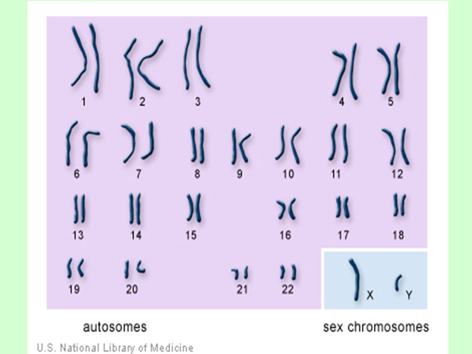
CHROMOSOMES In Humans, there are 23 pairs of chromosomes.
The first 22 pairs are called autosomes. The 23rd pair, the sex chromosome differs between males and females. Females have two X chromosomes, and males have one X and one Y chromosome.

10
VOCABULARY TO KNOW Chromatid – half of a chromosome
Centromere – the constriction region that divides the chromosome into two chromatids Sister chromatids – two identical DNA molecules attached at the centromere Homologous Chromosomes – a chromosome pair, one inherited from the mother and one from the father, containing genes for the SAME trait or characteristic.

11
VOCABULARY TO KNOW Chromatid, Centromere, Sister chromatids, Homologous Chromosomes

12
CHROMOSOMES During cell division the nuclear membrane disappears and the chromatin coils up and darkens. This creates chromosomes that are easily visible. The “doubled” chromosomes are produced during DNA replication. -happens in interphase.

13
CHROMOSOMES The doubled chromosomes are held together by a centromere.
Types of Centromeres:

14
CHROMOSOMES Chromosomes can be studied to determine genetic abnormalities Amniocentesis: a test done on pregnant women to get fetal cells to study. Amniotic fluid is drawn from the womb. Then a Karyotype is done to determine the amount and type of chromosomes present.

15
KARYOTYPE A Karyotype is the number and visual appearance of the chromosomes in the cell nuclei of an organism or species. Scientists take a picture of the cells while they are dividing and blow up the picture to see the chromosomes. The chromosomes are cut out and sorted. Karyotype

16
KARYOTYPE Chromosomes are aligned by size, centromere location, and banding patterns. Autosomes first, numbered Sex chromosomes last XX = female XY = male Chromosome errors can be spotted such as additions, deletions, and extra or less whole chromosomes (ex. Down’s Syndrome)
")
17
KARYOTYPE

18
KARYOTYPE Look at the Karyotypes given to you.
Answer the questions in Section B, #10. Then do Section B #11-13. Quiz Friday over page one of the notes and vocabulary.

19
KARYOTYPES

20
CELL CYCLE series of events of cell growth and division
all cells are on their own cycle - this is from the time of cell division through growth to the next division.

21
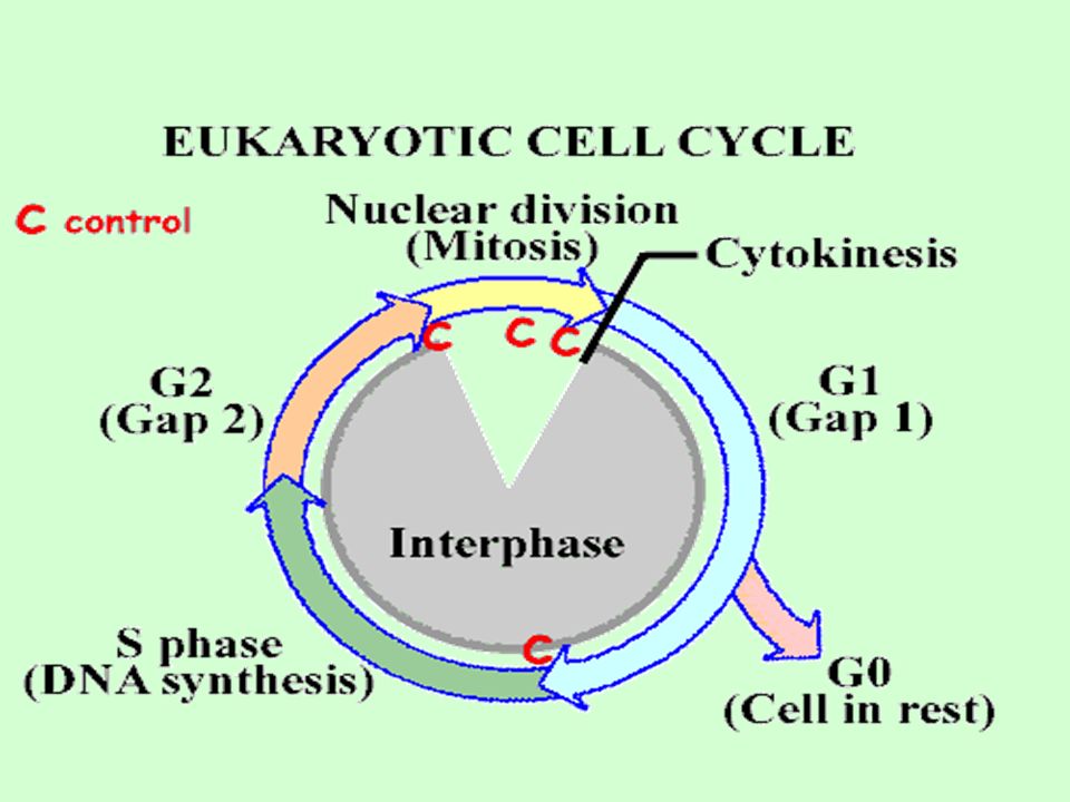
CELL CYCLE Steps of Cell Cycle: Interphase – G1, S, G2
M phase – mitosis or meiosis Cytokinesis

23
INTERPHASE 3 parts 90-95% of the life of the cell
G1: 1st growth phase -cell makes new proteins S: synthesis of DNA (DNA replication) G2: 2nd growth phase -cell makes new organelles Interphase:The cell is engaged in metabolic activity and performing its prepare for mitosis (the next four phases that lead up to and include nuclear division). Chromosomes are not clearly discerned in the nucleus, although a dark spot called the nucleolus may be visible. The cell may contain a pair of centrioles (or microtubule organizing centers in plants) both of which are organizational sites for microtubules.
 G2: 2nd growth phase -cell makes new organelles. Interphase:The cell is engaged in metabolic activity and performing its prepare for mitosis (the next four phases that lead up to and include nuclear division). Chromosomes are not clearly discerned in the nucleus, although a dark spot called the nucleolus may be visible. The cell may contain a pair of centrioles (or microtubule organizing centers in plants) both of which are organizational sites for microtubules.")
24
INTERPHASE During interphase, a cell: Increases in mass
Roughly doubles its organelles, enzymes, etc. Duplicates its DNA For most cells, it is the longest portion of the cell cycle.

25
INTERPHASE G1 Phase “G” stands for “Gap”. Longest phase
Builds proteins, carbohydrates, and lipids and more cytoplasm.

26
INTERPHASE S Phase “S” stands for “Synthesis”. More histones are made.
DNA is replicated.

27
INTERPHASE G2 Phase The cell synthesizes a variety of proteins.
More organelles are made and the cytoplasm expands. Mitochondria and chloroplasts divide to make more of themselves. Other organelles “bud” off parts.

28
INTERPHASE The advantage of having three phases in interphase is that it allows time for preparations for mitosis to happen in an orderly fashion. It also allows time to check that things are happening as they should.

29
CELL CYCLE Sometimes the cells exit the cell cycle and enter the G0 phase. In the G0 phase, cells are alive and metabolically active, but do not divide. Many cells in the human body, including those in heart muscle, eyes, and brain are in the G0 phase. If these cells are damaged, they cannot be replaced.

30
CELL CYCLE Some cells can be “called back” from the Go phase to the cell cycle by certain external cues such as growth factors.

31
TO DO ANIMATION: How the Cell Cycle Works Do Section C

32
CELL DIVISION Mitosis nucleus divides into 2 identical nuclei with identical genetic information In Eukaryotes It is the main process of growth and repair. It is the process by which unicellular and multicellular organisms asexually reproduce.

33
CELL DIVISION Mitosis takes place in somatic (body) cells.
Each cell is 2N or diploid -this means that there are 2 chromosomes of every type present. In humans there are 46 chromosomes. 2 daughter cells will be formed from every parent cell. Mitosis ensures genetic continuity (cloning).
.")
34
CELL REPRODUCTION Organelles involved in cell division are: -centrioles (animals only) -nucleus -spindle fibers
 -nucleus -spindle fibers.")
35
STEPS OF MITOSIS PROPHASE The longest phase of cell division
Chromatin condenses and becomes visible chromosomes. Centrioles move to opposite sides of the cell. Spindle fibers appear and each chromosome attaches to one fiber. Nuclear membrane disappears Chromatin in the nucleus begins to condense and becomes visible in the light microscope as chromosomes. The nucleolus disappears. Centrioles begin moving to opposite ends of the cell and fibers extend from the centromeres. Some fibers cross the cell to form the mitotic spindle.

36
STEPS OF MITOSIS Metaphase Fastest phase
Spindle fibers attach to centromeres. Chromosomes line up along the equator of the cell – midway. Spindle fibers align the chromosomes along the middle of the cell nucleus. This line is referred to as the metaphase plate. This organization helps to ensure that in the next phase, when the chromosomes are separated, each new nucleus will receive one copy of each chromosome.

37
STEPS OF MITOSIS Anaphase Doubled chromosomes pull apart
Sister chromatids are pulled to opposite sides of the cell by the spindle fibers. The paired chromosomes separate at the kinetochores and move to opposite sides of the cell. Motion results from a combination of kinetochore movement along the spindle microtubules and through the physical interaction of polar microtubules.

38
STEPS OF MITOSIS Telophase
All the events that happened in prophase have to be reversed. Chromosomes decondense (uncoil) to form chromatin Nuclear membrane reform – there are now two nuclei. Spindle fibers dissolve into the cytoplasm. Chromatids arrive at opposite poles of cell, and new membranes form around the daughter nuclei. The chromosomes disperse and are no longer visible under the light microscope. The spindle fibers disperse, and cytokinesis or the partitioning of the cell may also begin during this stage.
 to form chromatin. Nuclear membrane reform – there are now two nuclei. Spindle fibers dissolve into the cytoplasm. Chromatids arrive at opposite poles of cell, and new membranes form around the daughter nuclei. The chromosomes disperse and are no longer visible under the light microscope. The spindle fibers disperse, and cytokinesis or the partitioning of the cell may also begin during this stage.")
39
CYTOKINESIS Cytokinesis occurs. Cytoplasm divides.
Cell pinches in half to form two new cells.

40
CYTOKINESIS Animal cells: cytokinesis results when a fiber ring composed of a protein called actin around the center of the cell contracts pinching the cell into two daughter cells, each with one nucleus. Plant cells: the rigid wall requires that a cell plate be formed between the two daughter cells.

41
YIELD 2 diploid daughter cells genetically identical to parent

42
MITOSIS IN ONION ROOT TIP
PROPHASE INTERPHASE METAPHASE ANAPHASE TELOPHASE

43
TO DO VIDEO: Mitosis and Cytokinesis
Work on Cell Reproduction Practice packet – due next Friday.

44
DO NOW VIDEO: Mitosis COMPUTERS:
Section E: Cell Cycle Section F: Cell Division Section G: Online Onion Root Tips PRACTICE: Work on Cell Reproduction Practice Questions.

45
DO NOW Mitosis in Onion Root Tip Section H Section I
Work on Cell Reproduction Practice Questions

46
CELL CYCLE : CREATING GAMETES
Three Phases: Interphase, Meiosis I and II, Cytokinesis Meiosis: sex cell formation Reduces the chromosome number by half Meiosis

47
MEIOSIS

48
MEIOSIS Nucleus divides twice into 4 different nuclei with different genetic information. In Eukaryotes, it is the formation of gametes for sexual reproduction. Each gamete cell will contain one of the pairs of homologous chromosomes As a result: 4 cells form from 1 parent cell and they contain the haploid (N) number of chromosomes
 number of chromosomes.")
49
MEIOSIS Meiosis leads to genetic variation: mixing of the genes within an individual’s sex cells. Ex. Tall or short Curly hair or short hair

50
MEIOSIS I: PMAT I Prophase I
the same events that occur in mitosis except for synapsis- the pairing of homologous chromosomes to form a tetrad During this time crossing over of the chromosome occurs and this leads to even more variation

51
MEIOSIS I: PMAT I A single cell divides into 2 daughter cells that are not genetically identical. Crossing over: greater genetic variation

52
MEIOSIS I: PMAT 1 The homologous chromosomes come together and literally swap parts of themselves with each other. This process is called Crossing Over and ensures that the daughter cells produced after the first cytokinesis will not be genetically identical.

53
MEIOSIS I: PMAT 1 Metaphase I same as mitosis

54
MEIOSIS I: PMAT 1 Anaphase I
chromosome reduction centromeres stay in tact, chromosomes don’t separate into chromatids. Independent Assortment – each homologous chromosome is randomly assorted at the equator and into different gametes.

55
MEIOSIS I: PMAT 1 Telophase I
cell splits, but the nucleus and nucleolus don’t reform There is one more round of cell division

56
MEIOSIS II: PMAT 2 Metaphase II line up in the middle

57
MEIOSIS II: PMAT 2 Anaphase II
chromosomes separate at their chromatids

58
MEIOSIS II: PMAT 2 Telophase II nucleus reforms nucleolus reforms
membrane reforms cytokinesis occurs 4 haploid cells are formed

59
CYTOKINESIS division of the cytoplasm

60
YIELD 4 haploid daughter cells genetically different from parent

61
OOGENESIS VS. SPERMATOGENESIS
Oogenesis is formation of an egg Only one egg is formed (three polar bodies are also formed). Spermatogenesis is formation of sperm. 4 sperm cells are formed from 1 parent cell.
. Spermatogenesis is formation of sperm. 4 sperm cells are formed from 1 parent cell.")
62
OOGENESIS VS. SPERMATOGENESIS

63
TO DO Animation: How Meiosis Works Section J
Cell reproduction Practice is due Friday.

64
DO NOW Get out your notes and worksheet packet.
Cell reproduction Practice is due tomorrow. Control of the Cell Cycle

65
CELL CYCLE CONTROL A Molecular Control System
Several checkpoints act as built-in stop signs that halt the cell until they are over-ridden by go ahead signals. Three checkpoints exists in G1, G2, and M. Cells also have a predetermined lifespan.

66
CELL CYCLE CONTROL Three checkpoints exist during interphase to make sure that everything has gone as planned and fix errors if needed. G1-S checkpoint (end of the G1 phase) makes sure that the DNA is intact and that the cell has enough energy to enter the S phase. The S phase checkpoint makes sure that DNA is replicated correctly without any breakages. The G2-M checkpoint at the end of the G2 phase is another safeguard in case something happens to the DNA or cell before it undergoes the massive task of dividing.
 makes sure that the DNA is intact and that the cell has enough energy to enter the S phase. The S phase checkpoint makes sure that DNA is replicated correctly without any breakages. The G2-M checkpoint at the end of the G2 phase is another safeguard in case something happens to the DNA or cell before it undergoes the massive task of dividing.")
67
CELL CYCLE CONTROL Timing is controlled by regulatory proteins – cyclins and kinases. These proteins selectively access, activate and silence information in DNA. Contact Inhibition – Cells release chemicals to inhibit growth when they become too crowded.

68
CELL CYCLE CONTROL When things go wrong? Uncontrolled Cell Growth
Mutant genes cause tumors to form by disrupting normal cell cycle controls Altered cells grow and divide abnormally. Tumors may be cancerous

69
TO DO The Effect of Contact Inhibition on Cell Division
Mistakes in Meiosis Cancer and the Cell Cycle Cell reproduction Practice is due tomorrow.

70
DO NOW Turn in Cell Reproduction Practice packet. Pick up review.
Get out notes and worksheet. How to remember Mitosis versus Meiosis Mitosis and Meiosis

71
PROKARYOTIC CELL DIVISION
Prokaryotic fission, which is binary fission, is a form of asexual reproduction and cell division used by all prokaryotes (bacteria and archaebacteria) some organelles within eukaryotic organisms (e.g., mitochondria).
 some organelles within eukaryotic organisms (e.g., mitochondria).")
72
PROKARYOTIC CELL DIVISION
Most bacterial genes are located on a single bacterial chromosome (~4million base pairs) which consists of a circular DNA molecule and associated proteins.
 which consists of a circular DNA molecule and associated proteins.")
73
PROKARYOTIC CELL DIVISION
Bacteria do not have as many genes or DNA molecules as long as those in eukaryotes. Humans ~2.3 billion base pairs. Bacteria circular chromosome is still highly folded and coiled in the cell.

74
PROKARYOTIC CELL DIVISION
Prokaryotes reproduce by binary fission, not mitosis. In binary fission, chromosome replication begins at one point in the circular chromosome, the origin of replication site. Binary Fission

75
PROKARYOTIC CELL DIVISION

Similar presentations


. -Histones - help maintain the shape of the.>")


