Download presentation
Presentation is loading. Please wait.

1
THE LYMPHOID SYSTEM Kristina C. Erasmo, M.D.

2
Defense System of the Body
Non-Immune Immune

3
Non-Immune Defense System
INFLAMMATION Mediated by: Neutrophils Eosinophils Basophils NK cells Monocytes and macrophages

4
Immune Defense System Lymphocytes – principal effector cells

5
Lymphoid System Refers to the tissues and organs that participate in the immune defense system Components are distributed in various areas of the body

6
LYMPHOID SYSTEM Non-Encapsulated Lymphoid Tissue
Encapsulated Lymphoid Organs Tonsils

7
LYMPHOID SYSTEM Non-Encapsulated Lymphoid Tissue
Diffuse lymphoid tissue Lymphoid nodules (lymphoid follicles) Encapsulated Lymphoid Organs Lymph Node Thymus Spleen
 Encapsulated Lymphoid Organs. Lymph Node. Thymus. Spleen.")
8
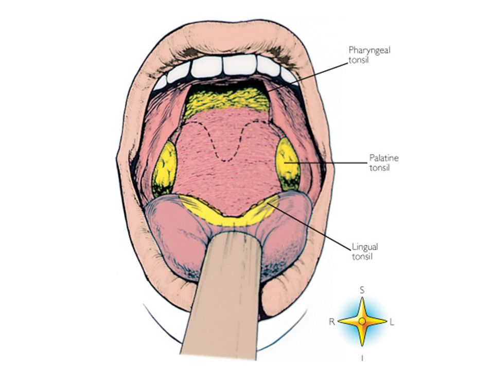
LYMPHOID SYSTEM Tonsils Palatine Pharyngeal Lingual Tubal

9
LYMPHOID SYSTEM Non-Encapsulated Lymphoid Tissue
Diffuse lymphoid tissue Lymphoid nodules (lymphoid follicles)
")
10
Diffuse Lymphoid Tissue
Histologic features: Stroma composed of network of reticular fibers and reticular cells Spaces occupied primarily by lymphocytes Some macrophages found

11
Diffuse Lymphoid Tissue
Location: Practically in all CT in the body Most prominent in lamina propria and submucosa of GIT, respiratory tract

12
Diffuse Lymphoid Tissue (Stomach)
")
13
Diffuse Lymphoid Tissue
Types: Loose lymphoid tissue – lymphocytes far apart Dense lymphoid tissue – lymphocytes tightly packed

14
Lymphoid Nodule LYMPHOID FOLLICLE
Lymphoid tissue where clustered lymphocytes form discrete masses or lumps Usually interspersed in areas of diffuse lymphoid tissue

15
Lymphoid Nodule Location: Lamina propria of GIT and respiratory tracts
Spleen Lymph nodes Tonsils

16
Lymphoid Nodule Types based on arrangement:
Solitary nodule – occur singly Aggregates – Peyer’s patches (submucosa of the ileum)
")
17
Peyer’s Patch

18
Peyer’s Patches

19
Lymphoid Nodule Types based on structure:
Primary nodule – cells evenly distributed throughout nodule, no mitotic figures seen Secondary nodule (germinal centers) – 2 distinct regions
 – 2 distinct regions.")
20
Lymphoid Nodule Secondary nodule
Germinal center (reaction center) – pale, central Forms the core of the nodule B lymphocytes proliferate and differentiate following exposure to antigen Many mitotic figures Very few T lymphocytes Corona – darker, peripheral
 – pale, central. Forms the core of the nodule. B lymphocytes proliferate and differentiate following exposure to antigen. Many mitotic figures. Very few T lymphocytes. Corona – darker, peripheral.")
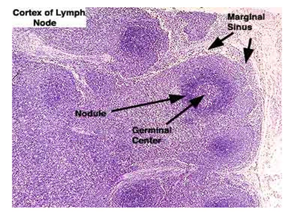
21
Secondary Lymphoid Follicle

22
Secondary Lymphoid Follicle (Lymph Node)
")
23
LYMPHOID SYSTEM Encapsulated Lymphoid Organs Lymph Node Thymus Spleen

24
Lymph Node Encapsulated, bean-shaped organ Location:
Popliteal, inguinal, axillary regions Sides of neck Along abdominal vessels In mesentery

25
Lymph Node

26
Lymph Node Hilus – the indented area of the lymph node, where blood vessels enter and leave the organ Efferent lymphatic vessels leave the lymph node on the hilar area Afferent lymphatic vessels enter the lymph node on the convex side

27
Lymph Node

29
Histologic Features of Lymph Node
Capsule – dense connective tissue covering Trabeculae – incompletely subdivide the organ into compartments Reticular cells and fibers – supporting meshwork

30
Lymph Node

32
Histologic Features of Lymph Node
Parenchyma: Cortex – outer Outer portion – composed of lymphoid nodules (primary and secondary), mostly B lymphocytes Inner – dense lymphoid tissue, no lymphoid nodules, mostly T lymphocytes Medulla – inner, paler-staining, contain medullary cords
, mostly B lymphocytes. Inner – dense lymphoid tissue, no lymphoid nodules, mostly T lymphocytes. Medulla – inner, paler-staining, contain medullary cords.")
33
Lymph Node

34
Lymph Node

35
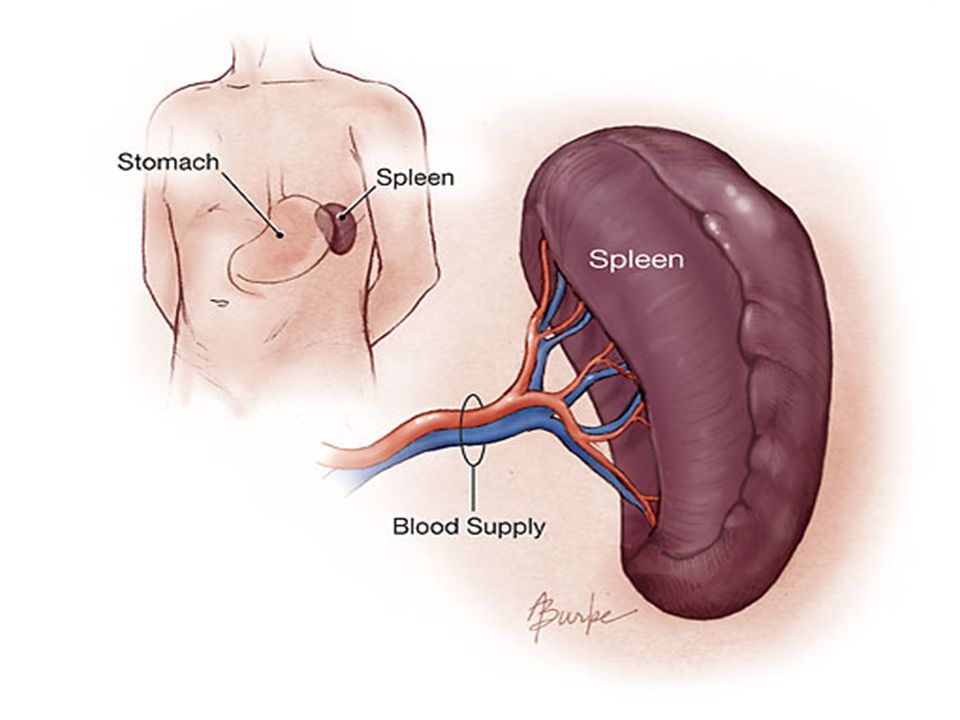
Spleen Largest lymphoid organ (7 x 12 cm)
Found in the upper left quadrant of the abdominal cavity Hilus – where splenic vessels enter and leave the spleen

37
Spleen Functions: Contain macrophages – destroy foreign substances, microorganisms, abnormal cells in blood Removes and destroys old RBCs and platelets from circulating blood Recycles iron contain in the RBCs

38
Spleen Functions: Storage area for blood
Where lymphocytes proliferate and differentiate into different types after being stimulated

39
Histologic Features of Spleen
Enveloped by peritoneum that blends with its capsule (mesothelium) Trabeculae – divide into compartments Reticular cells and fibers – supporting framework
 Trabeculae – divide into compartments. Reticular cells and fibers – supporting framework.")
40
Spleen

41
Histologic Features of Spleen
Splenic pulp – parenchyma of the spleen White pulp Red pulp Marginal zone

42
Histologic Features of Spleen
White pulp Consists of lymphoid nodules embedded in dense lymphoid tissue Lymphoid nodules – B lymphocytes Dense lymphoid tissue – T lymphocytes

43
Histologic Features of Spleen
Red pulp Bulk of the parenchyma Consists of large blood-filled sinusoids with strands of lymphoid tissue in-between (Billroth’s cords, splenic cords)
")
44
Histologic Features of Spleen
Marginal zone Poorly-delineated transitional area between red and white pulp Contains lymphocytes and plasma cells

45
Spleen

46
Red Pulp (Spleen)
")
47
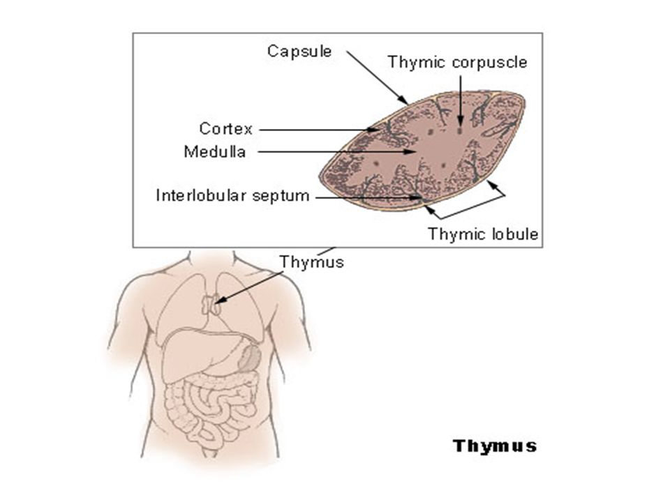
Thymus Located in the superior mediastinum
Composed of two pyramidal lobes fused together

49
Thymus Where T cell stem cells proliferate, differentiate, and transform into immunologically competent T lymphocytes Devoid of B lymphocytes

50
Histologic Features of Thymus
Capsule – thin loose connective tissue Trabeculae Reticular cells – supporting meshwork

51
Histologic Features of Thymus
Cortex – peripheral, darker-staining region Medulla – central, lighter-staining region Hassall’s corpuscles (Hassall’s bodies, thymic corpuscles) Composed of hyaline core surrounded by layers of flattened epithelioid cells
 Composed of hyaline core surrounded by layers of flattened epithelioid cells.")
52
Thymus

53
Thymus

54
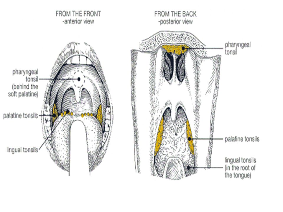
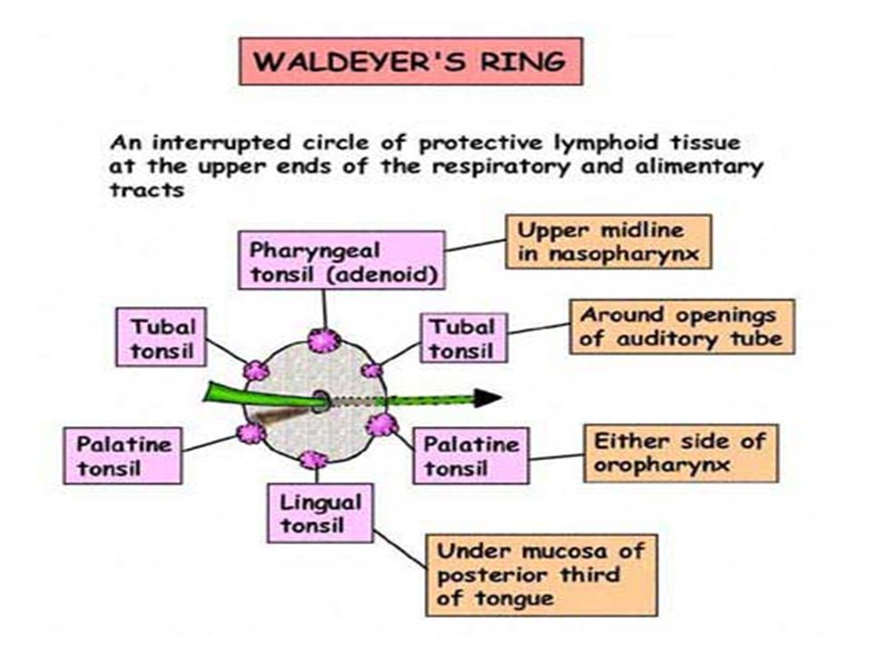
Tonsils Lymphoid organs that form a ring (Waldeyer’s ring) underneath the epithelium around the entrance to the respiratory and digestive passages
 underneath the epithelium around the entrance to the respiratory and digestive passages.")
58
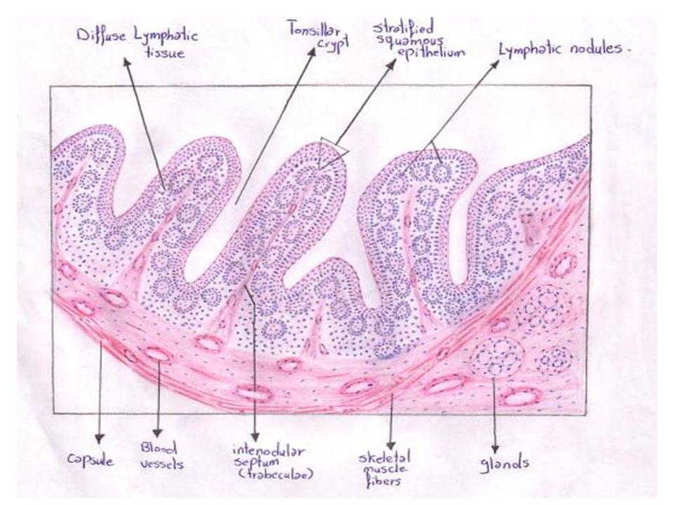
Tonsils Histologic feature:
Dense lymphoid tissue embedded with lymphoid nodules Types: Palatine, lingual, pharyngeal, tubal tonsils

59
Palatine Tonsils Located in the lateral aspect o the oropharynx (one on each side) Covered by stratified squamous non-keratinized epithelium Tonsillar crypts – deep invaginations of the epithelium (contain dead epithelial cells, lymphocytes, etc)
")
62
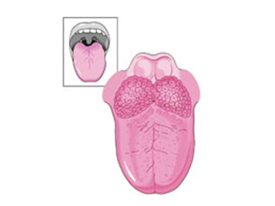
Lingual Tonsils Several discrete masses of lymphoid tissue located in the dorsum of the tongue Covered by stratified squamous non-keratinized epithelium May have single broad deep crypt into which ducts of mucus-secreting glands open

64
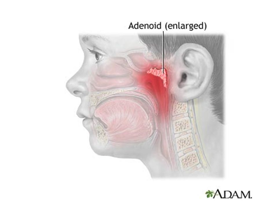
Pharyngeal Tonsils Central area of the posterior and superior nasopharyngeal wall Covered by ciliated pseudostratified columnar epithelium (“respiratory epithelium”) Some areas covered with stratified squamous non-keratinized epithelium Does not form crypts but form shallow folds Adenoids – enlarged pharyngeal tonsils
 Some areas covered with stratified squamous non-keratinized epithelium. Does not form crypts but form shallow folds. Adenoids – enlarged pharyngeal tonsils.")
66
Tubal Tonsils Masses of lymphoid tissue in the nasopharynx near the openings of the eustachian tube Some believe they are simple extensions of the pharyngeal tonsils Covered by respiratory epithelium

Similar presentations


 -mast cells -interdigitating.>")


