Download presentation
Presentation is loading. Please wait.

1
Lipoid proteinosis Presented by: Lale sayadi 851111638 Esfand 1390

2
Case: (Monday 1390/12/15) a 16 years old boy with hoarse cry from birth and hoarseness remains throughout his life.Initially, an inflammatory, vesicular, and crusted eruption appears on the face and extremities at 4 years old. Then verrucous papules and plaques arise the elbows, knees, and hands.now he had pox-like scars on his whole body.he has restricted motion in tongue. He has not had seizure.he does not have any other positive clue in examination.he has a younger brother with same problems but milder than him.

3
Aks:

6
Dx: lipoid proteinosis (by biopsy+clinical presentations)
")
7
Definition: Lipoid proteinosis is a rare,chronic and autosomal recessive disorder that presents in early infancy with hoarseness, followed by pox-like and acneiform scars, along with infiltration and thickening of the skin and certain mucous membranes.

8
Pathophysiology: loss of function mutations in the gene encoding extracellular matrix protein 1 (ECM1) on band 1q21 is identified as the cause of lipoid proteinosis. The ECM1 gene product is a glycoprotein with functional roles in skin physiology and homeostasis. ECM1 is involved in keratinocyte differentiation in the epidermis and in regulation of basement membrane integrity, interstitial collagen fibril macroassembly, and growth factor binding in the dermis

9
Epidemiology: Race: Patients of European ancestry are most commonly affected Sex: No sex predilection is reported. Age: Patients typically present in early childhood, but manifestations may be present at birth. Some cases may occur in adults Mortality/Morbidity: Life span is usually normal unless altered by complications. Mortality rates in infants and adults are slightly increased because of laryngeal obstruction.

10
Frequency: is listed as a "rare disease" by the Office of Rare Diseases (ORD) of the National Institutes of Health (NIH).rare disease
 of the National Institutes of Health (NIH).rare disease")
11
Clinical manifestation: The classic manifestation is onset in infancy with a hoarse cry due to laryngeal infiltration Cutaneous manifestations usually arise during the first 2 years of life including:variably sized vesicles, pustules, bullae, and hemorrhagic crusts. Resolution of the lesions occurs with permanent, poxlike atrophic scarring. Late findings are noted as the child ages; the skin develops a waxy, thickened, yellowish appearance due to dermal infiltration

12
Clinical manifestation(con … ) Skin: Papules, plaques, and nodules arise on the face, axillae, and scrotum. A pathognomonic sign is a row of beaded papules along the eyelid margins, resembling a string of pearls; this is termed moniliform blepharosis Hyperkeratotic, verrucous plaques may arise in sites of trauma, particularly the elbows, knees, and dorsum of the hands Scalp: patchy or diffuse hair loss.

14
Clinical manifestation(con … ) Oral cavity: woody firmness and impaired mobilityof tongue. Transient swelling and ulceration of the lips and tongue. Pebbling of the lip mucosa imparts a cobblestone appearance Hypoplasia or aplasia of the teeth. Recurrent parotitis may occur as a consequence of infiltration of the Stensen duct.

16
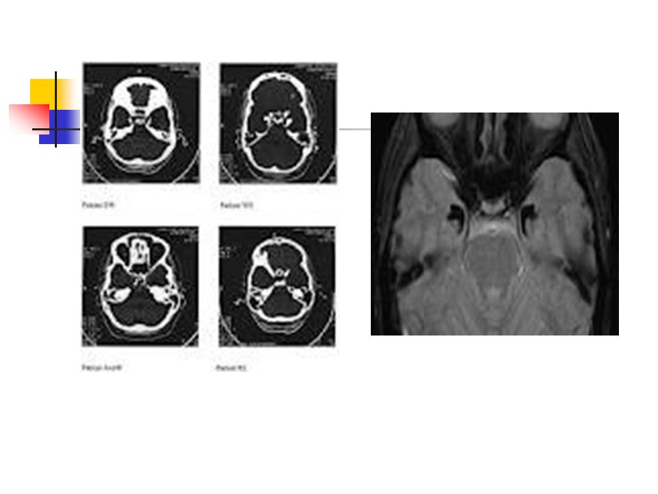
Clinical manifestation(con … ) Upper airway: Infiltration of the larynx, vocal cords, and surrounding structures may produce hoarseness, dysphagia,and airway obstruction Central nervous system: A classic and pathognomonic radiographic finding is bilateral, intracranial, bean-shaped suprasellar calcifications in the temporal lobe.
 Upper airway: Infiltration of the larynx, vocal cords, and surrounding structures may produce hoarseness, dysphagia,and airway obstruction Central nervous system: A classic and pathognomonic radiographic finding is bilateral, intracranial, bean-shaped suprasellar calcifications in the temporal lobe.")
17
Tests : Laboratory Studies: No laboratory findings are consistently abnormal. Increased ESR Increased production of alpha- and beta-globulins PCR of the ECM1 gene Immunolabeling of affected tissue with polyclonal antibodies against the ECM1 protein

18
Tests: Imaging Studies: A pathognomonic finding on plain radiographs and CT scans of the brain is bilateral, intracranial, bean-shaped calcifications within the hippocampal region of the temporal lobes.

20
Tests: Skin biopsy of affected cutaneous or mucosal sites

21
Histologic Findings: Early lesions have eosinophilic hyaline thickening of papillary dermal capillaries. hyperkeratosis papillary dermis is widened by hyaline material Hyaline deposits may be arranged around the hair follicles, eccrine glands, sebaceous glands, and arrector pili muscles and nerves in an onionskin arrangement

23
Differential diagnosis: Amyloidosis lichen Amyloidosis, Nodular Localized Cutaneous colloid milium Leprosy Lichen myxedematosus Xanthomas EPP

24
Complications: Laryngeal involvement may lead to airway obstruction. Vocal cord involvement may lead to impaired speech. Intracranial calcifications may result in seizures, behavioral changes, rage attacks, and dystonia. The deposition of hyaline in the small bowel is reported to cause gastrointestinal bleeding. pox-like and acneiform scars

25
Prognosis: Lipoid proteinosis has a stable or slowly progressive course. The presence of this disease is compatible with a normal life span unless altered by airway obstruction or fatal seizure activity.

26
Treatment: No cure is known According some clinical trials: D-penicillamine oral dimethyl sulfoxide (DMSO ) potent topical corticosteroids Etretinate Seizures if present : anticonvulsant
 potent topical corticosteroids Etretinate Seizures if present : anticonvulsant")
27
Patient Education The parents should be educated about the risk of having affected offspring

28
The end

Similar presentations