Download presentation
Presentation is loading. Please wait.

1
Low Angle X-ray Scattering (LAXS) for Tissue Characterization Dr M A Oghabian
 for Tissue Characterization Dr M A Oghabian")
2
Statement of Problem

3
Deficiencies of the Current Imaging Techniques Poor contrast between healthy and diseased soft tissues (eg; Breast tissues) Presence of scatter, which further degrades the contrast Low Specificity in Mammography (50%) No molecular and cellular changes is possible in early stages Only Bulk changes are visible Types of materials consisted in a specific tissue are not accessible
 Presence of scatter, which further degrades the contrast Low Specificity in Mammography (50%) No molecular and cellular changes is possible in early stages Only Bulk changes are visible Types of materials consisted in a specific tissue are not accessible")
4
What is the SOLUTION? Refractive/interference effects is about 1000 times larger than absorption If interference-related effects of scattering is properly exploited, much more intense information is obtained

6
Characteristic curve for a tissue contract, C without scatter, C ’ with scatter

7
2

8
Differential Thomson Cross Section per electron for Elastic Scattering Scattering from a single electron (regarded as a point charge) is defined as:
 is defined as:")
9
Coherent (Rayleigh) Differential Scatter Cross Section for Atomic Species When Photons excite more than one electron, Coherent Scatter from different electrons demonstrate interference effects: f 2 (x,Z) is Atomic form Factor, x is momentum transfer, Z atomic Number
 Differential Scatter Cross Section for Atomic Species When Photons excite more than one electron, Coherent Scatter from different electrons demonstrate interference effects: f 2 (x,Z) is Atomic form Factor, x is momentum transfer, Z atomic Number")
10
What is Atomic form factor? Atomic form factor, or atomic scattering factor, is a measure of the amplitude of a wave scattered from an isolated atom (scattering amplitude).Atomic form factor, or atomic scattering factor, is a measure of the amplitude of a wave scattered from an isolated atom (scattering amplitude).amplitudescattering amplitude scattering amplitude x-rays are scattered by the electron cloud of the atom and hence the scattering power of x-rays increases with the atomic number of the atoms in a sample.x-rays are scattered by the electron cloud of the atom and hence the scattering power of x-rays increases with the atomic number of the atoms in a sample.atomic numberatomic number The x-ray form factor is defined as the Fourier transform of the electron charge density of scaterer.The x-ray form factor is defined as the Fourier transform of the electron charge density of scaterer.Fourier transformelectroncharge densityFourier transformelectroncharge density
.Atomic form factor, or atomic scattering factor, is a measure of the amplitude of a wave scattered from an isolated atom (scattering amplitude).amplitudescattering amplitude scattering amplitude x-rays are scattered by the electron cloud of the atom and hence the scattering power of x-rays increases with the atomic number of the atoms in a sample.x-rays are scattered by the electron cloud of the atom and hence the scattering power of x-rays increases with the atomic number of the atoms in a sample.atomic numberatomic number The x-ray form factor is defined as the Fourier transform of the electron charge density of scaterer.The x-ray form factor is defined as the Fourier transform of the electron charge density of scaterer.Fourier transformelectroncharge densityFourier transformelectroncharge density.")
11
Coherent Differential Scattering Cross section of Condensed Material Atomic form factor gives a good description of scattering when phase relationship from different atoms is not constant (eg in gaseous samples) In Condensed state of Matters, the atomic form factor should be modified, to include the interference effects from neighboring atoms:
 In Condensed state of Matters, the atomic form factor should be modified, to include the interference effects from neighboring atoms:")
12
Molecular Form factor F 2 (x)=f 2 (x)(1+H(x)) f(x) is the IAM (independent atomic model) form factorf(x) is the IAM (independent atomic model) form factor H(x) an oscillatory structure function which accounts for the interference effect.H(x) an oscillatory structure function which accounts for the interference effect. For amorphous materials and liquids, H(x) shows a damped behavior around zero and vanishes for values of x>4-5 nm -1.For amorphous materials and liquids, H(x) shows a damped behavior around zero and vanishes for values of x>4-5 nm -1.
 shows a damped behavior around zero and vanishes for values of x>4-5 nm -1.For amorphous materials and liquids, H(x) shows a damped behavior around zero and vanishes for values of x>4-5 nm -1..")
13
Structure function data can be taken from experimental measurements.Structure function data can be taken from experimental measurements. If x ranges from about 4-5 to 10 10 nm -1, IAM is valid, and each atom is assumed to scatter independent of the others, therefore the Sum rule is applied:If x ranges from about 4-5 to 10 10 nm -1, IAM is valid, and each atom is assumed to scatter independent of the others, therefore the Sum rule is applied: w i is Mass fraction, and A i is Atomic Mass of ith element, M: molecular weightw i is Mass fraction, and A i is Atomic Mass of ith element, M: molecular weight Incoherent contribution is always considered in the IAM frame, that is:Incoherent contribution is always considered in the IAM frame, that is: S i and f i can be found from the extensive tabulation from the existing literature for all elementsS i and f i can be found from the extensive tabulation from the existing literature for all elements Form-factor of Multi-element materials

14
Form-factor of an amorphous material The form-factor for an amorphous material is given by: W i is atom fraction and f Ri (x) is Atomic form-factor of the ith element in the material. H(x) is a structure function that accounts for diffraction effects
 is a structure function that accounts for diffraction effects.")
15
Rayleigh cross-section of an amorphous material Rayleigh differential cross-section of an amorphous material r 0 is the classical electron radius, θ is the scattering angle, F R (x) is the form-factor x = sin ( θ /2)/ λ is the momentum transfer.
 is the form-factor x = sin ( θ /2)/ λ is the momentum transfer.")
16
Cross-section for Crystal material (Bragg cross-section) cross-section for Bragg scattering from a crystal: λ is the X-ray wavelength, N is the number of atoms in the crystal unit cell, V is the unit crystal volume, m i, multiplicity, d i, atomic plane spacing f i, Structure factor (form-factor) of the plane i.
 cross-section for Bragg scattering from a crystal: λ is the X-ray wavelength, N is the number of atoms in the crystal unit cell, V is the unit crystal volume, m i, multiplicity, d i, atomic plane spacing f i, Structure factor (form-factor) of the plane i.")
17
Total Linear differential Coefficient for Mono- molecular material Linear differential scattering coefficient which is the probability of a photon being scattered per unit length of beam path, and has unit of m -1 sr -1 : M: molecular weight NA: Avogadro number ρ : density of the material. F (x): Molecular form factor of sample material referring to coherent (Rayleigh) scattering S(x): incoherent (Compton) scattering function x: Momentum transfer x = sin(θ/2)/λ
: Molecular form factor of sample material referring to coherent (Rayleigh) scattering S(x): incoherent (Compton) scattering function x: Momentum transfer x = sin(θ/2)/λ.")
18
Bragg Diffraction Diffraction is a phenomenon of reinforced Coherent scattering. Coherent Scattering from all atoms in a material undergo reinforcement in a certain direction where they are in phase (Constructive interference), And cancel each other in other directions, where they are out of phase (Destructive interference) Diffraction can easily observed in material with Crystalline structure, because the atoms are rigidly fixed to one another
, And cancel each other in other directions, where they are out of phase (Destructive interference) Diffraction can easily observed in material with Crystalline structure, because the atoms are rigidly fixed to one another.")
19
Bragg ’ s Law When constructive interference occurs, we get diffracted beams in specific directions These directions are defined by the wavelength λ of the incident radiation and the nature of the crystalline sample (ie d) Bragg’s law relates the wavelength of the x-rays to the spacing of the atomic planes a a a and scattering angle n:an integral number λ:wavelength d:interplanar spacing Ө:angle between the incident wave & atomic planes nλ=2dsin(Ө/2)
 Bragg’s law relates the wavelength of the x-rays to the spacing of the atomic planes a a a and scattering angle n:an integral number λ:wavelength d:interplanar spacing Ө:angle between the incident wave & atomic planes nλ=2dsin(Ө/2)")
20
Bragg’s law and Form factor of Crystal Bragg’s law states that scattering can only occur when d =1/2x Therefore: δ(x) is the Dirac delta function x i =1/(2d i ) is momentum exchange corresponding to plane i. x = sin(θ/2)/λ
/λ.")
21
cross-sections and form-factors of mixed crystalline and amorphous materials For both Rayleigh scattering from amorphous materials and Bragg scattering from crystals, the total scattering cross- section of a mixture is: E is the photon energy α i is the mass fraction σ i is either the Rayleigh or Bragg cross-section of material i in the mixture. Similarly, the Rayleigh and Bragg form factors of a mixture are given by: F i (x) is the Rayleigh or Bragg form factor of the ith material.
 is the Rayleigh or Bragg form factor of the ith material..")
22
Calculated Linear differential scattering coefficients for fat, water, bone matrix (collagen), bone mineral, and dry bone (28.2% bone matrix, 71.8% bone mineral),.
, bone mineral, and dry bone (28.2% bone matrix, 71.8% bone mineral),.")
23
Diffraction patterns of free molecule, Independent free Atom, and Molecular form factor (condensed molecules)
")
26
Low Angle X-ray Scattering Signatures of carcinoma and normal breast tissue

27
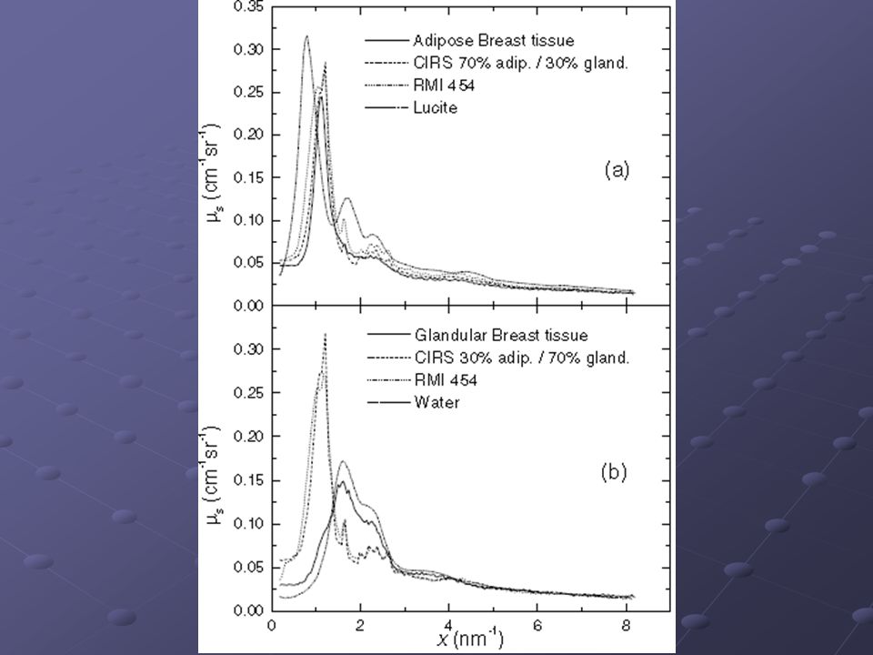
Diffraction Profile for different Amorphous tissues

28
Diffraction profile for Hydroxiapatite over scattering angle from 4 to 10 degrees

29
Low Angle x-ray Scattering (LAXS or SAXS) Interference effects occurring among the Low angle coherently scattered photons from a material due to the electron distribution This type of scattering leads to materials characterization
 Interference effects occurring among the Low angle coherently scattered photons from a material due to the electron distribution This type of scattering leads to materials characterization")
31
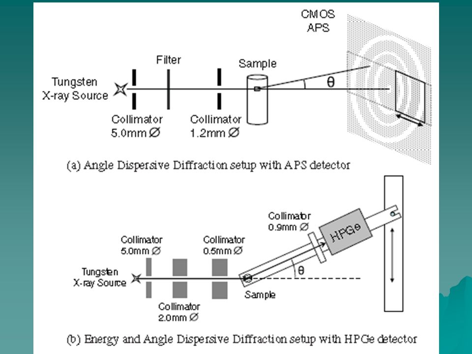
LAXS SET UP X-ray tube Primary collimator Sample Secondary collimator HP GE detector Amplifire MCA

32
The physical lay out of the LAXS experimental set up Sample Slits Scattered x-rays X-ray tube Incident x-rays detector

34
Coherent X-Ray Scatter for Non-Destructive

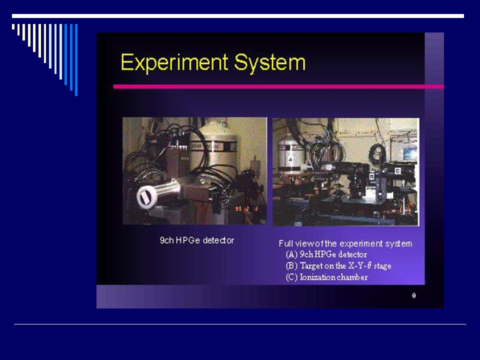
35
Dual Detector system for Breast Imaging

Similar presentations


 Quantum Physics.>")


 + University.>")
 Pure metal target (Cu) Electrons remover inner-shell electrons from target. Other electrons “fall”>")

 We also looked at internal ordering of atoms in 3-D structure (230 space.>")


 Characterization of Nanomaterials.>")

>")

>")
