Download presentation
Presentation is loading. Please wait.

1
Dr Pradeep Kumar, Professor department of physiology, KGMU, Lucknow

2
We need a range of different types of food

3
Food Composition Chemical substances found in the largest amounts in food: ◦ Water (found inside cells in plants and animals) ◦ Carbohydrates ◦ Fats ◦ Protein ◦ Miniral ◦ vitamins
 ◦ Carbohydrates ◦ Fats ◦ Protein ◦ Miniral ◦ vitamins")
4
Understand the composition of protein Understand the enzymes responsible for digestion of proteins What are sites for absorption Molecular basis of protein transportation Learn about diseases related to protein digestion

5
Proteins are a sequence of amino acids One amino acids is joined to the next by a PEPTIDE bond Provide energy substrate for metabolism (4 kcals/g). Protein load received by the gut is derived from two primary sources: 70-100 g dietary protein, and 35-200 g endogenous protein,

6
Of the 20 amino acids that exist, 9 are essential amino acids, and 11 are non-essential There are also 4 amino acids that can be considered conditionally essential: arginine, tyrosine, glutamine, and cysteine

7
Dipeptide – 2 amino acids Tripeptide – 3 amino acids Oligopeptides – 4-10 amino acids Polypeptide – more than 10 amino acids Proteins in the body and diet are long polypeptides (100s of amino acids)
")
8
Consist of a central carbon atom bonded to: a hydrogen, a carboxylic acid, an amino group, and an additional side group that is unique to each amino acid

10
Acid, alkaline, heat and alcohol, can disrupt the chemical forces that stabilize proteins and can cause them to lose their shape (denature) Denaturing of proteins happens during food preparation (cooking, whipping, adding acids) or digestion (in the stomach with hydrochloric acid)
 Denaturing of proteins happens during food preparation (cooking, whipping, adding acids) or digestion (in the stomach with hydrochloric acid)")
11
No digestion of protein takes place in the mouth, Hydrochloric acid denatures protein and also converts pepsinogen to pepsin Pepsin breaks the protein down into peptides of various lengths and some amino acids Pepsin act only at pH 1.6-3.2 Pepsin completes ~ 10-20% of digestion

12
Pepsin hydrolyses the bonds between aromatic amino acids(phenylanine or tyrosine) and a second amino acid So the product of pepsin hydrolysis is polypeptides of diverse sizes
 and a second amino acid So the product of pepsin hydrolysis is polypeptides of diverse sizes")
13
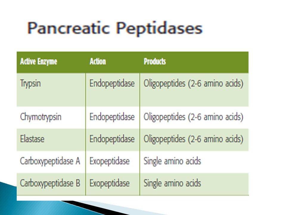
Trypsinogen and chymotrypsinogen (proenzymes) are secreted by pancreas in response to protein in the small intestine They will be activated to trypsin and chymotrypsin (now called proteases)
 are secreted by pancreas in response to protein in the small intestine They will be activated to trypsin and chymotrypsin (now called proteases)")
16
These enzymes can either cleave internal peptide bonds (i.e. endopeptidases) exopeptidases cleave off one amino acid at a time from either the –COOH or –NH2 terminal of the polypeptide (i.e. they are carboxypeptidases, and aminopeptidases, respectively)
 exopeptidases cleave off one amino acid at a time from either the –COOH or –NH2 terminal of the polypeptide (i.e. they are carboxypeptidases, and aminopeptidases, respectively).")
17
The endopeptidases cleave the large polypeptides to smaller oligopeptides, which can be acted upon by the exopeptidases to produce the final products of protein digestion, amino acids, di- and tripeptides, which are then absorbed by the enterocytes

18
By the action of endo and exopeptidases some free amino acids are liberated in the intestinal lumen, But others are liberated at the cell surface by the aminopeptidases, carboxypeptidases, endopeptidases, and dipeptidases in the brush border of the mucosal cells.

20
The di- and tripeptides are actively transported into enterocytes by a system known as peptide transporter 1) that requires H + instead of Na +
 that requires H + instead of Na +")
21
The movement of any one amino acid can occur through one or more amino acid transporters. At least five amino acid transporters are present in the basolateral membrane. Three amino acid transport processes on the basolateral membrane mediate amino acid exit from the cell into the blood Two other amino acid transporters mediate uptake from the blood for the purposes of cell nutrition.

22
Individual amino acids are transported across the basolateral membrane without the need for cotransport. Many different amino acid transporters are located on the basolateral membrane and provide specificity

23
Absorption of amino acids is rapid in the duodenum and jejunum. There is little absorption in the ileum in health There is little absorption in the ileum in health, because the majority of the free amino acids have already been assimilated at that point.

24
Approximately 50% of the digested protein comes from ingested food, 25% from proteins in digestive juices, and 25% from desquamated mucosal cells. Only 2–5% of the protein in the small intestine escapes digestion and absorption. Some of this is eventually digested by bacterial action in the colon.

25
During the postnatal period, intestinal epithelial cells absorb protein by endocytosis, a process that provides a mechanism for transfer of passive immunity from mother to child. The uptake of intact protein by the epithelial cell ceases by the sixth month

26
The adult intestine can absorb some amounts of intact protein and polypeptides by the process of endocytosis, may act as an allergens, most of this protein is degraded in lysosomes,

27
Acute Pancreatitis Premature activation of pancreatic proteolytic enzymes in the pancreas itself causes digestion of the secretory mucosa causing Acute pancreatitis. It is a life-threatening condition. In conditions of deficient pancreatic secretions like cystic Fibrosis, chronic pancreatitis or surgical removal of pancreas, the digestion and absorption of fats and proteins is left incomplete with the resultant appearance of lipids and undigested proteins in the feces. This condition is called Steatorrhea

28
Hartnup disease and cystinuria are hereditary disorders of amino acid transport across the apical membrane. These autosomal recessive disorders are associated with both small intestine and renal tubule abnormalities the absorption of neutral amino acids in the case of Hartnup disease and of cationic (i.e., basic) amino acids and cystine in the case of cystinuria.
 amino acids and cystine in the case of cystinuria..")
29
Lippincott’s Illustrated Reviews: Physiology (2013) Medical Physiology, UPDATED SECOND EDITION (Walter F. Boron, MD, PhD) BERNE & LEVY, PHYSIOLOGY, SIXTH EDITION, UPDATED EDITION Ganong’s Review of Medical Physiology, T W E N T Y -F O U R T H E D I T I O N
 BERNE & LEVY, PHYSIOLOGY, SIXTH EDITION, UPDATED EDITION Ganong’s Review of Medical Physiology, T W E N T Y -F O U R T H E D I T I O N.")
31
a. Alkaline PH b. Enterokinase c. Bile salts D. Biliverdin

32
a. Alkaline PH b. Enterokinase c. Bile salts D. Biliverdin

33
a. Enterokinase b. Alkaline PH c. Trypsin d. Bile salts

34
a. Enterokinase b. Alkaline PH c. Trypsin d. Bile salts

35
a. gastrin B. secretin b. Bile salts c. Enterokinase

36
a. gastrin B. secretin b. Bile salts c. Enterokinase

37
(A) Trypsinogen is activated by enterokinase. (B) Chymotrypsinogen is activated by trypsin (C) Pancreatic lipase is activated by alkaline pH (D) Bile salts causes emulsification of lipids
 Chymotrypsinogen is activated by trypsin (C) Pancreatic lipase is activated by alkaline pH (D) Bile salts causes emulsification of lipids.")
38
(A) Trypsinogen is activated by enterokinase. (B) Chymotrypsinogen is activated by trypsin (C) Pancreatic lipase is activated by alkaline pH (D) Bile salts causes emulsification of lipids
 Chymotrypsinogen is activated by trypsin (C) Pancreatic lipase is activated by alkaline pH (D) Bile salts causes emulsification of lipids.")
39
(A) Bile salts. (B) Secretin. (c) Acetylcholine. (D) Bile pigments
 Bile salts. (B) Secretin. (c) Acetylcholine. (D) Bile pigments")
40
(A) Bile salts. (B) Secretin. (c) Acetylcholine. (D) Bile pigments
 Bile salts. (B) Secretin. (c) Acetylcholine. (D) Bile pigments")
41
(A) Aminopeptidase. (B) Carboxypeptidase. (C) Nucleases. (D) Dipeptidase.
 Aminopeptidase. (B) Carboxypeptidase. (C) Nucleases. (D) Dipeptidase.")
42
(A) Aminopeptidase. (B) Carboxypeptidase. (C) Nucleases. (D) Dipeptidase.
 Aminopeptidase. (B) Carboxypeptidase. (C) Nucleases. (D) Dipeptidase.")
43
A. Apical fructose uptake B. Basolateral glucose transport C. Apical glucose uptake D. Basolateral amino acid transport

44
A. Apical fructose uptake B. Basolateral glucose transport C. Apical glucose uptake D. Basolateral amino acid transport

45
a) brunner’s gland b) Crypts of lieberkuhns c) Peyer’s patches d) Gut associated lymphoid tissues
 brunner’s gland b) Crypts of lieberkuhns c) Peyer’s patches d) Gut associated lymphoid tissues")
46
a) brunner’s gland b) Crypts of lieberkuhns c) Peyer’s patches d) Gut associated lymphoid tissues
 brunner’s gland b) Crypts of lieberkuhns c) Peyer’s patches d) Gut associated lymphoid tissues")
47
a) Bile salts b) CCK c) Secretin d) Gastrin
 Bile salts b) CCK c) Secretin d) Gastrin")
48
a) Bile salts b) CCK c) Secretin d) Gastrin
 Bile salts b) CCK c) Secretin d) Gastrin")
49
a) Secretin b) CCK c) Gastrin d) GIP
 Secretin b) CCK c) Gastrin d) GIP")
50
a) Secretin b) CCK c) Gastrin d) GIP
 Secretin b) CCK c) Gastrin d) GIP")
51
Thank you

52
Dr Pradeep Kumar, Professor department of physiology, KGMU, Lucknow

Similar presentations


>")

Cardia 2)Fundus 3)Body 4)Pylorus -Pyloric sphincter CARDIA BODY FUNDUS PYLORUS Pyloric sphincter Rugae of mucosa.>")
In stomach: passage of food into stomach stimulates gastric mucosa to secret a polypeptide hormone called: Gastrin.>")


.>")


>")

