Download presentation
Presentation is loading. Please wait.

1
SYNAPTIC TRANSMISSION

2
Introduction SYNAPTIC TRANSMISSION
The process by which neurons transfer information at a synapse Charles Sherrington (1897) : named ‘Synapse’ Chemical synapse vs. Electrical synapse Otto Loewi (1921) : Chemical synapses Edwin Furshpan and David Potter (1959) : Electrical synapses John Eccles (1951) : Glass microelectrode
 : named ‘Synapse’ Chemical synapse vs. Electrical synapse. Otto Loewi (1921) : Chemical synapses. Edwin Furshpan and David Potter (1959) : Electrical synapses. John Eccles (1951) : Glass microelectrode.")
3
Types of Synapses Electrical Synapses
Direct transfer of ionic current from one cell to the next Gap junction The membranes of two cells are held together by clusters of connexins Connexon A channel formed by six connexins Two connexons combine to from a gap junction channel Allows ions to pass from one cell to the other 1-2 nm wide : large enough for all the major cellular ions and many small organic molecules to pass

4
Types of Synapses Electrical Synapses
Cells connected by gap junctions are said to be “electrically coupled” Flow of ions from cytoplasm to cytoplasm bidirectionally Very fast, fail-safe transmission Almost simultaneous action potential generations Common in mammalian CNS as well as in invertebrates

5
Electrical synapses Postsynaptic potential (PSP)
Caused by a small amount of ionic current that flow into through the gap junction channels Bidirectional coupling PSP generated by a single electrical synapse is small (~1 mV) Several PSPs occuring simultaneously may excite a neuron to trigger an action potential
 Several PSPs occuring simultaneously may excite a neuron to trigger an action potential.")
6
Electrical synapses High temporal precision
Paired recording reveals synchronous voltage responses upon depolarizing or hyperpolarizing current injections Often found where normal function requires that the neighboring neurons be highly synchronized Oscillations, brain rhythm, state dependent…

7
Types of Synapses Chemical Synapses
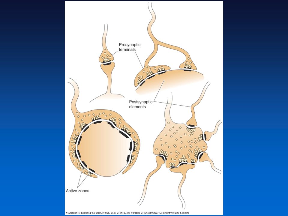
Synaptic cleft : nm wide (gap junctions : 3.5 nm) Adhere to each other by the help of a matrix of fibrous extracellular proteins in the synaptic cleft Presynaptic element (= axon terminal) contains Synaptic vesicles Secretory granules (~100nm) (=dense-core vesicles) Membrane differentiations Active zone Postsynaptic density
 Adhere to each other by the help of a matrix of fibrous extracellular proteins in the synaptic cleft. Presynaptic element (= axon terminal) contains. Synaptic vesicles. Secretory granules (~100nm) (=dense-core vesicles) Membrane differentiations. Active zone. Postsynaptic density.")
8
Chemical Synapses vs Electrical synapses

9
Types of Synapses CNS Synapses Axoaxonic: Axon to axon
Axodendritic: Axon to dendrite Axosomatic: Axon to cell body Axoaxonic: Axon to axon Dendrodendritic: Dendrite to dendrite

11
Types of Synapses CNS Synapses Gray’s Type I: Asymmetrical, excitatory
Gray’s Type II: Symmetrical, inhibitory

12
Types of Synapses The Neuromuscular Junction (NMJ)
Synapses between the axons of motor neurons of the spinal cord and skeletal muscle Studies of NMJ established principles of synaptic transmission Fast and reliable synaptic transmission(AP of motor neuron always generates AP in the muscle cell it innervates) thanks to the specialized structural features The largest synapse in the body Precise alignment of synaptic terminals with junctional folds
 thanks to the specialized structural features. The largest synapse in the body. Precise alignment of synaptic terminals with junctional folds.")
13
Principles of Chemical Synaptic Transmission
Basic Steps Neurotransmitter synthesis Load neurotransmitter into synaptic vesicles Vesicles fuse to presynaptic terminal Neurotransmitter spills into synaptic cleft Binds to postsynaptic receptors Biochemical/Electrical response elicited in postsynaptic cell Removal of neurotransmitter from synaptic cleft Must happen RAPIDLY!

14
Principles of Chemical Synaptic Transmission
Neurotransmitters Amino acids Amines Peptides

15
Principles of Chemical Synaptic Transmission
Neurotransmitters Amino acids and amines are stored in synaptic vesicles Peptides are stored in and released from secretory granules Often coexist in the same axon terminals Fast synaptic transmission and slower synaptic transmission

16
Principles of Chemical Synaptic Transmission
Neurotransmitter Synthesis and Storage Natural building blocks vs specialized neurotransmitters

17
Principles of Chemical Synaptic Transmission
Neurotransmitter Release Voltage-gated calcium channels open - rapid increase from mM to greater than 0.1 mM Exocytosis can occur very rapidly (within 0.2 msec) because Ca2+ enters directly into active zone ‘Docked’ vesicles are rapidly fused with plasma membrane Protein-protein interactions regulate the process (e.g. SNAREs) of ‘docking’ as well as Ca2+- induced membrane fusion Vesicle membrane recovered by endocytosis
 because Ca2+ enters directly into active zone. ‘Docked’ vesicles are rapidly fused with plasma membrane. Protein-protein interactions regulate the process (e.g. SNAREs) of ‘docking’ as well as Ca2+- induced membrane fusion. Vesicle membrane recovered by endocytosis.")
19
Principles of Chemical Synaptic Transmission
Neurotransmitter Release Reserve pool and Readily releasable pool (RRP)
")
20
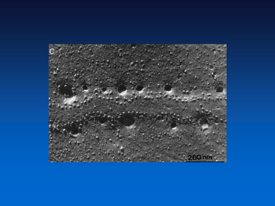
Fig. 1. Scattered distribution of RRP vesicles
S. O. Rizzoli et al., Science 303, (2004) Published by AAAS
 Published by AAAS.")
21
Principles of Chemical Synaptic Transmission
Neurotransmitter Release Secretory granules Released from membranes that are away from the active zones Requires high-frequency trains of action potentials to be released Ca2+ needs to be build up throughout the axon terminal Leisurely process (50 msec)
")
22
Principles of Chemical Synaptic Transmission
Neurotransmitter receptors: Ionotropic: Transmitter-gated ion channels Ligand-binding causes a slight conformational change that leads to the opening of channels Not as selective to ions as voltage-gated channels Depending on the ions that can pass through, channels are either excitatory or inhibitory Reversal potential

23
Principles of Chemical Synaptic Transmission
Excitatory and Inhibitory Postsynaptic Potentials: EPSP:Transient postsynaptic membrane depolarization by presynaptic release of neurotransmitter Ach- and glutamate-gated channels cause EPSPs

24
Principles of Chemical Synaptic Transmission
Excitatory and Inhibitory Postsynaptic Potentials: IPSP: Transient hyperpolarization of postsynaptic membrane potential caused by presynaptic release of neurotransmitter Glycine- and GABA-gated channels cause IPSPs

25
Principles of Chemical Synaptic Transmission
Metabotropic: G-protein-coupled receptors Trigger slower, longer-lasting and more diverse postsynaptic actions Same neurotransmitter could exert different actions depending on what receptors it bind to Autoreceptors: present on the presynaptic terminal Typically, G-protein coupled receptors Commonly, inhibit the release or synthesis of neurotransmitter Negative feedback Effector proteins

26
Principles of Chemical Synaptic Transmission
Neurotransmitter Recovery and Degradation Clearing of neurotransmitter is necessary for the next round of synaptic transmission Simple Diffusion Reuptake aids the diffusion Neurotransmitter re-enters presynaptic axon terminal or enters glial cells through transporter proteins The transporters are to be distinguished from the vesicular forms Enzymatic destruction In the synaptic cleft Acetylcholinesterase (AchE) Desensitization: Channels close despite the continued presence of ligand Can last several seconds after the neurotransmitter is cleared Nerve gases (e.g. sarin) inhibit AchE - increased Ach - AchR desensitization - muscle paralysis
 Desensitization: Channels close despite the continued presence of ligand. Can last several seconds after the neurotransmitter is cleared. Nerve gases (e.g. sarin) inhibit AchE - increased Ach - AchR desensitization - muscle paralysis.")
27
Principles of Chemical Synaptic Transmission
Neuropharmacology The study of effect of drugs on nervous system tissue Receptor antagonists: Inhibitors of neurotransmitter receptors e.g. Curare binds tightly to Ach receptors of skeletal muscle Receptor agonists: Mimic actions of naturally occurring neurotransmitters E.g. Nicotine binds and activates the Ach receptors of skeletal muscle (nicotinic Ach receptors) Toxins and venoms Defective neurotransmission: Root cause of neurological and psychiatric disorders
 Toxins and venoms. Defective neurotransmission: Root cause of neurological and psychiatric disorders.")
28
Principles of Synaptic Integration
Process by which multiple synaptic potentials combine within one postsynaptic neuron Basic principle of neural computation The Integration of EPSPs + - - + + -

29
Principles of Synaptic Integration
The integration of EPSPs Quantal Analysis of EPSPs Synaptic vesicles: Elementary units of synaptic transmission Contains the same number of transmitter molecules (several thousands) Postsynaptic EPSPs at a given synapse is quantized = The amplitude of EPSP is an integer multiple of the quantum Quantum: An indivisible unit determined by the number of transmitter molecules in a synaptic vesicle the number of postsynaptic receptors available at the synapse Miniature postsynaptic potential (“mini”) is generated by spontaneous, un-stimulated exocytosis of synaptic vesicles Quantal analysis: Used to determine number of vesicles that release during neurotransmission Neuromuscular junction: About 200 synaptic vesicles, EPSP of 40mV or more CNS synapse: Single vesicle, EPSP of few tenths of a millivolt
 Postsynaptic EPSPs at a given synapse is quantized = The amplitude of EPSP is an integer multiple of the quantum. Quantum: An indivisible unit determined by. the number of transmitter molecules in a synaptic vesicle. the number of postsynaptic receptors available at the synapse. Miniature postsynaptic potential ( mini ) is generated by spontaneous, un-stimulated exocytosis of synaptic vesicles. Quantal analysis: Used to determine number of vesicles that release during neurotransmission. Neuromuscular junction: About 200 synaptic vesicles, EPSP of 40mV or more. CNS synapse: Single vesicle, EPSP of few tenths of a millivolt.")
30
Principles of Synaptic Integration
EPSP Summation Allows for neurons to perform sophisticated computations Integration: EPSPs added together to produce significant postsynaptic depolarization Spatial summation : adding together of EPSPs generated simultaneously at different synapses Temporal summation : adding together of EPSPs generated at the same synapse in rapid succession (within msec of one another)
")
31
Principles of Synaptic Integration
The Contribution of Dendritic Properties to Synaptic Integration Dendrite as a straight cable : EPSPs have to travel down to spike-initiation zone to generate action potential Membrane depolarization falls off exponentially with increasing distance Vx = Vo/ex/ Vo : depolarization at the origin : Dendritic length constant Distance where the depolarization is 37% of origin (V= 0.37 Vo) In reality, dendrites have branches, changing diameter..
 In reality, dendrites have branches, changing diameter..")
32
Principles of Synaptic Integration
The Contribution of Dendritic Properties to Synaptic Integration Length constant () An index of how far depolarization can spread down a dendrite or an axon Depends on two factors internal resistance (ri) : the resistance to current flowing longitudinally down the dendrite membrane resistance (rm) : the resistance to current flowing across the membrane While ri is relatively constant (largely determined by the diameter of dendrite and electrical property of cytoplasm) in a mature neuron, rm changes from moment to moment (depends on the number of opne channels) Excitable Dendrites Dendrites of neurons having voltage-gated sodium, calcium, and potassium channels Can act as amplifiers (vs. passive) : EPSPs that are large enough to open voltage-gated channels can reach the soma by the boost offered by added currents through VGSCs Dendritic sodium channels: May carry electrical signals in opposite direction, from soma outward along dendrites : back-propagating action potential might inform the dendrites that an action potential has been generated I added this slide, it may be helpful to include the figure hear for the instructor
 An index of how far depolarization can spread down a dendrite or an axon. Depends on two factors. internal resistance (ri) : the resistance to current flowing longitudinally down the dendrite. membrane resistance (rm) : the resistance to current flowing across the membrane. While ri is relatively constant (largely determined by the diameter of dendrite and electrical property of cytoplasm) in a mature neuron, rm changes from moment to moment (depends on the number of opne channels) Excitable Dendrites. Dendrites of neurons having voltage-gated sodium, calcium, and potassium channels. Can act as amplifiers (vs. passive) : EPSPs that are large enough to open voltage-gated channels can reach the soma by the boost offered by added currents through VGSCs. Dendritic sodium channels: May carry electrical signals in opposite direction, from soma outward along dendrites : back-propagating action potential might inform the dendrites that an action potential has been generated. I added this slide, it may be helpful to include the figure hear for the instructor.")
33
Principles of Synaptic Integration
Inhibition Action of synapses to take membrane potential away from action potential threshold IPSPs and Shunting Inhibition Excitatory vs. inhibitory synapses: Bind different neurotransmitters, allow different ions to pass through channels GABA or glycine :: Cl- Ecl : -65 mV, at resting membrane potential no IPSP is visible

34
Principles of Synaptic Integration
Shunting Inhibition Inhibiting current flow from soma to axon hillock The Geometry of Excitatory and Inhibitory Synapses Inhibitory synapses Gray’s type II morphology Clustered on soma and near axon hillock Powerful position to influence the activity of the postsynaptic neuron

35
Principles of Synaptic Integration
Modulation Synaptic transmission that does not directly evoke EPSPs and IPSPs but instead modifies the effectiveness of EPSPs generated by other synapses with transmitter-gated ion channels Mediated by G-protein-coupled neurotransmitter receptors Example: Activating NE β receptor Suggest inserting figure 5.21 of NE example

Similar presentations


 REQUIRED READING: Kandel text, Chapter 12 At neuromuscular synapse, single axonal action.>")


 Nervous system functions Structure of a neuron Sensory, motor, inter- neurons Membrane potential Sodium.>")

 1. arrival of action potential at terminal bulb triggers opening of voltage-gated Ca ++ channels.>")


