Download presentation
Presentation is loading. Please wait.

1
Fluid and Electrolyte Balance during Injury Zohair Al Aseri. MD. FCEM(UK).FRCPC (EM&CCM) Chairman,National Emergency Medicine Committee Coordinatoor MPH-DME Master Consultant, ICU Department of Critical Care Chairman, Department of Emergency Medicine College of Medicine, King Saud University Medical City. Riyadh, KSA. zalaseri@ksu.edu.sa http://fac.ksu.edu.sa/zalaseri
.FRCPC (EM&CCM) Chairman,National Emergency Medicine Committee Coordinatoor MPH-DME Master Consultant, ICU Department of Critical Care Chairman, Department of Emergency Medicine College of Medicine, King Saud University Medical City. Riyadh, KSA.")
2
Fluid and Electrolyte Balance during Injury Objectives Understand normal regulation of fluid balance Fluid Imbalance In Shock State Fluid Therapy (Types) & Indication Electrolyte disturbances in trauma and surgery Acid base in surgery patients
 & Indication Electrolyte disturbances in trauma and surgery Acid base in surgery patients")
3
Case 1 39 year old male involved in MVC brought to ED by EMT, he is unconscious, heart rate 120 beat per minute blood pressure of 80/50, intubated in the scene, what is your immediate action a) Take further history b) Start him on dextrose 5% with NS c) Start him in colloid d) Start him in Normal saline
 Take further history b) Start him on dextrose 5% with NS c) Start him in colloid d) Start him in Normal saline")
4
Case2 A recovery nurse calling you to see a 70 year old male, 7 hours post appendicictomy, because he is drowsy and unresponsive, his vital signs are normal and oxygen saturation 92% on room air? What is the most likely diagnosis? a) Intracranial bleeding b) Stroke c) Acute renal failure d) Respiratory failure
 Intracranial bleeding b) Stroke c) Acute renal failure d) Respiratory failure.")
5
Case3 70 year old male, admitted for elective hernia repair, kept NPO and started in D5 ½ normal saline 24 hour ago, his current electrolyte showed k of 5 mmol and Na of 128 mmol What is the most likely diagnosis? a) DI b) SAIDH c) Acute renal failure d) Iatrogenic hyponatremia
 DI b) SAIDH c) Acute renal failure d) Iatrogenic hyponatremia.")
6
Hypovolaemia Reduced circulating volume Causes Loss of blood, electrolyte-containing fluid or water. Third-space loss due to increased vascular permeability. Fluid and Electrolyte Balance during Injury

7
Hypovoluemia will reduce oxygen and nutrient delivery Leads to increase healing and recovery times.

8
Fluid-conserving measures Oliguria and sodium and water retention Due to the release ADH and aldosterone after major surgery or injury May persist even after normal circulating volume has been restored. Fluid and Electrolyte Balance during Injury

9
Fluid-conserving measures: Increased ADH Secretion from the posterior pituitary is response to Afferent nerve impulses from the site of injury Atrial stretch receptors (responding to reduced volume) & aortic and carotid baroreceptors (responding to reduced pressure) Increased plasma osmolality (principally the result of an increase in sodium ions) detected by hypothalamic osmoreceptors. Input from higher centres in the brain (responding to pain, emotion and anxiety). Fluid and Electrolyte Balance during Injury
. Fluid and Electrolyte Balance during Injury.")
10
Fluid-conserving measures: ADH promotes retention of free water (without electrolytes) by cells of the distal renal tubules and collecting ducts. Fluid and Electrolyte Balance during Injury

11
Aldosterone secretion from the adrenal cortex is increased by: Activation of the renin-angiotensin system. Renin is released from afferent arteriolar cells in the kidney in response to reduced blood pressure and activation of the renal sympathetic nerves. Fluid and Electrolyte Balance during Injury

12
Aldosterone secretion from the adrenal cortex is increased by: Renin converts circulating angiotensinogen to angiotensin AT-1. AT-1 is converted by angiotsion converting enzyme (ACE) in plasma and tissues (particularly the lung) to AT-2 which cause arteriolar vasoconstriction and aldosterone secretion Fluid and Electrolyte Balance during Injury
 in plasma and tissues (particularly the lung) to AT-2 which cause arteriolar vasoconstriction and aldosterone secretion Fluid and Electrolyte Balance during Injury.")
13
Adrenocorticotropic hormone (ACTH) Increased by the anterior pituitary in response to hypovolaemia and hypotension via afferent nerve impulses from stretch receptors in the atria, aorta and carotid arteries. lt is also raised by ADH. Direct stimulation of the adrenal cortex by hyponatraemia or hyperkalaemia. Fluid and Electrolyte Balance during Injury

14
Aldosterone increases the reabsorption of both sodium and water by distal renal tubular + simultaneous excretion of hydrogen and potassium ions into the urine. Fluid and Electrolyte Balance during Injury

15
Duration of increased ADH and aldosterone secretion following injury? Usually lasts 48-72 hours during which time urine volume is reduced and osmolality increased. Fluid and Electrolyte Balance during Injury

16
Urinary sodium excretion decreases to 10-20 mmol /24hrs (normal 50- 80mmol /24 hrs) Urinary potassium excretion increases to > 100 mmol/24 hrs (normal 50- 80mmol /24 hrs). Fluid and Electrolyte Balance during Injury

17
Blood flow-conserving measures: Hypovolaemia Reduces cardiac preload which leads to a fall in cardiac output. Increased sympathetic activity results in a compensatory increase in cardiac output, peripheral vasoconstriction and a rise in blood pressure. Fluid and Electrolyte Balance during Injury

18
FLUID AND ELECTROLYTE BALANCE: May be altered in the surgical patient for several reasons: ADH and aldosterone secretion as described above. Loss from the GI tract (e.g. bowel preparation, ileus, stomas, fistulae). Reduced oral fluid intake in the perioperative period
. Reduced oral fluid intake in the perioperative period.")
19
Fluid and Electrolyte Balance during Injury FLUID AND ELECTROLYTE BALANCE: Insensible losses (e.g. sweating secondary to fever). Third space losses. Surgical drains. Medications (e.g. diuretics). Underlying chronic illness (e.g. cardiac failure, portal hypertension).
. Third space losses. Surgical drains. Medications (e.g. diuretics). Underlying chronic illness (e.g. cardiac failure, portal hypertension)..")
20
Normal water and electrolyte balance: Water forms about 60% of total body weight in men and 55% in women. Approximately two-thirds is intracellular, one- third extracellular. Extracellular water is distributed between the plasma and the interstitial space Fluid and Electrolyte Balance during Injury

21
Regulation of Fluid Balance TOTAL BODY FLUID (40) liters;60%TBW Red cell volume (2 liters) Plasma volume (3 liters,5 %) Extracellular (15 liters,20%) Blood volume (5 liters) Intracellular (25 liters,40%) The intracellular and extracellular compartments are separated by water-permeable cell membranes.
 liters;60%TBW Red cell volume (2 liters) Plasma volume (3 liters,5 %) Extracellular (15 liters,20%) Blood volume (5 liters) Intracellular (25 liters,40%) The intracellular and extracellular compartments are separated by water-permeable cell membranes.")
22
IC. WATER ECF 2/3 intrest. 1/3 blood 25 150 15 0.01 2 6 50 Na K Mg Ca Cl Hco3 Phos 140 4.5 1.2 2.4 100 25 1.2 Fluid & Electrolyte Balance

23
Regulation of Fluid Balance ECC Osmolarity ECF Volume Prevent swelling or shrinking of the cells Maintain BP

24
Osmolality of extracellular fluid normally 275-295 mOsmol/kg determined primarily by sodium and chloride ion concentrations. Fluid and Electrolyte Balance during Injury

25
Normal water and electrolyte balance: Plasma oncotic pressure is primarily determined by albumin. Fluid and Electrolyte Balance during Injury

26
Normal water and electrolyte balance: In adults, Normal daily fluid requirement is 30-35ml / kg (-2500 ml /day). In newborn babies and children Contain proportionately more water than adults. Daily maintenance fluid requirement at birth is about 75ml/ kg, increasing to 150 ml/ kg during the first weeks of life. Fluid and Electrolyte Balance during Injury

27
Normal water and electrolyte balance: After first month of life, fluid requirements decrease and the '4/2/1' formula can be used to estimate maintenance fluid requirements: first l0 kg of body weight requires 4ml /kg/h; the next 10kg 2ml /kg/ h; thereafter each kg of body requires 1ml/kg/h. Fluid and Electrolyte Balance during Injury

28
The estimated maintenance fluid requirements of a 35 kg child would therefore be: (10 X 4) + (10 X 2) + (15 X 1) = 75 mljh.150. The daily requirement for both sodium and potassium in children is about 2-3mmol/kg. Fluid and Electrolyte Balance during Injury

29
Assessing losses in the surgical patient: Fluid and Electrolyte Balance during Injury

30
Assessing losses in the surgical patient: Normal Daily Losses and requirements for Fluid and Electrolytes Volume (ml) Na+ (mmol)K+ (mmol) Urine20008060 Insensible losses from skin and respiratory tract 700-- Faeces300-10 Less water created from metabolism 300-- Total27008070
 Na+ (mmol)K+ (mmol) Urine Insensible losses from skin and respiratory tract Faeces Less water created from metabolism Total")
31
Typical Losses per 24hrs Factors Modifying Volume Insensible Losses 700-2000ml Losses associated with pyrexia, sweating and use of non-humidified Urine 1000- 2500ml With aldosterone and DH secretion; With diuretic Therapy Gut300-1000ml Losses with obstruction, ileus, fistulae and diarrhea (may increase substantially) Third spaces Losses 0-4000ml Losses with greater extent of surgery and tissue trauma Fluid and Electrolyte Balance during Injury Source of Fluid Loss in Surgical Patients
 Third spaces Losses ml Losses with greater extent of surgery and tissue trauma Fluid and Electrolyte Balance during Injury Source of Fluid Loss in Surgical Patients")
32
Insensible fluid losses: Hyperventilation increases insensible water loss is not usually large unless the normal mechanisms for humidifying inhaled air (the nasal passages and upper airways) are compromised. this occurs in intubated patients or in those receiving non humidified high-flow oxygen. In these situations inspired gases should be humidified routinely. Fluid and Electrolyte Balance during Injury

33
Insensible fluid losses: Pyrexia 200ml/day for each 1°C rise in temperature. Sweating May increase fluid loss by up to 1 litre/hour Sweat also contains significant amounts of sodium (20- 70mmol/l) and potassium (10mmol/l). Fluid and Electrolyte Balance during Injury
 and potassium (10mmol/l). Fluid and Electrolyte Balance during Injury.")
34
The effect of surgery: The stress response ADH leads to water retention and a reduction in urine volume for 2-3days following major surgery. Aldosterone conserves both sodium and water, further contributing to oliguria. Urinary sodium excretion falls while urinary potassium excretion increases, predisposing to hypokalaemia. Fluid and Electrolyte Balance during Injury

35
'Third-space' losses: if tissue injury is severe, widespread and/or prolonged then the loss of water, electrolytes and colloid particles into the interstitial space can amount to many litres and can significantly decrease circulating blood volume following trauma and surgery. Fluid and Electrolyte Balance during Injury

36
Regulation of Fluid Balance Q=K[(Pc-Pi)-@(Oc-Oi)] 17 Mm Hg Oncotic P=25 37 mm Hg VenuleArteriole Pnet =(37-1)+(0-25)=11 Interstitial Hydrostatic P=1 Pnet =(17-1)+(0-25)=-9
+(0-25)=11 Interstitial Hydrostatic P=1 Pnet =(17-1)+(0-25)=-9")
37
'Third-space' losses: Colloid oncotic pressure throughout the lumen of the capillary is 25mmHg. The hydrostatic pressure is 1 mmHg in the interstitium. Hydrostatic pressure on the arteriolar side of the capillary falls from 37 mmHg to 17 mmHg on the venular side. net outward pressure on the arteriolar side (37 - 1 - 25 = 11) net inward pressure (25 -17-1= 9) on the venular side. Fluid and Electrolyte Balance during Injury
 net inward pressure ( = 9) on the venular side. Fluid and Electrolyte Balance during Injury.")
38
Hydrostaticpressure Oncoticpressure tending to move fluid out of the capillaries tending to keep fluid within the capillaries THE STARLING EQUATION Excess fluid filtered is collected through the lymphatic circulation and returned to the Systemic circulation Regulation of Fluid Balance

39
Third-space' losses: Oedema is formed if 1. hydrostatic pressure increases on the venular side as in heart failure or 2. colloid oncotic pressure falls due to liver or kidney disease or 3. permeability is increased as in sepsis and /or injury. Fluid and Electrolyte Balance during Injury

40
Loss from the gastrointestinal tract The magnitude and content of fluid losses depends on the site of loss or lntestinal obstruction. ln general, the higher an obstruction occurs in the intestine, the greater the fluid loss Fluid and Electrolyte Balance during Injury

41
VolumeNa+K+Cl-HCO3 Plasma-140510025 Gastric Secretions 250050108040 Intestinal Fluid (Upper) 30001401010025 Bile and Pancreatic secretions 150014058060 Mature ileostomy5005052025 Diarrhoea (Inflammatory) -1104010040 Mixed Gastric Aspirate -12010-- The approximate daily volumes (ml) and electrolyte concentrations (mmol/l) of various gastrointestinal fluids.
 Bile and Pancreatic secretions Mature ileostomy Diarrhoea (Inflammatory) Mixed Gastric Aspirate The approximate daily volumes (ml) and electrolyte concentrations (mmol/l) of various gastrointestinal fluids.")
42
Loss from the gastrointestinal tract Paralytic ileus. Propulsion in the small intestine ceases, has numerous causes. Resolves within 1-2days of the operation. Fluid and Electrolyte Balance during Injury

43
Loss from the gastrointestinal tract: Intestinal fistula associated with the greatest fluid and electrolyte losses. Fluid and Electrolyte Balance during Injury

44
Loss from the gastrointestinal tract: Diarrhoea. Patients may present with diarrhoea or develop it during the perioperative period. Fluid and electrolyte losses may be considerable. Fluid and Electrolyte Balance during Injury

45
Intravenous fluid administration: When choosing and administering intravenous fluids it is important to consider: what fluid deficiencies are present. fluid compartments requiring replacement. electrolyte disturbances present. which fluid is most appropriate. Fluid and Electrolyte Balance during Injury

46
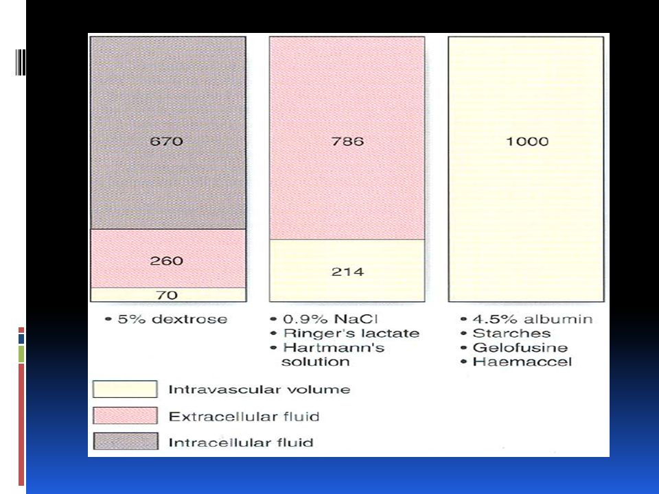
Types of intravenous fluid: Dextrose After the IV administration of 1000 ml 5% dextrose solution, about 670ml of water will be added to the lFC and about 330 ml of water to the EFC, of which about 70ml will be intravascular. therefore Dextrose solutions are of little value as resuscitation fluids to expand intravascular volume Fluid and Electrolyte Balance during Injury

48
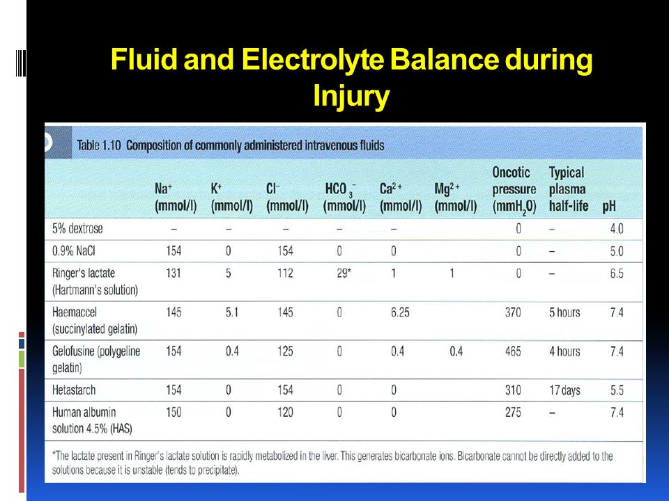
Crystalloids Sodium chloride 0.9%(NS) and Hartmann' s solution (Ringer) are isotonic solutions Sodium chloride NS 0.9 contains 9g of sodium chloride dissolved in l000ml of water. (Ringer's lactate) has a more composition, containing lactate, potassium and calcium addition to sodium and chloride ions. Fluid and Electrolyte Balance during Injury
 has a more composition, containing lactate, potassium and calcium addition to sodium and chloride ions. Fluid and Electrolyte Balance during Injury.")
49
Crystalloids Both normal saline and Ringer solution have an osmolality similar to extracellular fluid (about 300m0sm/l) distribute rapidly to ECF compartment after venous administration. Fluid and Electrolyte Balance during Injury

50
One liter of isotonic saline contains 154 meq of sodium and an equivalent number of chloride ions. Fluid and Electrolyte Balance during Injury

51
Balanced solutions, such as Ringer's lactate closely match the composition of extracellular fluid by providing physiological concentrations of sodium and lactate in place of bicarbonate. After administration the lactate is metabolized, resulting in bicarbonate generation. Fluid and Electrolyte Balance during Injury

52
Balanced solutions, such as Ringer's lactate Decrease the risk of hyperchloraermia, which can occur following large volumes of fluids NS. Fluid and Electrolyte Balance during Injury

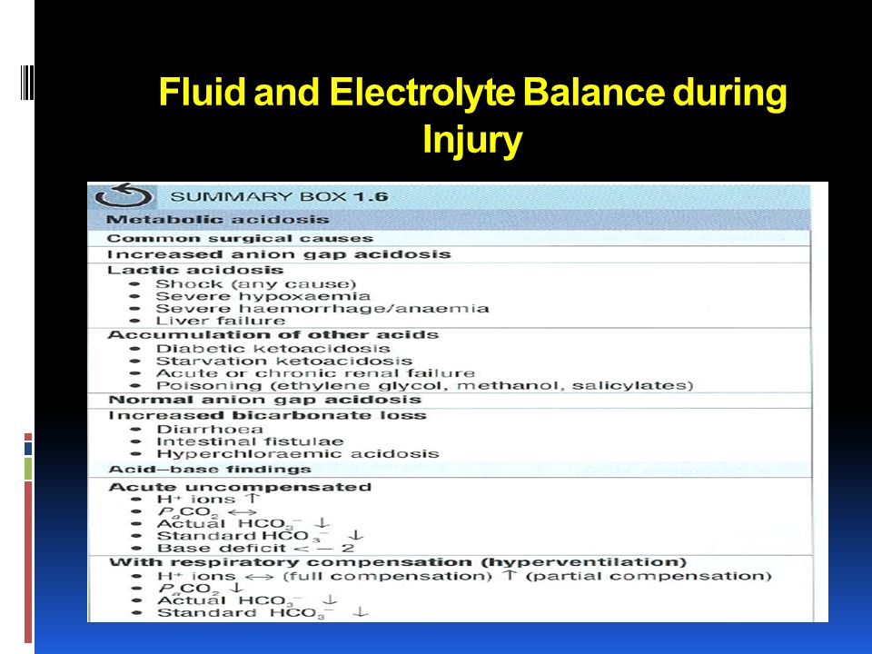
53
Hypertonic saline solutions Induce a shift of fluid from the IFC to the EFC Reducing brain water and increasing intra vascular volume and serum sodium concentration. Potential indications cerebral oedema raised intracranial pressure hyponatraemic seizures 'small volume' resuscitation of hypovolaemic shock. Fluid and Electrolyte Balance during Injury

54
Dextrose saline solutions Commercially available 5% dextrose with 0.9% normal saline is a hypertonic solution (twice the osmolarity of plasma) and should be used with caution. Fluid and Electrolyte Balance during Injury

55
Colloids: albumin or be synthetically modified (e.g. gelatins, hydroxyethyl starches [HES], dextrans). When administered, colloid remains largely within the intravascular space until the colloid particles are removed by the reticuloendothelial system. Fluid and Electrolyte Balance during Injury
. When administered, colloid remains largely within the intravascular space until the colloid particles are removed by the reticuloendothelial system. Fluid and Electrolyte Balance during Injury.")
56
Colloids: The intravascular half-life is usually between 6 and 24 hours and such solutions are therefore appropriate for fluid resuscitation. Electrolyte-containing solution distributes throughout the EFC. But No Evidence Fluid and Electrolyte Balance during Injury

57
Colloids: Synthetic colloids are more expensive than crystalloids and have variable side effect profiles. Recognized risks Coagulopathy Reticuloendothelial system dysfunction Pruritus and anaphylactic reactions Renal failure when used for resuscitation in patients with septic shock. Fluid and Electrolyte Balance during Injury

59
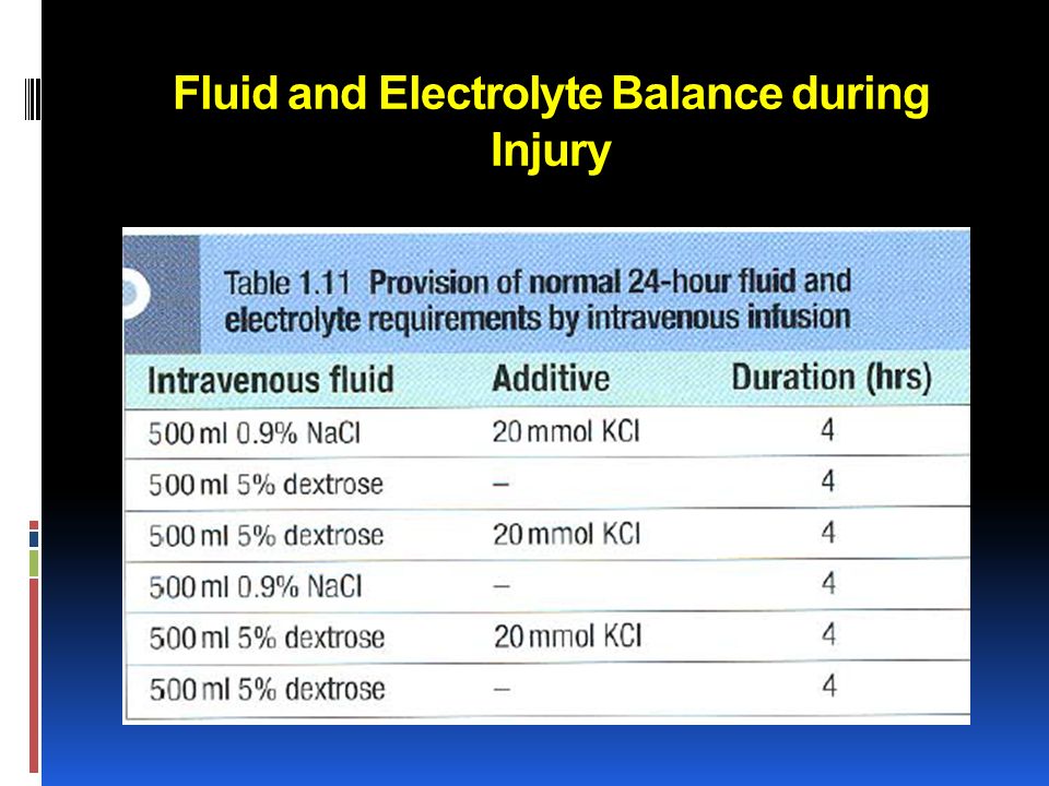
Maintenance fluid requirements under normal conditions, adult daily sodium requirements (80mmol) may be provided by the administration of 500- 1000ml of 0.9% sodium chloride. The remaining requirement to maintain fluid balance (2000-2500ml).., typically provided as 5% dextrose. Fluid and Electrolyte Balance during Injury
.., typically provided as 5% dextrose. Fluid and Electrolyte Balance during Injury.")
60
Maintenance fluid requirements Daily potassium requirements (60-80mmol) are usually met by adding potassium chloride to maintenance fluids, but the amount added can be titrated to measured plasma concentrations. “potassium should not be administered at a rate greater than 10-20 mmol/h except in severe potassium deficiency. Fluid and Electrolyte Balance during Injury

61
Maintenance fluid requirement: The provision of total parenteral nutrition should also be considered in this situation. Fluid and Electrolyte Balance during Injury

62
Treatment of postoperative hypovolaemia and/or hypotension: Hpovolaemia is common in the postoperative period and may present with one or more of the following: tachycardia, pallor, clammy skin, collapsed peripheral pulses oliguria and / or hypotension. Fluid and Electrolyte Balance during Injury

63
Treatment of postoperative hypovolaemia and/or hypotension: Intravascular volume should be rapidly restored with a series of fluid boluses (e.g. 250-500 ml) with the clinical response being assessed after each bolus. Fluid and Electrolyte Balance during Injury
 with the clinical response being assessed after each bolus. Fluid and Electrolyte Balance during Injury.")
64
Crystalloids Versus Colloids Crystalloids or colloids in fluid therapy??

65
Fluid and Electrolyte Balance during Injury

67
Water depletion Pure water depletion is common in surgical practice, and is usually combined with sodium loss. The most frequent causes are inadequate intake or excessive gastrointestinal losses. Fluid and Electrolyte Balance during Injury

68
Water excess common in patients who receive large volumes of intravenous 5% dextrose in the early postoperative period. Such patients have an increased extracellular volume and are commonly hyponatraemic. Fluid and Electrolyte Balance during Injury

69
Water Excess: Difficult to detect clinically Patients with water excess usually remain well Oedema may not be evident until the extracellular volume has increased by more than 4 litres. Fluid and Electrolyte Balance during Injury

70
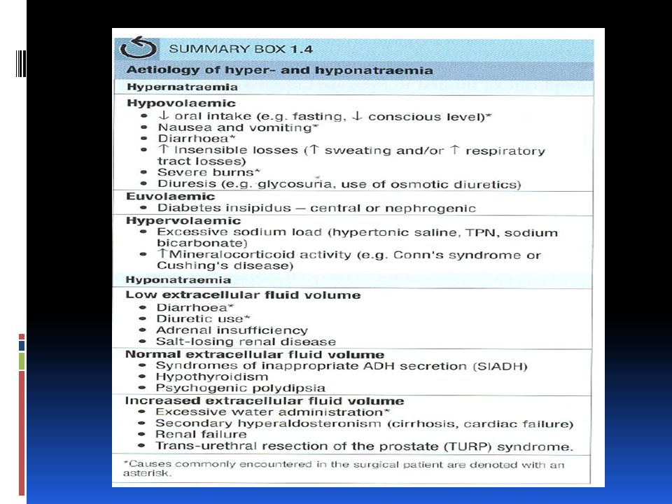
Hypernatraemia: Normal sodium levels are in range 136- 144mmol/l. Hypernatraemia (>145mmol) results from either water or hypotonic fluid loss or sodium gain. Fluid and Electrolyte Balance during Injury
 results from either water or hypotonic fluid loss or sodium gain. Fluid and Electrolyte Balance during Injury.")
71
Hypernatraemia: Water loss is commonly caused by reduced water intake vomiting, diarrhea diuresis, burns sweating and insensible losses from the respiratory tract diabetes insipidus. Fluid and Electrolyte Balance during Injury Typically associated with hypovolaemia

72
Hypernatraemia: Sodium gain is usually caused by excess sodium administration in hypertonic intravenous fluids Typically associated with hypervolaemia. Fluid and Electrolyte Balance during Injury

73
Hypovolaemic Hypernatraemia is treated with isotonic crystalloid followed by the more gradual administration of water to correct the relative water deficit. We can use 5%dextrose,1/2 NS or 1/4 NS Fluid and Electrolyte Balance during Injury

74
Cells, particularly brain cells, adapt to a high sodium concentration in extracellular fluid, and once this adaptation has occurred, rapid correction of severe hypernatraemia can result in a rapid rise in intracellular volume, cerebral oedema, seizures and permanent neurological injury. Fluid and Electrolyte Balance during Injury

75
Hyponatraemia (Na- < 135mmolfl) can occur with high, low or normal extracellular volume. The commonest cause is the administration of hypotonic intravenous fluids (as intravenous 5% dextrose) is administered in the postoperative period (dilutional hyponatraemia). Fluid and Electrolyte Balance during Injury
 is administered in the postoperative period (dilutional hyponatraemia). Fluid and Electrolyte Balance during Injury.")
76
Hyponatraemia Other causes include diuretic use and (SIADH) Co-morbidities associated with secondary hyperaldosteronism, such as cirrhosis and congestive cardiac failure. Fluid and Electrolyte Balance during Injury

77
Sodium deficit This can be calculated as follows: 140- measured sodium x 0.2 x weight in kg where 0.2 refers to the 20% extracellular space which represents the compartment in which sodium is the main cation. Fluid and Electrolyte Balance during Injury

78
Hyponatraemia Treatment depends on correct identification of the cause: If ECF volume is normal or increased, the most likely cause is excessive intravenous water administration and this will correct spontaneously if water intake is reduced. Fluid and Electrolyte Balance during Injury

79
Hyponatraemia Although less common in surgical patients, SIADH promotes the renal tubular reabsorption of water independently of sodium concentration, resulting in inappropriately concentrated urine (osmolality> 100m0sm / l) in the face of hypotonic plasma (osmolality< 290m0sm/ l). Fluid and Electrolyte Balance during Injury

80
Hyponatraemia The urine osmolality helps to distinguish inappropriate ADH secretion from excessive water administration. 'Spot‘ measurement of urine sodium will be high. Fluid and Electrolyte Balance during Injury

81
Hyponatraemia In patients with decreased ECF volume, hyponatracmia usually indicates combined water and sodium deficiency. This is most frequently the result of Diuresis Diarrhea Adrenal insufficiency Treatment by 0.9 sodium chloride Fluid and Electrolyte Balance during Injury

82
Hyponatraemia severe hyponatraemia (< 120mmol/ l) associated with confusion, seizures and coma. Fluid and Electrolyte Balance during Injury

83
Hyponatraemia Rapid correction of sodium concentration can precipitate an irreversible demyelinating condition known as central pontine myelinolysis Sodium concentration should not increase by more than 0.5 mmol/h. Fluid and Electrolyte Balance during Injury

84
Hyponatraemia This can usually be achieved by the cautious administration of isotonic (0.9%) sodium chloride, occasionally combined with the use of a loop diuretic (e.g. furosemide). Hypertonic saline solutions only for sever hyponatremia with CNS manifestation like seizure Fluid and Electrolyte Balance during Injury
. Hypertonic saline solutions only for sever hyponatremia with CNS manifestation like seizure Fluid and Electrolyte Balance during Injury.")
86
Potassium 98% of total body potassium (around 3500mmol) is intracellular serum potassium concentration (normally 3.5- 5 mmol/ l) is a poor indicator of total body potassium. Fluid and Electrolyte Balance during Injury

87
Potassium no absolute formula to determine K deficit. When the serum K is < 2.5mmol/ l about 100- 200mmol of KCL will be needed in a 70kg adult. Serial monitoring of serum K is necessary to prevent overcorrection Fluid and Electrolyte Balance during Injury

88
Potassium Once the serum K comes above 3.0 mmol /1, K supplements can be reduced. Acidosis reduces Na+/kATpase activity and results in a net efflux of potassium from cells and hyperkalaemia. Conversely, alkalosis results in an influx of potassium into cells and hypokalaemia. Fluid and Electrolyte Balance during Injury

89
Hyperkalaemia This is a potentially life-threatening condition. caused by exogenous administration of K release of K from cells (transcellular shift) as a result of tissue damage or changes in the Na / K-ATPase function impaired renal excretion. Fluid and Electrolyte Balance during Injury
 as a result of tissue damage or changes in the Na / K-ATPase function impaired renal excretion. Fluid and Electrolyte Balance during Injury.")
90
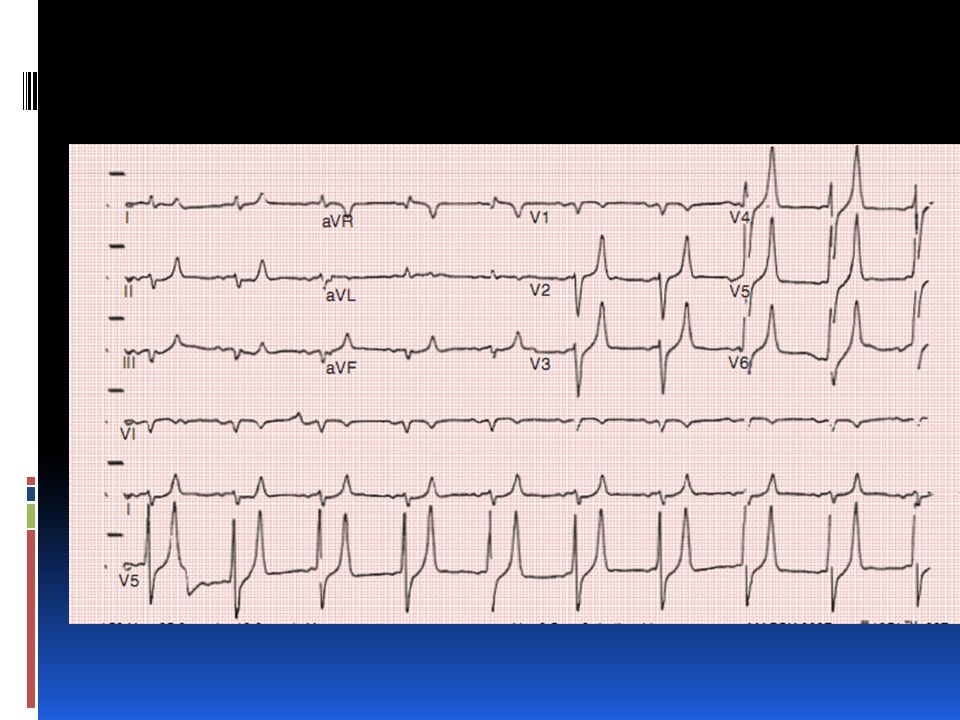
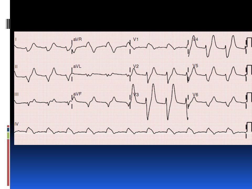
Hyperkalaemia Mild hyperkalaemia (K" < 6mmol/l) is often asymptomatic. High K cause progressive slowing of electrical conduction in the heart and the development of significant cardiac arrhythmias. So ECG is mandatory in all suspected hyperkalaemia Fluid and Electrolyte Balance during Injury

91
Hyperkalaemia ECG Finding Tall 'tented' T-waves in the precordial leads are the earliest flattening (or loss) of the P waves prolonged PR interval widening of the QRS asystole. Fluid and Electrolyte Balance during Injury

94
Hyperkalaemia Severe hyperkalaemia (K > 7m mmol/l) requires immediate treatment Fluid and Electrolyte Balance during Injury
 requires immediate treatment Fluid and Electrolyte Balance during Injury")
95
Treatment of hyperkalaemia

96
Hypokalaemia common in surgical patients. Dietary intake of k is normally 60-80 mmol / day. Under normal conditions, the majority of k loss (> 85%) is via the kidneys Maintenance of K balance largely depends on normal renal tubular regulation. Fluid and Electrolyte Balance during Injury
 is via the kidneys Maintenance of K balance largely depends on normal renal tubular regulation. Fluid and Electrolyte Balance during Injury.")
97
Hypokalaemia K excretion is increased by Metabolic alkalosis Diuresis Increased aldosterone release Increased losses from the GI tract. Fluid and Electrolyte Balance during Injury

98
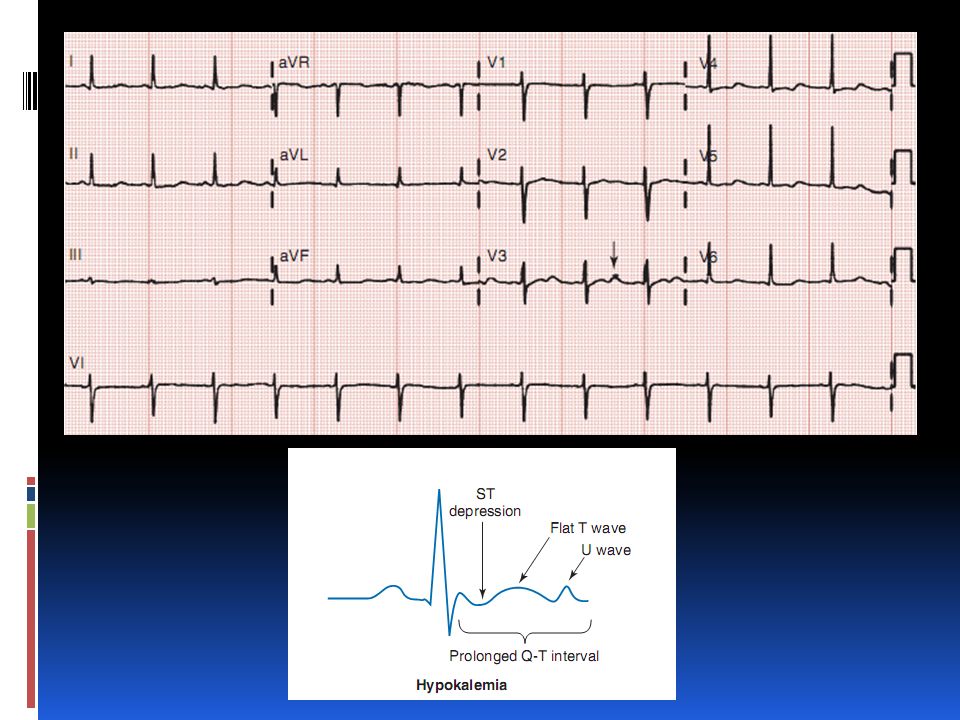
Hypokalaemia Diagnostic features Muscle weakness Paralytic ileus Flattening of T waves Prominent u waves Fluid and Electrolyte Balance during Injury

100
Hypokalaemia For every 3 K ions that come out from the intracellular compartment, one H and two Na ions are exchanged causing extracellular alkalosis and intracellular acidosis. Fluid and Electrolyte Balance during Injury

101
Hypokalaemia Treatment Oral or NG potassium replacement in mild hypokalaemia. Severe (K" < 2.5 mmol/1) or symptomatic hypokalaemia requires IV replacement. Fluid and Electrolyte Balance during Injury
 or symptomatic hypokalaemia requires IV replacement. Fluid and Electrolyte Balance during Injury.")
103
Calcium Clinically significant abnormalities in endocrine surgery. Fluid and Electrolyte Balance during Injury

104
Magnesium Hypomagnesaemia is common in restricted oral intake intravenous fluids for several days Fluid and Electrolyte Balance during Injury

105
Magnesium It is frequently associated with other electrolyte abnormalities, notably hypokalaemia, hypocalcaemia and hypophosphataemia. Fluid and Electrolyte Balance during Injury

106
Hypomagnesaemia associated with arrhythmias (most notably torsades de pointes (polymorphic ventricular tachycardia) and atrial fibrillation) Manifestations of are nonspecific (muscle weakness, muscle cramps, altered mentation, tremors, hyperreflexia and generalized seizures). Fluid and Electrolyte Balance during Injury

107
When hypokalaemia and hypomagnesaemia coexist it may be difficult to correct the former without correcting the latter. Fluid and Electrolyte Balance during Injury Hypomagnesaemia

108
Phosphate Phosphate is a critical component in many biochemical processes such as ATP synthesis, cell signaling and nucleic acid synthesis. Fluid and Electrolyte Balance during Injury

109
Hypophosphataemia common in surgical patients Severe (< 0.4 mmol/1) causes widespread cell dysfunction, muscle weakness, impaired myocardial contractility, reduced cardiac output altered sensorium. Fluid and Electrolyte Balance during Injury

110
Hypophosphataemia most commonly occurs in malnourished and/ or alcoholic patients commencing enteral or parenteral nutrition. Sepsis is another situation in which marked hypophosphataemia can be seen Fluid and Electrolyte Balance during Injury

111
Hypophosphataemia refeeding syndrome Hypophosphataemia accompanied by fluid retention and an increase in ECF volume To avoid it feeding should be established gradually with measurement and supplementation of serum electrolytes (phosphate, magnesium and potassium ). Fluid and Electrolyte Balance during Injury

112
Hypophosphataemia Treatment Phosphate can be supplemented orally or by slow intravenous infusion. Fluid and Electrolyte Balance during Injury

113
Acid-base balance Acidosis ('acidaemia' if plasma pH< 7.35) Alkalosis ('alkalaemia' if plasma pH> 7.45). Both acidosis and alkalosis may be respiratory or metabolic in origin. Fluid and Electrolyte Balance during Injury

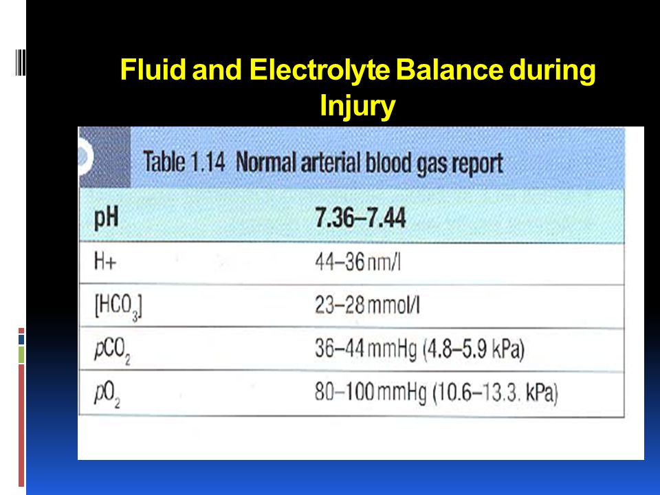
114
Acid-base balance VBG is good, ABG is more accurate coupled with measurement of blood lactate concentration Fluid and Electrolyte Balance during Injury

117
Acid-base balance Acid-base abnormalities are tackled by the body by means of blood buffers respiratory system kidneys. Fluid and Electrolyte Balance during Injury

118
Acid-base balance When the cause is metabolic, respiratory compensation is the most rapid (minutes) followed by the buffering systems hours) and kidneys (days). Fluid and Electrolyte Balance during Injury

119
Acid-base balance Bicarbonate buffer is the most important in the blood (65%) followed by the protein buffers (30%). Fluid and Electrolyte Balance during Injury

120
Acid-base balance Bicarbonate buffer (buffer is an acid-base combination where the acid is only partially dissociated) moves from left to right or vice versa depending on the addition of or loss of acid load with an aim to keep the HCO 3 / H 2 CO 3 ratio at 20:1. Fluid and Electrolyte Balance during Injury

121
Metabolic acidosis increase in plasma hydrogen ions in conjunction with a decrease in bicarbonate concentration. A rise in plasma hydrogen ion concentratlon stimulates chemoreceptors in the medulla resulting in a compensatory respiratory alkalosis an increase in minute volume and a fall in PaCO 2 Fluid and Electrolyte Balance during Injury

122
Metabolic acidosis Causes Endogenous acid (e.g. lactic acid or ketone bodies) referred to as 'increased anion gap acidosis' or Increased loss of bicarbonate (e.g. intestinal fistula, hyperchloraemic acidosis) which leads to 'normal anion gap acidosis'. Fluid and Electrolyte Balance during Injury
 referred to as increased anion gap acidosis or Increased loss of bicarbonate (e.g. intestinal fistula, hyperchloraemic acidosis) which leads to normal anion gap acidosis . Fluid and Electrolyte Balance during Injury.")
123
Metabolic acidosis Anion gap 12-l5mmol/l. Na - (Cl + HCO3) Fluid and Electrolyte Balance during Injury
 Fluid and Electrolyte Balance during Injury.")
124
Metabolic acidosis In surgery or trauma lactic acidosis is the communist cause Fluid and Electrolyte Balance during Injury

125
Metabolic acidosis Base deficit is a measure of the amount of bicarbonate required to correct acidosis and is calculated as follows: Base deficit= normal bicarbonate- measured bicarbonate x0.2 x weight in kg. where 0.2 refers to the extracellular compartment. Fluid and Electrolyte Balance during Injury

126
Metabolic acidosis Treatment is directed towards restoring circulating blood volume and tissue perfusion. Blood gas analysis should be repeated every 4-6 hours to assess the requirement for further corrections Fluid and Electrolyte Balance during Injury

128
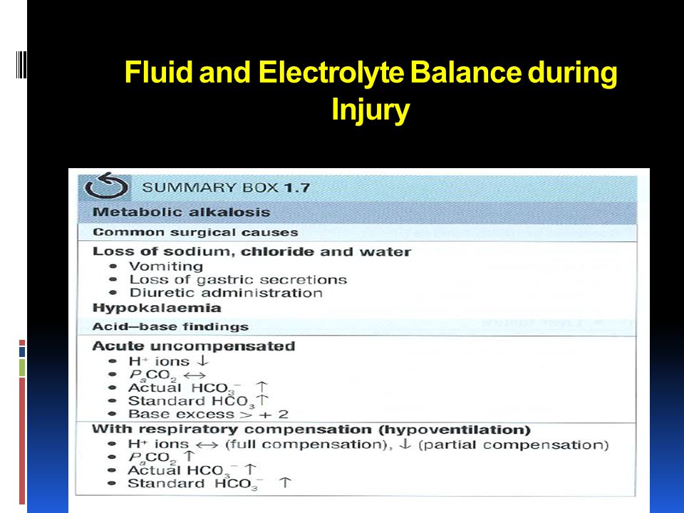
Metabolic alkalosis A decrease in plasma hydrogen ion concentration and an increase in bicarbonate concentration. A rise in PaCO 2 occurs as a consequence of the rise in bicarbonate concentration, resulting in a compensatory respiratory acidosis. Fluid and Electrolyte Balance during Injury

129
Metabolic alkalosis in surgery Mainly due to hypokalaemia and hypochloraemia. The kidney has an enormous capacity to generate bicarbonate ions and this is stimulated by chloride loss. Fluid and Electrolyte Balance during Injury

130
Metabolic alkalosis Example Metabolic alkalosis seen following significant (chloride-rich) losses from the GI tract when combined with loss of acid from conditions such as gastric outlet obstruction. Fluid and Electrolyte Balance during Injury

131
Metabolic alkalosis Treatment Adequate fluid replacement Correction of electrolyte disturbances, notably hypokalaemia and hypochloraemia Treatment of the primary cause. Fluid and Electrolyte Balance during Injury

133
Respiratory acidosis common postoperative problem Increased PC0 2 and plasma bicarbonate concentrations. Hypoventilation Fluid and Electrolyte Balance during Injury Examples general anaesthesia excessive opiate administration

134
Respiratory acidosis Hypoventilation respiratory acidosis require ventilatory support Fluid and Electrolyte Balance during Injury

136
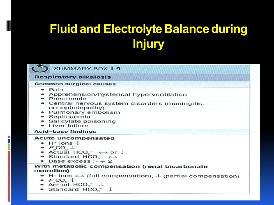
Respiratory alkalosis Respiratory alkalosis is caused by excessive excretion of CO 2 as a result of hyperventilation. PCO 2 and hydrogen ion-concentration decrease. Respiratory alkalosis is rarely chronic and usually does not need specific treatment. Fluid and Electrolyte Balance during Injury

138
Mixed patterns of acid-base imbalance Mixed patterns of acid-base disturbance are common, particularly in very sick patients. Fluid and Electrolyte Balance during Injury

139
Case 1 39 year old male involved in MVC brought to ED by EMT, he is unconscious, hr 120 bp 80/50, intubated in the scene, what is your immediate action a) Take further history b) Start him on dextrose 5% with NS c) Start him in colloid d) Start him in Normal saline
 Take further history b) Start him on dextrose 5% with NS c) Start him in colloid d) Start him in Normal saline")
140
Case2 A recovery nurse calling you to see a 70 year old male, 7 hours post appendicictomy, because he is drowsy and unresponsive, his vital signs are normal and oxygen saturation 92% on room air? What is the most likely diagnosis? a) Intracranial bleeding b) Stroke c) Acute renal failure d) Respiratory failure
 Intracranial bleeding b) Stroke c) Acute renal failure d) Respiratory failure.")
141
Case3 70 year old male, admitted for elective hernia repair, kept NPO and started in D5 ½ normal saline 24 hour ago, his current electrolyte showed k of 5 mmol and Na of 128 mmol What is the most likely diagnosis? a) DI b) SAIDH c) Acute renal failure d) Iatrogenic hyponatremia
 DI b) SAIDH c) Acute renal failure d) Iatrogenic hyponatremia.")
142
Fluid and Electrolyte Balance during Injury Summary Understand of The Normal Regulation of Fluid Balance Fluid Imbalance In Shock State Early Hemodynamic Optimization Fluid Therapy (Types) & Indication Electrolyte disturbances in trauma and surgery Acid base in surgery patients
 & Indication Electrolyte disturbances in trauma and surgery Acid base in surgery patients")
143
Thank you ??

Similar presentations