Download presentation
Presentation is loading. Please wait.

5
1. AP Projection. 2. Lateral Projection.

6
In general: 1. Ensure the removal of artifacts that may superimpose the anatomy of interest. 2. Only request the patient move into position if the possibility of spinal injury has been ruled out. 3. Take care to ensure no rotation of either the head, neck or torso. Exposure Factors: AP: 75 - 80 kVp, 15 mAs. Lateral: 90 kVp, 55 mAs. Cassette (IR) Size: 24 x 30 cm or 10 x 12 inches. Cassette Orientation: Portrait. FFD / SID: 100cm, (40 in). Always using Bucky (Grid). Collimation: Four sides of collimation, Collimate near IR margins.
 Size: 24 x 30 cm or 10 x 12 inches. Cassette Orientation: Portrait. FFD / SID: 100cm, (40 in). Always using Bucky (Grid). Collimation: Four sides of collimation, Collimate near IR margins..")
7
Positioning: Supine: 1.Patient supine on table. 2.Center the Midsagittal Plane to center of grid. Central Point: (2 in) Superior to Pubic Symphysis. Central Ray: 15º Cephalic.
 Superior to Pubic Symphysis. Central Ray: 15º Cephalic..")
9
Positioning: Supine: 1.Position Patient lateral with hips and knees flexed. 2.Support Body to place long axis of Spine Horizontal. Central Point: to level of ASIS and at a point 3.5 in Posteriorly. Central Ray: Perpendicular to IR.

12
1. AP Projection. 2. Lateral Projection.

13
In general: 1. Ensure the removal of artifacts that may superimpose the anatomy of interest. 2. Only request the patient move into position if the possibility of spinal injury has been ruled out. 3. Take care to ensure no rotation of either the head, neck or torso. Respiration: Suspended Respiration. Exposure Factors: 75 - 90 kVp, mAs 8 - 15. Cassette (IR) Size: 24 x 30 cm or 10 x 12 inches. Cassette Orientation: Portrait. FFD / SID: 100cm, (40 in). Always using Bucky (Grid). Collimation: Four sides of collimation, Collimate near IR margins.
 Size: 24 x 30 cm or 10 x 12 inches. Cassette Orientation: Portrait. FFD / SID: 100cm, (40 in). Always using Bucky (Grid). Collimation: Four sides of collimation, Collimate near IR margins..")
14
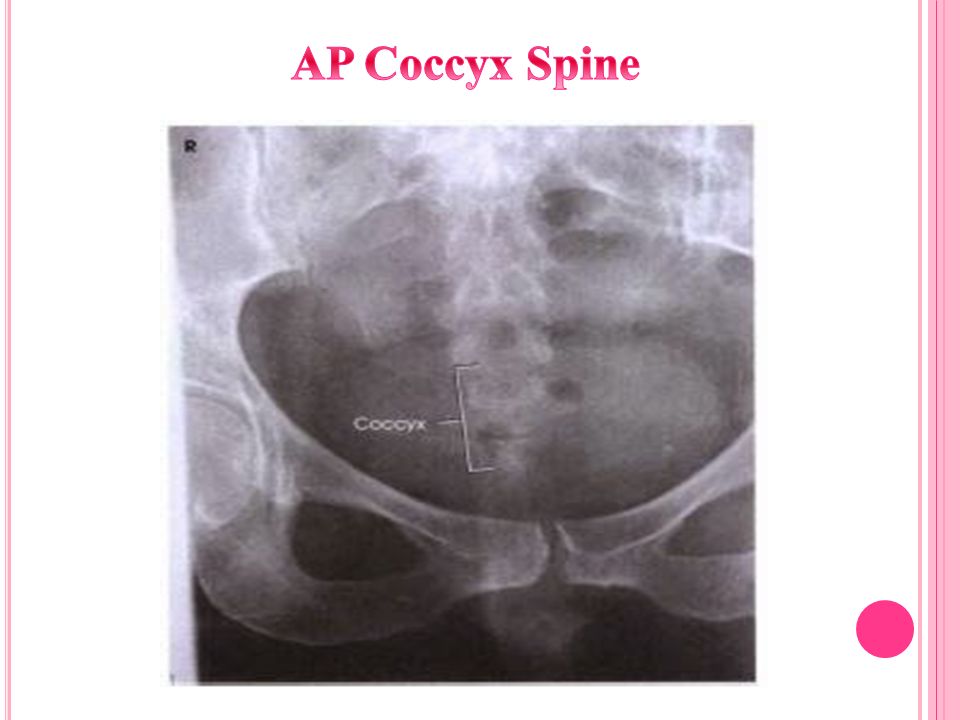
Positioning: 1.Patient is in supine position, provide pillow for head support and legs are extended, with support under knees for patient comfort. 2.Midsagittal plane is align to table or grid. 3.Ensure that the pelvis is not rotated. Central Point: (2 in) Superior to Pubic Symphysis. Central Ray: 10º Caudal.
 Superior to Pubic Symphysis. Central Ray: 10º Caudal..")
16
Positioning: Supine: 1.Position Patient lateral with hips and knees flexed. 2.Support Body to place long axis of Spine Horizontal. Central Point: to level of Pubic Symphysis (2 in Inferior to ASIS) and at a point 3.5 in Posteriorly. Central Ray: Perpendicular to IR.
 and at a point 3.5 in Posteriorly. Central Ray: Perpendicular to IR..")
Similar presentations


. leg Basic projections AP Lateral AP leg Exposure Factors KvmAsFFD (cm)GridFocusCassette 655100NoFine24 x 30 cm Patient Position ٍ Supine.>")
.>")


.>")


