Download presentation
Presentation is loading. Please wait.

2
Deep fascia of the Forearm The forearm by extensions of deep fascia which are called Med. & Lat. intermuscular septum and interosseus membraine divided in to two compartments ( Anterior & Posterior ).
..")
3
Deep fascia of the FOREARM

4
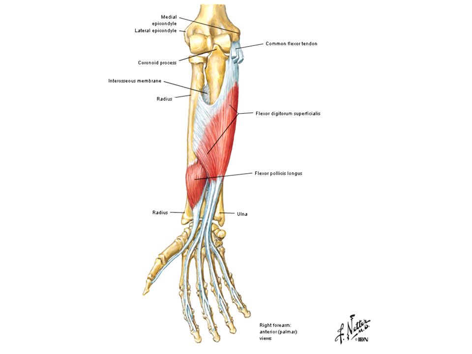
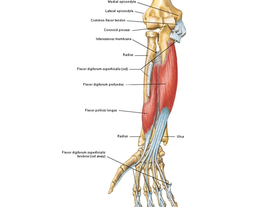
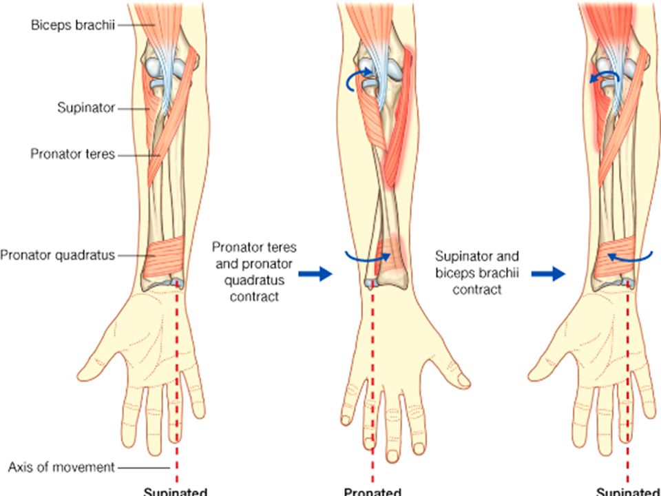
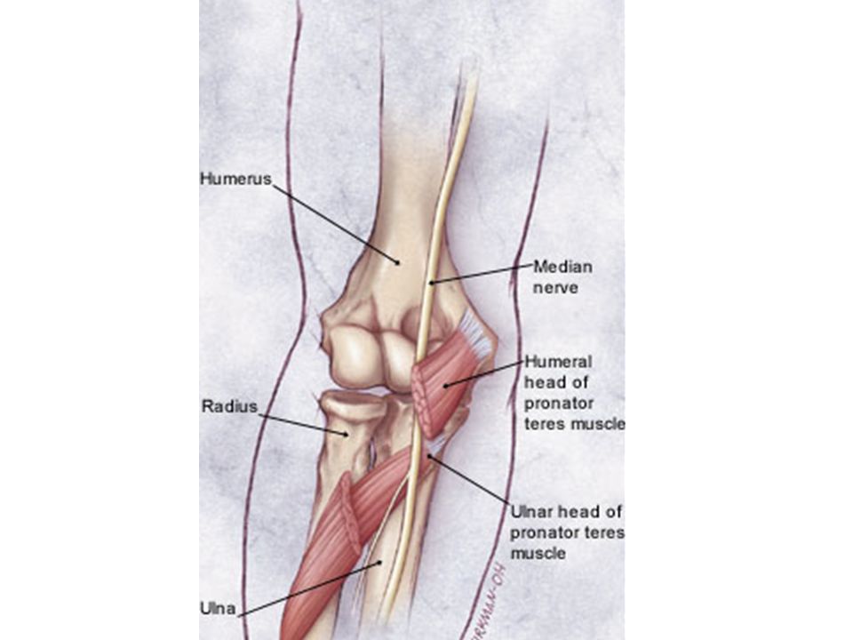
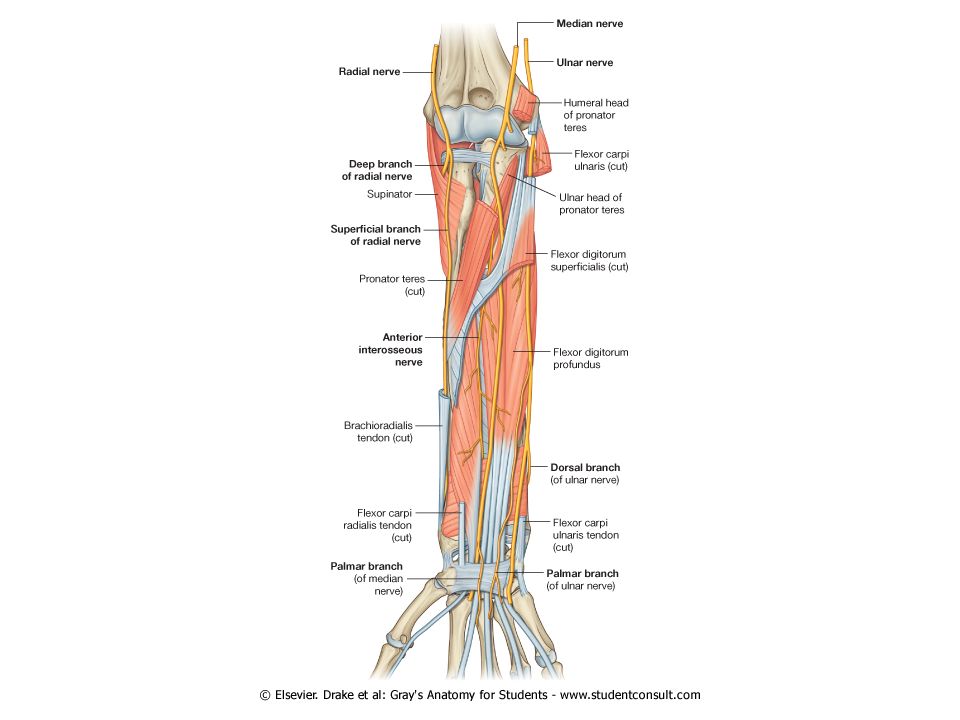
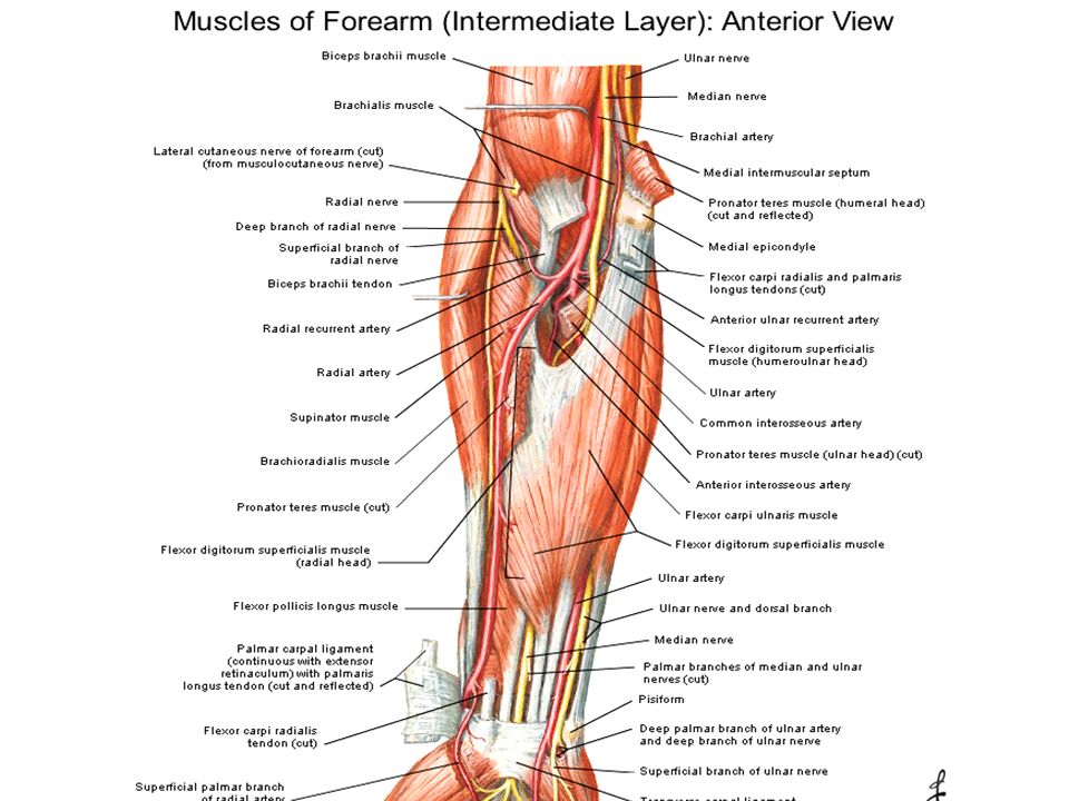
Ant.Compartment Muscles Arterial Supply Nerve Supply Superficial: Pronator Teres Flexor Carpi Radialis Palmaris Longus Flexor Digitorum Superficialis Flexor Carpi Ulnaris Deep: Flexor Digitorum Profondus Flexor Pollicis Longus Pronator Quadratus

10
Ant.Compartment Muscles Arterial Supply Nerve Supply Radial Artery Ulnar Artery Median Nerve & Ant.Interosseus Nerve Ulnar Nerve

11
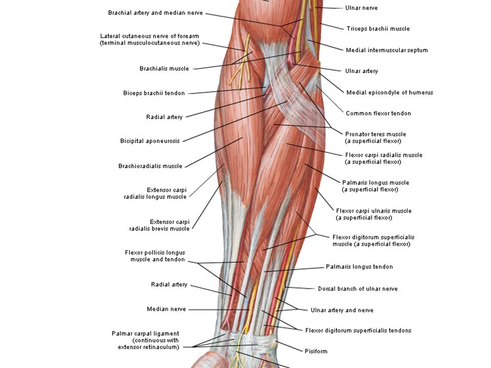
Muscles of the Anterior Compartment of the Forearm Superficial: Pronator Teres Flexor Carpi Radialis Palmaris Longus Flexor Digitorum Superficialis Flexor Carpi Ulnaris Superficial: Pronator Teres Flexor Carpi Radialis Palmaris Longus Flexor Digitorum Superficialis Flexor Carpi Ulnaris Deep: Flexor Digitorum Profondus Flexor Pollicis Longus Pronator Quadratus

15
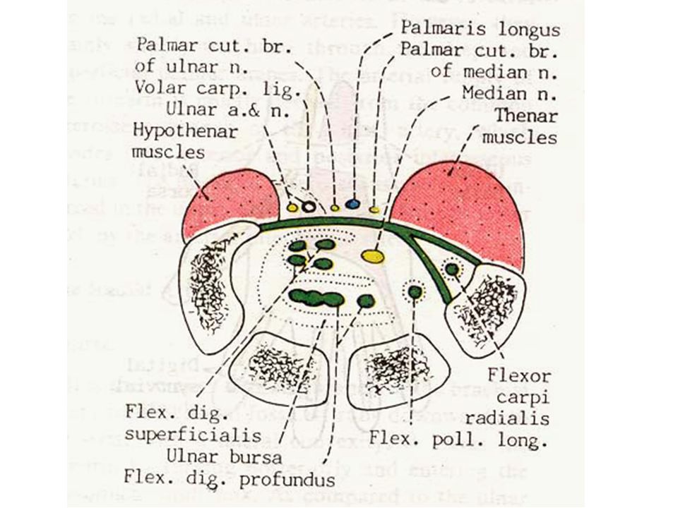
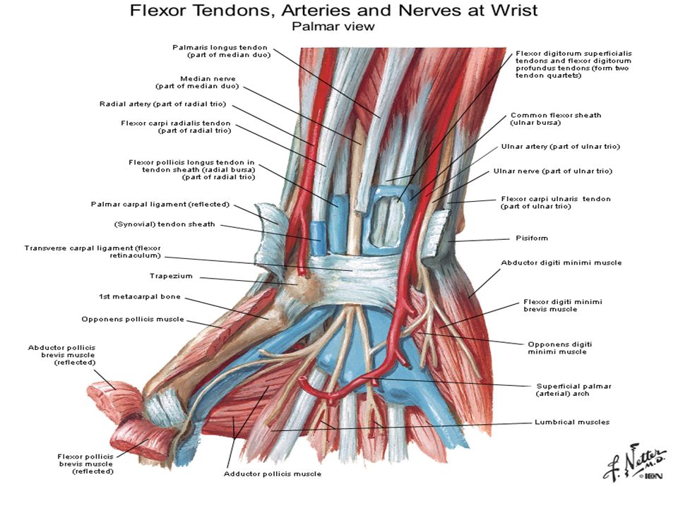
Is a strong fibrous band which bridges the anterior concavity of the carpus 1. Laterally : Tubercle of scaphoid Crest of trapezium 2. Medially : Pisiform Hook of hamat Situation : Situation : Attachments : Attachments : FLEXOR RETINACULUM

20
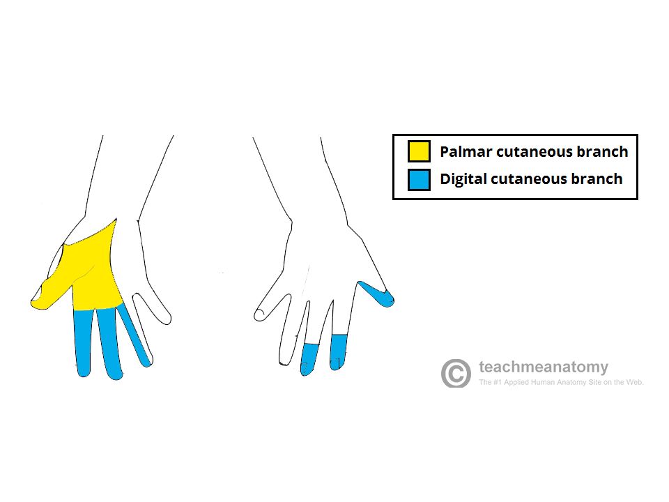
Tendon of palmaris longus, Tendon of flex.carpi ulnaris, Palmar cutaneus br.of Median nerve, Palmar cutaneus br.of Ulnar nerve, Ulnar vessels, Ulnar nerve Median nerve, Tendons of Flexor digitorum superficialis, Tendons of Flexor digitorum profondus, Tendons of Flexor pollicis longus, Ulnar bursa, Radial bursa ** The tendon of Flexor carpi radialis lies between the retinaculm and its deep slip, in the groove of trapezium** Structures passing superficial to the F. R : Structures passing superficial to the F. R : FLEXOR RETINACULUM Structures passing deep to the F. R : Structures passing deep to the F. R :

23
Cubital Fossa: Is the triangular hollow and lying in front of the elbow joint 1. Superioly ( Base) : Imaginary line between lat. & med. Epicondyles of humerus. 2. Laterally: Medial border of Brachioradialis muscle. 3. Medially : Lateral border of Pronator teres muscle. 4. Floor : Brachialis and Supinator muscle. 5. Roof : Skin, Superficial & Deep fascia and Bicipital aponeurosis. 6. Apex : Overlap of Brachioradialis & Peonator teres muscles. Situation : Situation : Boundaries : Boundaries :
 : Imaginary line between lat. & med. Epicondyles of humerus. 2. Laterally: Medial border of Brachioradialis muscle. 3. Medially : Lateral border of Pronator teres muscle. 4. Floor : Brachialis and Supinator muscle. 5. Roof : Skin, Superficial & Deep fascia and Bicipital aponeurosis. 6. Apex : Overlap of Brachioradialis & Peonator teres muscles. Situation : Situation : Boundaries : Boundaries :.")
25
Cubital Fossa: A ) Superficial : 1. Median cubital vein 2. Lateral cutaneus nerve of forearm. 3. Medial cutaneus nerve of forearm. B ) Deep : 1. Median Nerve 2. Brachial Artery 3. Tendon of Biceps brachii 4. Radial Nerve Content : Content :
 Deep : 1. Median Nerve 2. Brachial Artery 3. Tendon of Biceps brachii 4. Radial Nerve Content : Content :.")
Similar presentations