Download presentation
Presentation is loading. Please wait.

1
Cardiovascular practical Block
Practical II

2
Cardiovascular practical Block
8-Chronic venous congestion of the liver

3
NUTMEG LIVER Section of liver showing alternating pale and dark areas with a nutmeg like appearance possibly due to passive congestion secondary to right sided heart failure.

4
Cardiovascular Block

5
Cardiovascular Block

6
Chronic venous congestion of the liver: Section of liver shows:
The central portion of liver lobules shows: Congestion and dilatation of central veins and blood sinusoids, Atrophy and necrosis of liver cells. Kupffer cells contain few brown haemosiderin pigment granules.

7
Cardiovascular practical Block
9-Chronic venous congestion of the lung

8
Left ventricular hypertrophy.
Thickened left ventricular wall most likely due to hypertension.

10
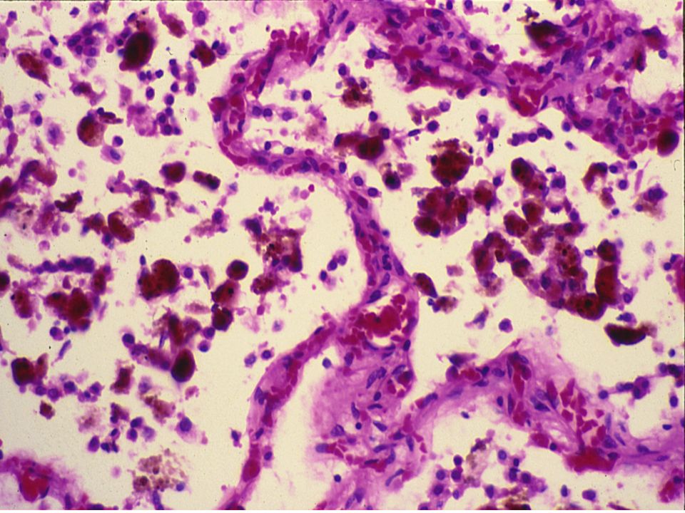
Chronic venous congestion of the lung: Section of lung shows:
The alveolar walls are thickened by dilated and engorged capillaries. The alveoli are dilated and contain edema fluid, red blood cells and large alveolar macrophages (heart failure cells), which are filled with haemosiderin pigment derived from red cells breakdown. The likely cause of CVC lung is Heart Failure.
, which are filled with haemosiderin pigment derived from red cells breakdown. The likely cause of CVC lung is Heart Failure.")
11
10-Thromboangitis oblitrans (Buerger disease)
Cardiovascular Block 10-Thromboangitis oblitrans (Buerger disease)
")
12
Black discoloration of fingers consistent with early gangrene.
10-Thromboangitis oblitrans (Buerger`s disease) Black discoloration of fingers consistent with early gangrene.
 Black discoloration of fingers consistent with early gangrene.")
13
Thromboangiitis obliterans
Also known as Buerger's disease, is a recurring segmental thrombosing (clotting), acute and chronic inflammation of small and medium arteries and veins of the hands and feet. It is strongly associated with use of tobacco products.
, acute and chronic inflammation of small and medium arteries and veins of the hands and feet. It is strongly associated with use of tobacco products.")
14
Cardiovascular Block

15
BUERGER’S DISEASE

16
Thromboagitis obliterans (Buerger’s disease):
Thrombosis and luminal obliteration of the blood vessels Segmental inflammatory infiltration in the blood vessel wall (arteries and veins) comprising of mainly neutrophills
 comprising of mainly neutrophills.")
17
Cardiovascular practical Block
11-Giant cell ( temporal ) arteritis
 arteritis.")
18
Giant cell arteritis Tender and thickened scalp veins

19
Giant cell arteritis Superficial temporal artery biopsy - intimal thickening and medial damage, giant cells with inflammatory cell infiltration in the internal elastic lamina

20
Giant cell (temporal) arteritis:
Segmental inflammatory lesions with intimal thickening , medial granulomatous inflammation with giant cells and chronic inflammatory cells and internal elastic lamina fragmentation . Giant cell (temporal) arteritis: Disruptions of the elastic lamina with inflammation and giant cells.
 arteritis: Disruptions of the elastic lamina with inflammation and giant cells.")
21
Cardiovascular practical Block
12-Leukocytoclastic / hypersensitivity vasculitis ( microscopic polyangitis)/ Pauci immune vasculitis
/ Pauci immune vasculitis.")
22
This condition might be complicated by glomerulonephritis and hemoptysis due to pulmonary capillaritis .

23
Leukocytoclastic vasculitis, foot
Leukocytoclastic vasculitis, foot. The purpuric eruption (Subcutaneous bleeding patches) tends to be most pronounced on dependent areas.
 tends to be most pronounced on dependent areas.")
24
Vasculitis, leukocytoclastic, (low power)
")
25
Section of the skin shows fibrinoid necrosis of blood vessels with extravasation of RBCs , neutrphilic infiltration with debris (leukocytoclasis ) .
 .")
Similar presentations

 Chapter: Hemodynamic disorders, Thrombosis and Shock - Edema - Hemorrhage - Hyperemia.>")
 Occurs when the right ventricle fails as an effective forward pump, causing back-pressure of blood into the systemic.>")


>")


 Aneurysms & Dissections Veins & Lymphatics Tumors.>")


 Decreased.>")