Download presentation
Presentation is loading. Please wait.

1
Department of Radiology
Thorax David S. Hartman, M.D. Department of Radiology

2
Identify all objects indicated by arrows on the radiolgical images
Learning Objectives: Identify all objects indicated by arrows on the radiolgical images in this presentation

3
Cases can be viewed by any computer in the institution
Cases in MDL \\hersheymed.net\files\MMS\Public\education\radiology\lab-thorax pps Cases can be viewed by any computer in the institution

4
PA and Lateral Chest Radiograph

5
Heart Chambers Most anterior Most posterior Right boarder Left boarder
Right ventricle Left atrium Right atrium Left atrium/ ventricle Most superior Left atrium

6
Heart from the Front Left atrium Left ventricle Pulmonary artery Aorta
Right Atrium Right Ventricle 204

7
Right Atrium Left atrium Ventricle Right ventricle
201

8
Right Atrium Left Atrium Vent- ricle

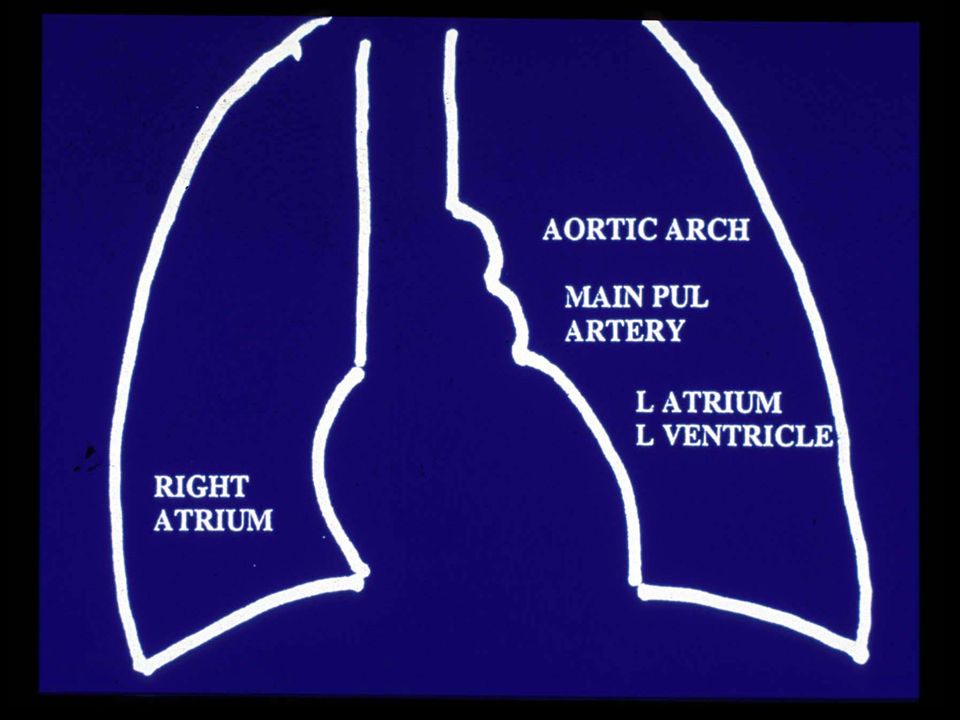
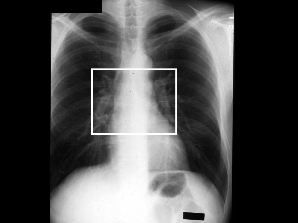
11
Aortic arch Main pulmonary artery Left atrium, ventricle

12
Trachea Clavicles Cardiophrenic angles Costophrenic angles

13
Hilum (pleural: hila) Root of the lung Pulmonary artery
Upper lobe pul vein Bronchus (Lymph nodes)
")
15
Right hilum Left hilum

16
Right ventricle Left atrium Left ventricle Cardiophrenic angle
Costophrenic angle Left Diaphragm 219

17
Right ventricle Left atrium left ventricle Diaphragm Cardiophrenic angle Costophrenic angles

18
Trachea Thoracic spine

19
Pulmonary Arteriogram

20
Pulmonary Arteries Main PA (pulmonary trunk) off right ventricle (anterior) Right main PA longer than left main PA and runs horizontaly superior to left atrium Left main PA is short and runs over left main stem bronchus

21
Right main PA Left main PA Main PA (trunk) Aorta RA RV LV
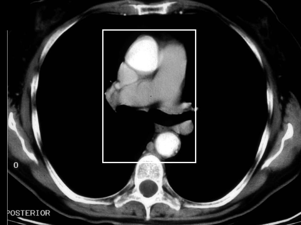
LA RA RV LV 194

22
Left inferior pulmonary artery
Right main pulmonary artery Left inferior pulmonary artery Catheter in main PA

23
Arch Aortogram

24
Left common carotid art
Ascending aorta Aortic arch Descending aorta Brachiocephalic art Left common carotid art Left subclavian art Aortic Arch 225

25
Arch Aortogram Aortic arch Brachiocephalic art Left common carotid art Left subclavian artery R L

26
CT of the Heart and Mediastinum (axial images)
")
27
View as though facing the patient
Axial section Right Left

28
Axial CT image with contrast By convention we look from below
Anterior Heart Liver Right Left Lung Posterior By convention we look from below

30
Right Sternum Scapula Spine Ribs

31
Vascular Arch

32
Vascular Arch

33
Vascular Arch

34
Vascular Arch

35
3 Vascular Arches Arch Structure below arch Aorta Left pulmonary
artery 3. Azygos vein Pulmonary artery, vein Left main bronchus Right main bronchus

36
The brachocephalic vein runs anterior to the branches of the aorta

37
Frontal of the brachiocephalic vein and aortic arch
195

38
Brachiocephalic artery Left common carotid artery
Aortic arch Brachiocephalic artery Left common carotid artery Right brachio-cephalic vein Left brachio-cephalic vein Superior vena cava Trachea 195

39
The trachea is just ventral to the esophagus

40
Brachiocephalic artery Left common carotid artery
Trachea Brachiocephalic artery Left common carotid artery Left subclavian artery Esophagus Branches of the aorta 196

41
Origin of the great vessels above the aortic arch
195

42
Origin of the great vessels above the aortic arch
Right

43
Left brachio-cephalic vein
Right brachio-cephalic vein Brachiocephalic artery Left common carotid artery Left subclavian artery Tracheal area Esophagus Right

44
The azygos arch passes over the right main-stem bronchus

45
Superior vena cava Right atrium Right ventricle Inferior vena cava
DIAPHRAGM 208

46
Right main pulmonary art Right pulmonary veins Right atrium
Superior vena cava Azygos arch Right bronchus Right main pulmonary art Right pulmonary veins Right atrium Azygos vein 208

47
Level of the aortic arch
195

48
Level of the aortic arch

49
Aortic arch Superior vena cava Trachea Azygos arch Esophagus Vertebra Level of the aortic arch

50
The left pulmonary artery passes over the left main-stem bronchus

51
Left main pulmonary art Left atrium Left ventricle
Aortic arch Left main pulmonary art Left atrium Left ventricle LEFT DIAPHRAGM 219

52
Left main pulmonary artery Left mainstem bronchus Left pulmonary veins
Aorta Left main pulmonary artery Left mainstem bronchus Left pulmonary veins Left atrium 219

53
Level below the aortic arch (top of the left pulmonary artery
SVC ASCENDING AORTA PULMONARY ARTERY 201

54
Level below the aortic arch (top of the left pulmonary artery

55
Top of left pulmonary artery Descending aorta Azygos vein
Ascending aorta Main pulmonary artery Superior vena cava Right pul art Right bronchus Left bronchus Top of left pulmonary artery Descending aorta Azygos vein BACK WALL OF BRONCHUS

56
Level at the root of the aorta and main pulmonary artery
Top of the right atrium Ascending Aorta Pulmonary artery 201

58
Ascending aorta Main pulmonary artery Right atrium Rt pulmonary artery
Right bronchus Left bronchus Lt lower lobe PA Descending aorta Azygos vein Back of the left bronchus

59
The pulmonary trunk is anterior to the aorta root

60
The arota arises from “center” of heart

61
Heart from the Front Left atrium Aorta Pulmonary artery Left ventricle
Right Atrium Aorta Pulmonary artery Right Ventricle Left ventricle 204

62
Level at the top of the ventricles
Right Atrium Left Ventricle Right ventricle 201

63
Level at the top of the ventricles

64
Right ventricle Right atrium Ascending aorta Left ventricle Left atrium Descending aorta Azygos vein Level at the top of the ventricles

65
Heart Chambers Most anterior Most posterior Right boarder Left boarder
Right ventricle Left atrium Right atrium Left atrium/ ventricle Most superior Left atrium

66
Level through all 4 heart chambers
Right ventricle Right atrium Left ventricle Left atrium Pulmonary veins Descending aorta Azygos vein Level through all 4 heart chambers 230

67
Level through all 4 heart chambers
Right Atrium Left Ventricle Right ventricle 201

69
Right ventricle Right atrium Left ventricle Left atrium Aorta Azygos vein

70
Level at the bottom of the heart (below left atrium)
Right Atrium Left Ventricle Right ventricle 201

71
Level at the bottom of the heart (below left atrium)
")
72
Right ventricle Inferior vena cava Left ventricle Aorta Azygos vein Level at the bottom of the heart (below left atrium)
")
73
Follow a drop of blood from the left arm to the head

74
TO THE LEFT BRACIOCEPHALIC VEIN
X MS 211 CT 7 NORMAL CHEST CT

75
INTO THE SVC X

76
SVC X

77
SVC X

78
SVC X

79
SVC X

80
SVC X

81
SVC X

82
SVC X

83
SVC X

84
TOP OF THE RA X

85
RA X

86
RA X

87
INTO THE RV X

88
RV X

89
RV X

90
RV X

91
RV X

92
RV X

93
RV X

94
RV X

95
RV OUTFLOW X

96
RV OUTFLOW X

97
RV INTO MAIN PA X

98
MAIN PA X

99
RT AND LT PA’S X

100
RT AND LT PA’S X X

101
FROM LUNG TO LA X

102
LA X

103
LV X

104
LV TO OUTFLOW X X X

105
INTO ASCENDING AORTA X

106
ASCENDING AORTA X

107
ASCENDING AORTA X

108
ASCENDING AORTA X

109
INTO AORTIC ARCH X

110
AORTIC ARCH X

111
AORTIC ARCH X

112
INTO BRACHIOCEPHALIC ARTERY
X

113
INTO THE CAROTIDS X X

114
Department of Radiology
Thorax David S. Hartman, M.D. Department of Radiology

Similar presentations