Download presentation
Presentation is loading. Please wait.

1
Cardiac Cath and Angiocardiography Adult II FINAL 2/2015

2
Definition of Cardiac Catheterization An invasive imaging procedure that involves inserting a catheter into a blood vessel in the arm or leg, and guiding it to the heart with the aid of a special x-ray machine. Contrast dye is injected through the catheter so that the valves, coronary arteries and heart chambers are can be visualized.

3
3 Definition of Cardiac Catheterization Diagnostic –Collects data to evaluate PT’s condition Therapeutic –To intervene by mechanical means to treat disorders of the vascular and conduction systems within the heart

4
Principles of Cardiac Catheterization

9
9 Indications Suspected or known coronary heart disease Myocardial infarction Valvular heart disease Congenital heart disease Aortic dissection Pericardial constriction Cardiomyopathy Initial and follow up assessment for heart transplant

10
10 Contraindications Active GI bleed Renal failure Recent stroke Fever from infection Electrolyte imbalance Anemia Short life expectancy Digitalis intoxication PT refusal Uncontrolled hypertension Bleeding disorders Pulmonary edema Uncontrolled ventricular arrhythmias Allergic to contrast

11
11 Complications and Risks Death Myocardial infarction CVA Arrhythmia Hemorrhage Contrast Hemodynamic Perforation

12
Angiographic Supplies and Equipment Catheters Contrast Media Pressure Injector

13
13 Catheters For LT cardiac cath similar to those for angio RT cath requires specialized catheters –Typically flow directed catheters –With manifolds

14
14 Contrast Media High Osmolar Ionic –Sometimes causes ECG changes Widely used –Non-ionic –Ionic low osmolar Restricted costs causes limited use of low osmolar contrast agents.

15
15 Pressure injector

16
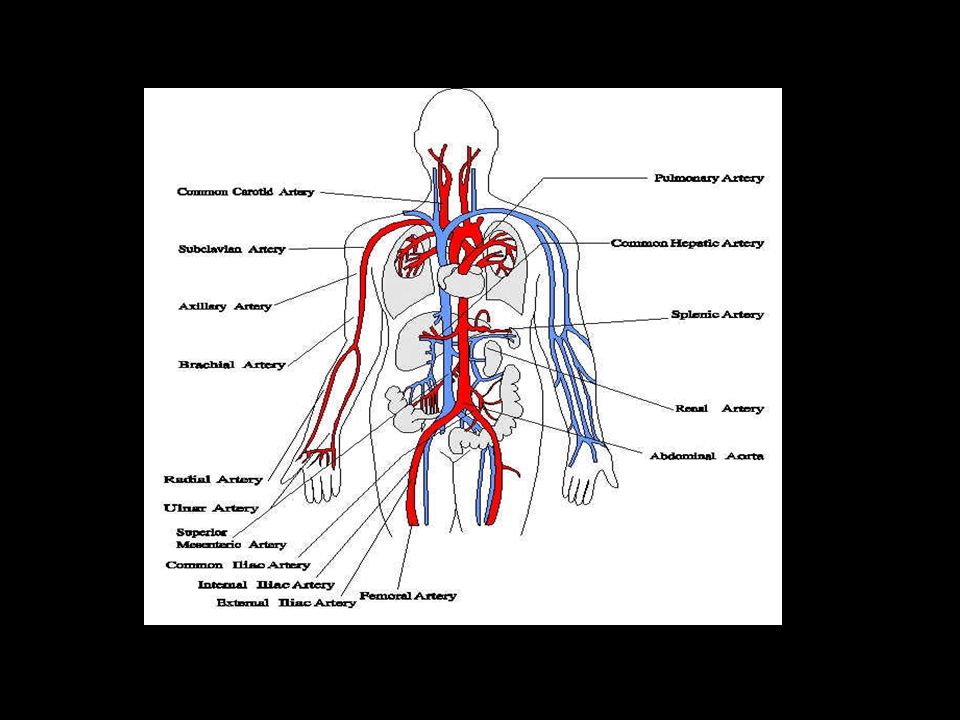
Imaging Image chain Digital Angiography imaging equipment

17
17 Digital Angiography Imaging equipment Long term storage of large amounts of digital files has benefited from advances in computer technology

18
Ancillary Equipment and Supplies Physiologic Equipment Other equipment

19
19 Physiologic Equipment Equipment to monitor –ECG –Hemodynamic pressures Vital signs to record PT function

20
20 Other Equipment Crash cart Oxygen and suction Defibrillator Temporary pacemaker Pulse oximeter Blood pressure cuff Equipment to perform cardiac output studies Activated clotting time (ACT) equipment
 equipment")
21
21 Patient Positioning for Cardiac Catheterization PT must be positioning so that they will not have to be moved during procedure Must be positioned so anatomic structures of interest are demonstrated PT is supine with shielding as appropriate

22
Catheterization Methods and Techniques

23
23 Pre-Catheterization Care Informed consent obtained PT history Physical exam CXR Blood work ECG Echocardiogram Exercise stress test

24
24 Pre-Catheterization Care IV started –Sedation. Nothing to eat 4-6 hours before procedure Records of procedure –PT hemodynamic data –Fluoro times –Medications administered –Supplies used –Other pertinent information

25
25 Catheter Introduction Prepare catheter introduction site with aseptic technique –Shaved and cleaned Can be at femoral (most common), brachial, radial, axillary, jugular and subclavian areas Selinger technique used
, brachial, radial, axillary, jugular and subclavian areas Selinger technique used")
26
26 Selinger Technique Needle with cannula inserted Needle withdrawn until there is blood flow Inner cannula removed & guidewire inserted Needle removed Catheter over guidewire Guidewire removed leaving catheter in artery

27
27 Data Collection Physiologic data unusually collected –Hemodynamic parameters Includes blood pressure Cardiac output Vascular pressures (inside & outside the heart) –ECG –Oximetry readings –Cardiac output –Blood samples to measure oxygen saturations levels in various parts of the heart
 –ECG –Oximetry readings –Cardiac output –Blood samples to measure oxygen saturations levels in various parts of the heart")
28
Nursing Management after cardiac catheterization Catheter Site is observed for bleeding or hematoma. Temperature and color of the affected extremity are evaluated. Dysrhythmias are carefully assessed by observing the cardiac monitor. Bed rest must be maintained for 2to 6 hours after the procedure. Observe for contrast agent induced renal failure.

Similar presentations


, awareness of heartbeat.>")

 IS A TECHNIQUE THAT TEMPORARILY TAKES OVER THE FUNCTION.>")


