Download presentation
Presentation is loading. Please wait.

1
CARDIOVASCULAR SYSTEM HEART

2
General Closed System -Blood Remains in Blood Vessels & Heart Double System -Four Chambers (Separation of Oxygenated & Deoxygenated Blood) -Each Side Supplies Different Circuit: *Pulmonary Circuit (R. Heart Lungs Heart) *Systemic Circuit (L. Heart Body Tissues Heart)
 *Systemic Circuit (L. Heart Body Tissues Heart).")
4
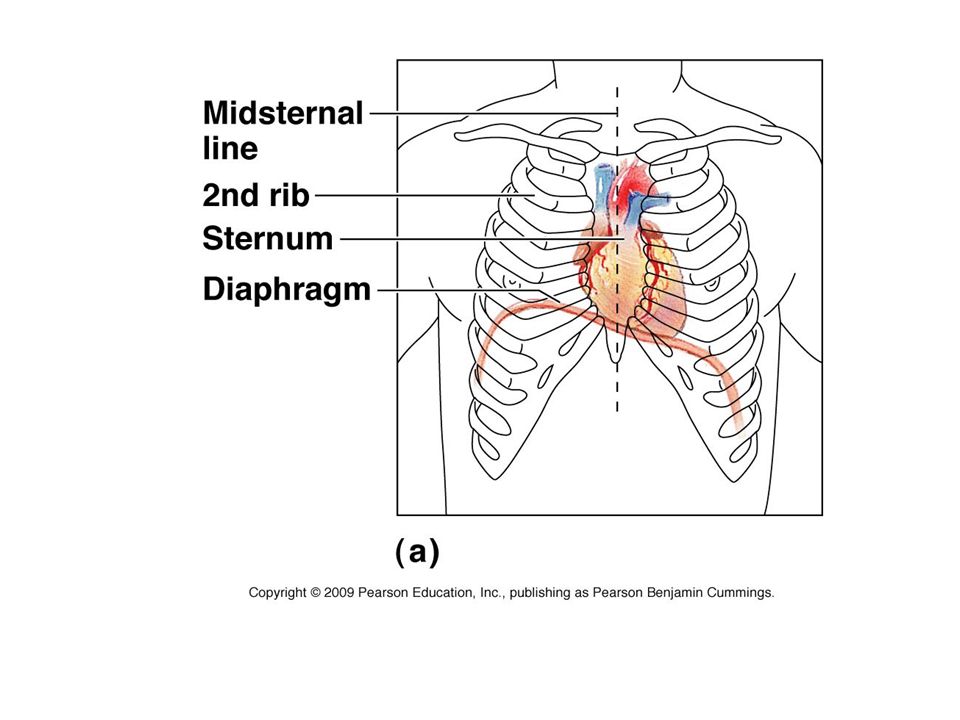
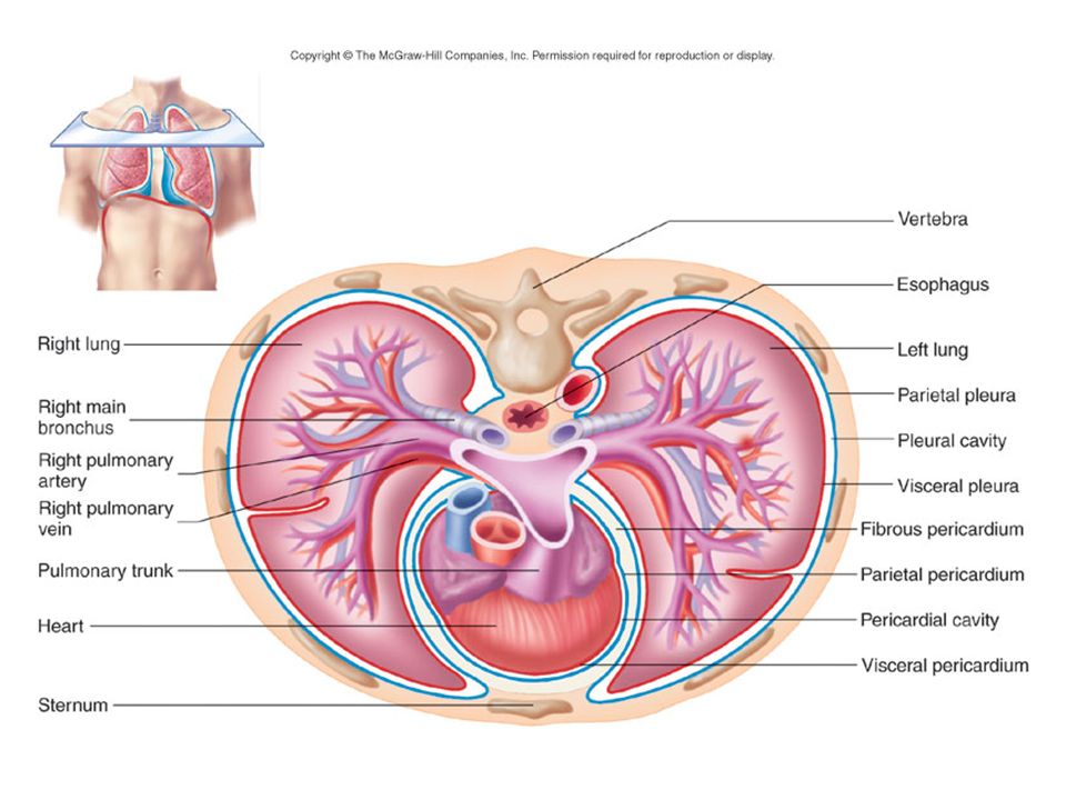
Heart – Size & Location Size: Adult Clenched Fist Position: Mediastinum, 2/3 to Left of Midline Relationships to Other Organs: -Deep to Sternum -Superior to Diaphragm -Medial to Lungs -Anterior to Esophagus, Descending Aorta

7
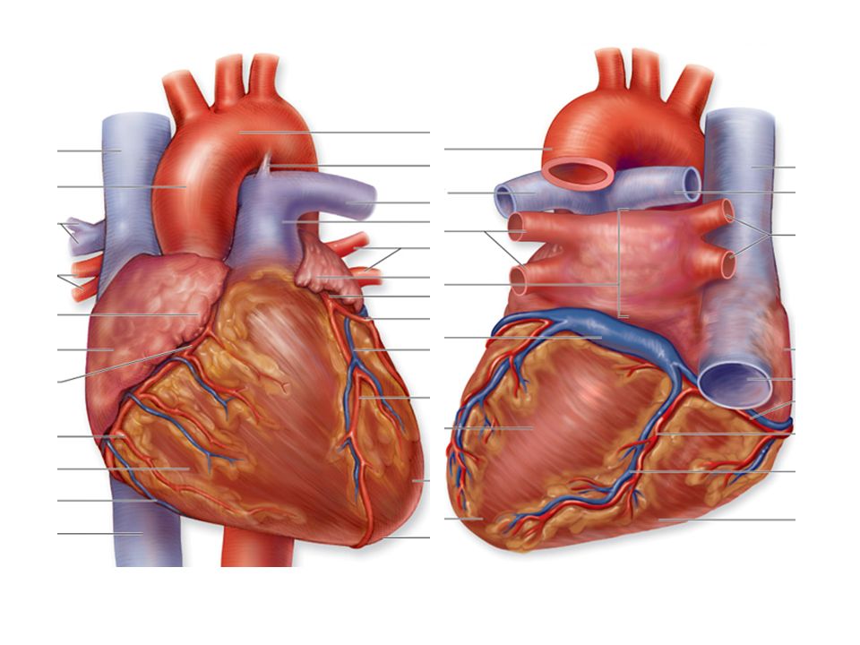
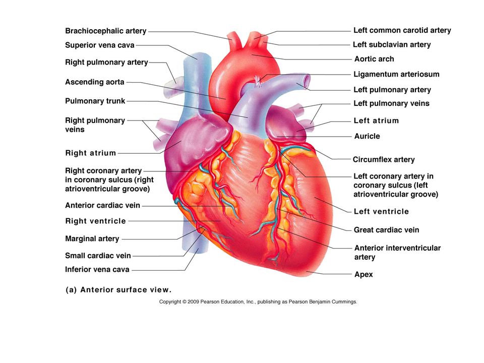
Heart – Surface Anatomy Sulci -Grooves, Indicate Boundaries of Chambers -Coronary Sulcus (Between Atria & Ventricles) -Interventricular Sulci (Between Ventricles) Apex (Inferior, Pointed Tip) Base (Posterior surface; formed by atria, mostly left)
 -Interventricular Sulci (Between Ventricles) Apex (Inferior, Pointed Tip) Base (Posterior surface; formed by atria, mostly left)")
9
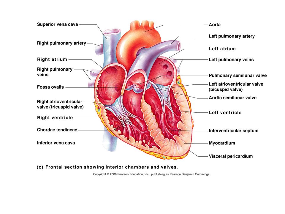
Heart - Chambers Atria (2) -Superior Receiving Chambers (From Veins) -Auricles (ear-like flaps) -Right Atrium: *O 2 Poor Blood From Venae Cavae & Coronary Sinus -Left Atrium: *O 2 Rich Blood From Pulmonary Veins of Lungs
 -Superior Receiving Chambers (From Veins) -Auricles (ear-like flaps) -Right Atrium: *O 2 Poor Blood From Venae Cavae & Coronary Sinus -Left Atrium: *O 2 Rich Blood From Pulmonary Veins of Lungs")
12
Heart – Chambers continued Ventricles (2) -Inferior Pumping Chambers (to Arteries) -Receive from Atria -Right Ventricle: *O 2 Poor Blood from Rt Atrium to Pulmonary Arteries (Pulmonary Circ.) -Left Ventricle: *O 2 Rich Blood from Left Atrium to Aorta (Systemic Circ.)
 -Inferior Pumping Chambers (to Arteries) -Receive from Atria -Right Ventricle: *O 2 Poor Blood from Rt Atrium to Pulmonary Arteries (Pulmonary Circ.) -Left Ventricle: *O 2 Rich Blood from Left Atrium to Aorta (Systemic Circ.)")
14
Heart - Septa Internal Walls Separate Chambers 2 Septa: -Interatrial Septum -(Between the Atria) -Interventricular Septum -(Between the Ventricles)
 -Interventricular Septum -(Between the Ventricles)")
15
Heart - Valves One-Way, Direct Blood Flow Passive; blood pushes them open & closed 2 Pairs: -Atrioventricular (Cuspid) Valves *Between Atria & Ventricles *Prevent blood flowing back into atria Tricuspid (Right) Bicuspid (Left)
 Valves *Between Atria & Ventricles *Prevent blood flowing back into atria Tricuspid (Right) Bicuspid (Left)")
17
Atrioventricular Valves cont. *AV Valves are anchored to structures in the ventricles: Chordae Tendineae Strong, Fibrous Strings Prevent Cusp Eversion Papillary Muscle Conical Extensions of Myocardium in Ventricles Contract & pull on chordae tendineae

19
Heart - Valves -Semilunar Valves *Between Ventricles & Arteries *Prevent blood from back flow into ventricles Pulmonary Semilunar Valve (Right) Aortic Semilunar Valve (Left)
 Aortic Semilunar Valve (Left)")
21
Heart - Wall 3 Layers: -Endocardium (Inner) *Endothelium *Lines Chambers, Valves, Septa, Blood Vessels -Myocardium (Middle) *Thickest Layer (Esp. in Left Ventricle!) *Cardiac Muscle Tissue *Inherent Rhythmicity
 *Cardiac Muscle Tissue *Inherent Rhythmicity.")
22
Heart Wall

23
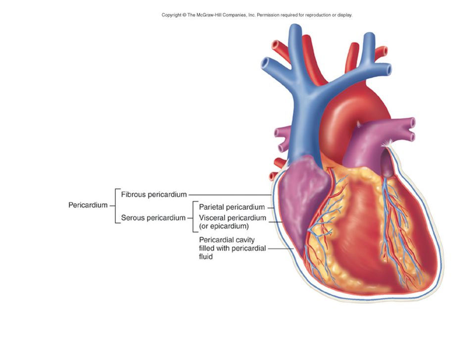
Heart – Wall continued -Epicardium (Outer) *Visceral Pericardium *Serous Membrane *Thin, Fibrous, Transparent
 *Visceral Pericardium *Serous Membrane *Thin, Fibrous, Transparent")
24
Fibrous Skeleton Between atria & ventricles Dense regular C.T. Encircle valves & great arteries Attachment for cardiac muscle Electrical insulation between atria & ventricles Stabilizes valves

25
Heart – Pericardial Sac Encloses Heart & Bases of Great Vessels Composed of 2 layers: -Parietal Pericardium *Inner serous coat of sac *Secretes lubricating pericardial fluid into *Pericardial cavity (space between serous layers)
")
28
Heart – Pericardial Sac continued -Fibrous Pericardium *Outer, fibrous coat of sac *Tough, protective *Reinforces serous coat

29
CARDIAC CYCLE Events of one heartbeat: -Systole *Atria Contract, Ventricles Fill *Ventricles Contract, Blood Forced into Aorta and Pulmonary Trunk -Diastole *Atria Relax & Fill *Ventricles Passively Receive Blood from the Atria

31
Cardiac Output Stroke Volume (SV) -Volume of blood pumped out by ventricle each heartbeat -70ml/beat Cardiac Output (CO) -Volume pumped out by ventricle in 1 minute -CO = Heart Rate x Stroke Volume -75 b/min x 70 ml/beat = over 5,000 ml/min
 -Volume of blood pumped out by ventricle each heartbeat -70ml/beat Cardiac Output (CO) -Volume pumped out by ventricle in 1 minute -CO = Heart Rate x Stroke Volume -75 b/min x 70 ml/beat = over 5,000 ml/min")
32
Factors Influencing Cardiac Output Venous Return directly effects Stroke Volume (& CO) Heart Rate (& CO) is effected by drugs, hormones & ions
 Heart Rate (& CO) is effected by drugs, hormones & ions")
33
BLOOD PRESSURE BP = pressure blood exerts on inner blood vessel walls BP rises & falls in response to heart contraction & relaxation BP keeps blood moving between heart contractions

34
BLOOD PRESSURE Systole (contraction of the heart) -Contraction of ventricles causes arterial pressure to rise -Systolic pressure (SBP) is the maximum pressure during contraction
 -Contraction of ventricles causes arterial pressure to rise -Systolic pressure (SBP) is the maximum pressure during contraction")
35
BLOOD PRESSURE CONTINUED Diastole -Relaxation and refilling of ventricles while semilunar valves are closed -Arterial pressure drops as blood flows “downstream” -Diastolic pressure (DBP) is the minimum pressure just before the next systole
 is the minimum pressure just before the next systole")
37
Average BP = less than 120/80 Arterial surge in pressure = Pulse Pulse rate usually = heart rate Average adult pulse = 60-80 BPM

38
CONDUCTION SYSTEM OF THE HEART Specialized Cardiac Muscle Tissue Capable of Generating & Conducting Action Potentials Stimulates Contraction of Myocardial Tissue Sets basic rhythm of the heart

39
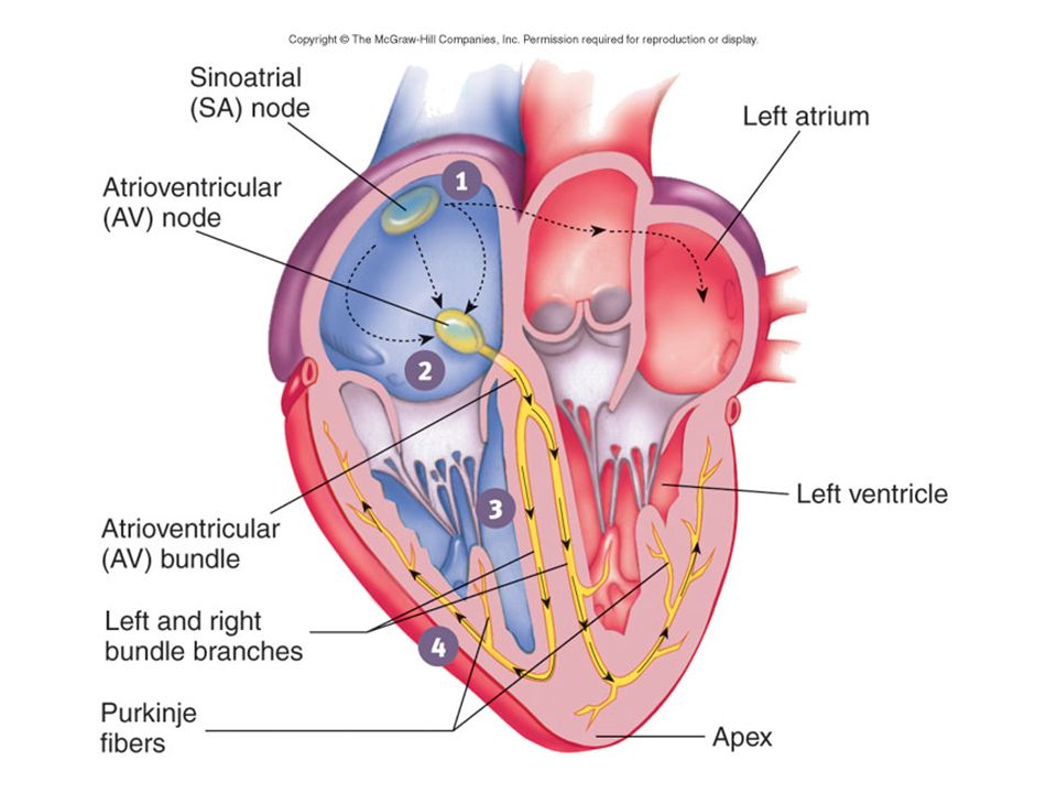
CONDUCTION SYSTEM OF THE HEART continued 5 Components: -Sinoatrial Node (Pacemaker) *Right Atrium *Spontaneously Depolarizes *Activates Atrial Contraction *Origin of Heart Beat *Action Potential Spreads to Atrial Myocardium &:
 *Right Atrium *Spontaneously Depolarizes *Activates Atrial Contraction *Origin of Heart Beat *Action Potential Spreads to Atrial Myocardium &:")
40
CONDUCTION SYSTEM OF THE HEART continued -Atrioventricular (AV) Node *Rt. atrium (interatrial septum) *Action potential spreads to: -Atrioventricular Bundle (Bundle of His) *Only electrical pathway between atria & ventricles (C.T. Block) *Carries action potential through interventricular septum to:
 *Action potential spreads to: -Atrioventricular Bundle (Bundle of His) *Only electrical pathway between atria & ventricles (C.T. Block) *Carries action potential through interventricular septum to:.")
41
CONDUCTION SYSTEM OF THE HEART continued -Bundle Branches (Left & Right ) *Interventricular septum *Carries action potential toward respective ventricles -Purkinje Fibers *Myocardium of ventricles *Conduct action potentials to ventricular myocardium
 *Interventricular septum *Carries action potential toward respective ventricles -Purkinje Fibers *Myocardium of ventricles *Conduct action potentials to ventricular myocardium")
43
C. T. Blocks Action Potentials Between Atria & Ventricles

44
Moderator Band (Septomarginal Trabecula)
")
45
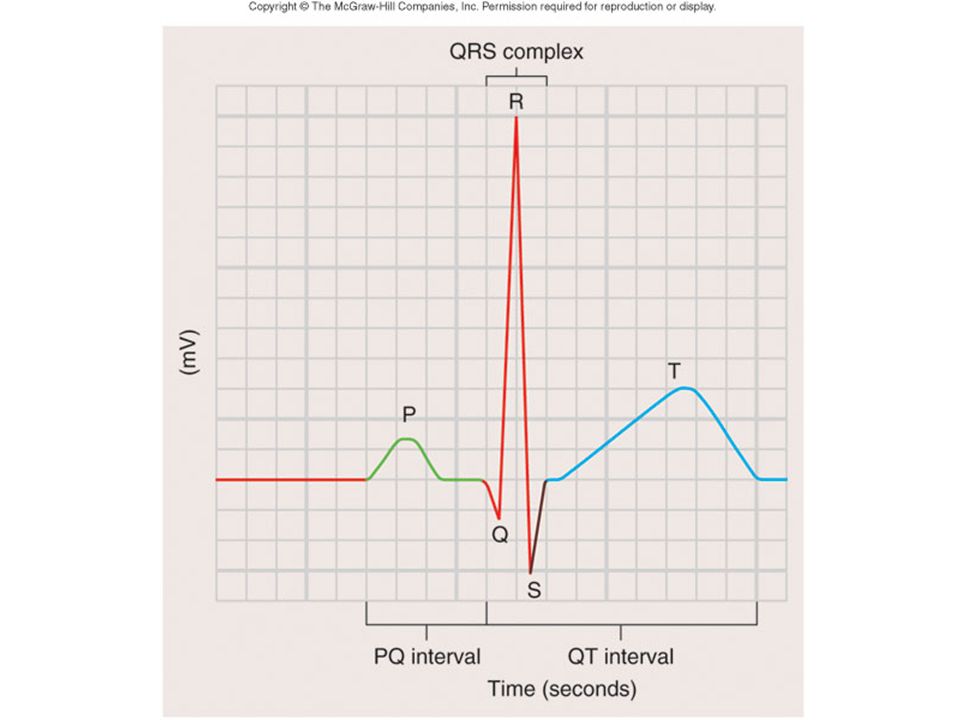
ELECTROCARDIOGRAM (ECG/EKG) Record of Electrical Changes in Heart Muscle Electrical Changes due to Depolarization & Repolarization of Cardiac Muscle Fibers Metal Electrodes Applied to Skin, Attached to Physiograph, Pick-up Electrical Activity Normal Cardiac Cycle Gives Rise to Characteristic “Waves”
 Record of Electrical Changes in Heart Muscle Electrical Changes due to Depolarization & Repolarization of Cardiac Muscle Fibers Metal Electrodes Applied to Skin, Attached to Physiograph, Pick-up Electrical Activity Normal Cardiac Cycle Gives Rise to Characteristic Waves")
46
ELECTROCARIDOGRAM (ECG/EKG) continued P Wave -SA Node Stimulates Atrial Depolarization -Occurs Prior to Atrial Contraction QRS Complex -Ventricular Depolarization -More Tissue, More Electrical Activity, Larger Wave -Occurs Prior to Ventricular Contraction T Wave -Ventricular Repolarization
 continued P Wave -SA Node Stimulates Atrial Depolarization -Occurs Prior to Atrial Contraction QRS Complex -Ventricular Depolarization -More Tissue, More Electrical Activity, Larger Wave -Occurs Prior to Ventricular Contraction T Wave -Ventricular Repolarization")
48
EKG ABNORMALITIES Tachycardia (BPM>100) Bradycardia (BPM<60) Premature Beats (Ectopic Source) Flutter (BPM 250-350) Fibrillation (Rapid, Uncoordinated)
 Bradycardia (BPM<60) Premature Beats (Ectopic Source) Flutter (BPM ) Fibrillation (Rapid, Uncoordinated)")
49
HEART SOUNDS Caused by Closing of Heart Valves AV Valves -prevent blood from flowing backwards into atria -“Lubb” Semilunar Valves -prevent blood from flowing backwards into ventricles -“Dupp”

50
HEART MURMURS Abnormal sound Often indicates valve disorder Causes: -Congenital defects -Scarring -Insufficiency/Backflow

Similar presentations


 in thorax, in inferior mediastinum>")


