Download presentation
Presentation is loading. Please wait.

1
Early pregnancy assessment (first trimester scan) Dr Shuhaila Ahmad Associate Professor Feto-Maternal Unit UKM Medical Centre 12/7/2015
 Dr Shuhaila Ahmad Associate Professor Feto-Maternal Unit UKM Medical Centre 12/7/2015")
2
Scope of the lecture Importance of early pregnancy scan Viable pregnancy Non-viable pregnancy – Miscarriage – Ectopic – Molar pregnancy Others – Multiple pregnancy – Nuchal translucency 12/7/2015

3
Importance of EP scan Determine: – Site of pregnancy Site of pregnancy – Gestational age Gestational age – Viability Viability – Fetal number Fetal number – Pathology in the uterus and adnexae Pathology in the uterus and adnexae 12/7/2015

4
Ectopic pregnancy Intrauterine pregnancy 12/7/2015

8
Pregnancy with corpus luteal cyst 12/7/2015

9
Viable pregnancy Calculate gestational age using LMP Scan the whole uterus and adnexae systematically Identify the IUGS – Anechoic area within the uterine cavity – TVS : 4.5 – 5 weeks – Ensure not pseudogestational sac: Must see the throphoblastic rim around the GS Identify the yolk sac 12/7/2015

10
Gestational sac Trophoblastic rim TVS TAS

11
If TVS: – fetal echo should be present by 16mm – Presence of yolk sac does not guarantee viable fetus. 12/7/2015 Yolk sac Fetal echo

12
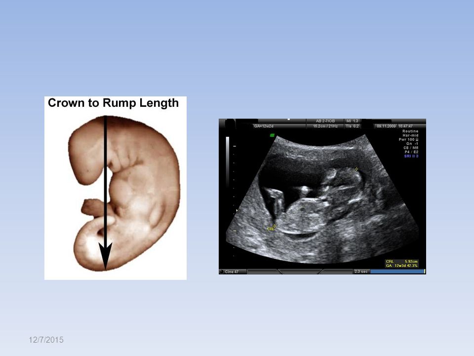
Gestational age Most accurate is the crown rump length Least biological variation Early gestation is better as at late gestation, the fetus assumes flexion attitude Beware not to include yolk sac into the measurement Wrong measurement may cause wrong EDD 12/7/2015

13
Gestational age Crown-rump length – Get the sagittal view in longest image of the fetus – Enlarge the image to nearly 70% of the monitor – Ensure the fetus in neutral position – Measure from the highest point in the head to the bottom end. – Do not include the legs – Do at least twice 12/7/2015

14
Which image is good for measuring CRL? 12/7/2015 Longest image but not sagittal Longest axis, sagital view but the fetus is in flexion

15
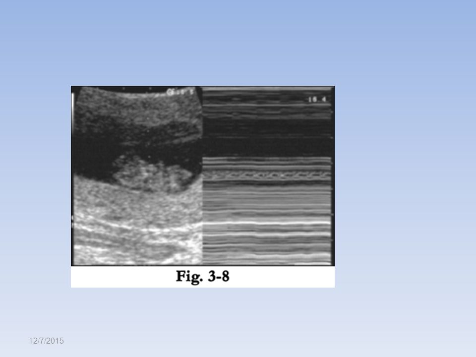
Fetal viability Must be able to see pulsation – If unsure, use M mode or colour doppler – Should be present if CRL is 2-4mm – If > 5mm, no FH activity may mean missed miscarriage 12/7/2015

16
Important discriminatory values 12/7/2015 Parameters suggestive of viable pregnancy TASTVS Serum βHCG and presence of IUGS 6000 IU/L2000 IU/L Mean diameter of GC and presence of yolk sac NA8mm Mean diameter of GC and presence of fetal echo 25mm16mm Mean fetal length (CRL) and presence of fetal heart activity 10mm5mm
 and presence of fetal heart activity 10mm5mm")
17
12/7/2015 TVSGS IUGS <16mm Repeat US 1 week IUGS >16mm Fetal echo +ve CRL >5mm FHA +ve Viable pregnancy FHA -ve Missed miscarriage CRL< 5mm FHA -ve Repeat scan 1 week Fetal echo -ve Blighted ovum No GS HCG <2000 Repeat HCG (48 hours) Doubling Repeat scan 1 week Not doubling EctopicMiscarriage HCG >2000 TRO ectopic
 Doubling Repeat scan 1 week Not doubling EctopicMiscarriage HCG >2000 TRO ectopic")
18
Ectopic pregnancy Definition: – Pregnancy that is not present within the inner lining of the uterus – 95% in the fallopian tubes 12/7/2015

19
Ultrasonographic features Empty uterus Free fluid the POD # complex mass / GS in the adnexae 12/7/2015 U

20
Molar pregnancy Excessive pregnancy symptoms Uterus maybe palpable in first trimester Usually no fetal echo Cavity filled up with multiple sonolucent areas = snow storm appearance Presence of multiple theca lutein cysts in the ovaries 12/7/2015

21
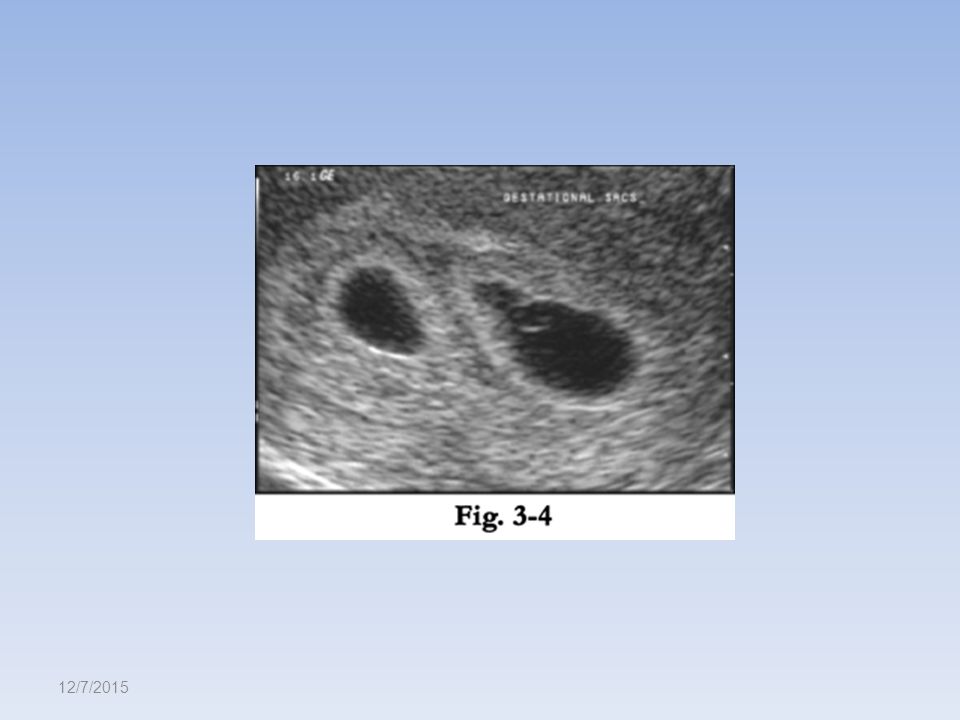
Multiple pregnancies Determine number of fetuses Determine chorionicity – MC : “T” sign – DC : “λ” sign Determine viability Determine gestational age 12/7/2015

22
Number of fetuses 12/7/2015

23
Chorionicity 12/7/2015

24
Nuchal translucency Between 11 to 13 +6 weeks About CRL of 45-84mm Mid sagittal view of the head Just chest and the head 12/7/2015

25
Key points Determine LMP and gestational age Identify uterus, adnexae and cervix Identify site if gestational sac Confirm viability and numbers Determine gestational age by measuring the fetus – Identification by TAS is delayed by I week compared to TVS Do not hesitate to proceed to TVS if needed It will NOT increase bleeding or risk of miscarriage 12/7/2015

26
Thank you and Good Luck for the hands on session 12/7/2015

Similar presentations

 of less than 5 mm located within the thickened,>")


, Javam M. (B.Sc), Ahmadi F. (MD)>")


