Download presentation
Presentation is loading. Please wait.

1
The Cardiac Cycle

2
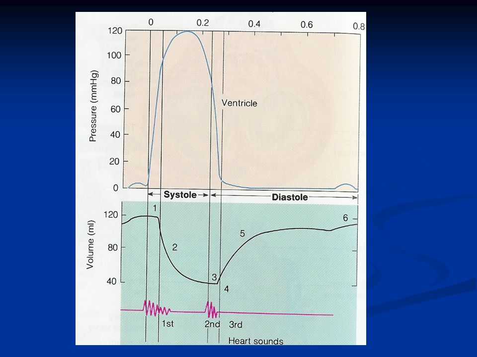
The repeating pattern of contraction (systole) and relaxation (diastole) of the heart The repeating pattern of contraction (systole) and relaxation (diastole) of the heart Duration of cardiac cycle = 0.8 seconds Duration of cardiac cycle = 0.8 seconds Diastole longer than systole Diastole longer than systole Ventricular contraction follows atrial contraction (0.1 to 0.2 second later) remember the delay from AV node that’s why Ventricular contraction follows atrial contraction (0.1 to 0.2 second later) remember the delay from AV node that’s why
 and relaxation (diastole) of the heart The repeating pattern of contraction (systole) and relaxation (diastole) of the heart Duration of cardiac cycle = 0.8 seconds Duration of cardiac cycle = 0.8 seconds Diastole longer than systole Diastole longer than systole Ventricular contraction follows atrial contraction (0.1 to 0.2 second later) remember the delay from AV node that’s why Ventricular contraction follows atrial contraction (0.1 to 0.2 second later) remember the delay from AV node that’s why")
3
The end diastolic volume: the total volume of blood in the ventricles at the end of diastole (120 ml) The end diastolic volume: the total volume of blood in the ventricles at the end of diastole (120 ml) Stroke volume is the volume of blood pumped by each ventricle per beat (70 ml) Stroke volume is the volume of blood pumped by each ventricle per beat (70 ml) Residual volume: amount of blood left in each ventricle at the end of systole (50 ml) Residual volume: amount of blood left in each ventricle at the end of systole (50 ml) The Cardiac Cycle
 The end diastolic volume: the total volume of blood in the ventricles at the end of diastole (120 ml) Stroke volume is the volume of blood pumped by each ventricle per beat (70 ml) Stroke volume is the volume of blood pumped by each ventricle per beat (70 ml) Residual volume: amount of blood left in each ventricle at the end of systole (50 ml) Residual volume: amount of blood left in each ventricle at the end of systole (50 ml) The Cardiac Cycle")
4
Ventricles contract Ventricles contract Ventricular pressure: increasing Ventricular pressure: increasing Ventricular volume: no change Ventricular volume: no change AV valves: closed.. prevent backflow of blood AV valves: closed.. prevent backflow of blood Semilunar valves: closed (P in ventricles < P in vessels) Semilunar valves: closed (P in ventricles < P in vessels) The Cardiac Cycle Isovolumetric ventricular contraction
 Semilunar valves: closed (P in ventricles < P in vessels) The Cardiac Cycle Isovolumetric ventricular contraction.")
6
Ventricular pressure: increasing > the pressure in the aortic and pulmonary vessels Ventricular pressure: increasing > the pressure in the aortic and pulmonary vessels Left ventricular pressure up to 120 mmHg (thicker wall, aorta artery higher pressure than polmunary) Left ventricular pressure up to 120 mmHg (thicker wall, aorta artery higher pressure than polmunary) Right ventricular pressure up to 25 mmHg Right ventricular pressure up to 25 mmHg Ventricular volume: decreasing Ventricular volume: decreasing Semilunar valves: open Semilunar valves: open AV valves: closed.. prevent backflow of blood AV valves: closed.. prevent backflow of blood The Cardiac Cycle Ejection phase

7
Ventricles relax Ventricles relax Ventricular pressure: decreasing Ventricular pressure: decreasing Ventricular volume: no change Ventricular volume: no change AV valves: closed AV valves: closed Semilunar valves: closed Semilunar valves: closed The Cardiac Cycle Isovolumetric relaxation

8
Ventricular pressure: below atrial pressure ( slightly above zero) Ventricular pressure: below atrial pressure ( slightly above zero) Ventricular volume: increasing Ventricular volume: increasing AV valves: open when pressure in the atria> the pressure in the ventricles AV valves: open when pressure in the atria> the pressure in the ventricles Semilunar valves: closed Semilunar valves: closed Passive ventricular filling via AV valves (80%) Passive ventricular filling via AV valves (80%) The Cardiac Cycle Rapid filling of the ventricles
 Ventricular pressure: below atrial pressure ( slightly above zero) Ventricular volume: increasing Ventricular volume: increasing AV valves: open when pressure in the atria> the pressure in the ventricles AV valves: open when pressure in the atria> the pressure in the ventricles Semilunar valves: closed Semilunar valves: closed Passive ventricular filling via AV valves (80%) Passive ventricular filling via AV valves (80%) The Cardiac Cycle Rapid filling of the ventricles")
9
Active filling of the ventricles (20%) Active filling of the ventricles (20%) Ventricular volume: slight rise Ventricular volume: slight rise Ventricular pressure: slight rise Ventricular pressure: slight rise Semilunar valves: closed Semilunar valves: closed AV valves: open AV valves: open The Cardiac Cycle Atrial systole
 Active filling of the ventricles (20%) Ventricular volume: slight rise Ventricular volume: slight rise Ventricular pressure: slight rise Ventricular pressure: slight rise Semilunar valves: closed Semilunar valves: closed AV valves: open AV valves: open The Cardiac Cycle Atrial systole")
10
The Cardiac Cycle 1. Isovolumetric contraction 2. Ejection phase 3. Isovolumetric relaxation 4. Rapid filling of the ventricles 5. Atrial systole

11
Heart Sounds The first heart sound: The first heart sound: Cause: closure of the AV valves Cause: closure of the AV valves The second heart sound: The second heart sound: Cause: closure of the semilunar valves Cause: closure of the semilunar valves

12
Cardiac Output Cardiac output is the volume of blood pumped by each ventricle per minute Cardiac output is the volume of blood pumped by each ventricle per minute CO= Stroke volume x Heart rate CO= Stroke volume x Heart rate (L/min)(ml/beat) (beat/min) = 70 X 70 = 4900 ml/min = 5 L/min Normal cardiac output (CO) = 5 L/min Normal cardiac output (CO) = 5 L/min
(ml/beat) (beat/min) = 70 X 70 = 4900 ml/min = 5 L/min Normal cardiac output (CO) = 5 L/min Normal cardiac output (CO) = 5 L/min")
13
Cardiac Output

14
Sympathetic stimulation Sympathetic stimulation HR (positive chronotropic effect) HR (positive chronotropic effect) CO CO Parasympathetic stimulation Parasympathetic stimulation HR HR CO CO Cardiac centers in the medulla oblangata Cardiac centers in the medulla oblangata Cardiac Output Regulation of Heart Rate
 HR (positive chronotropic effect) CO CO Parasympathetic stimulation Parasympathetic stimulation HR HR CO CO Cardiac centers in the medulla oblangata Cardiac centers in the medulla oblangata Cardiac Output Regulation of Heart Rate")
15
End Diastolic Volume (EDV) Frank- Starling Law of the Heart Frank- Starling Law of the Heart venous return EDV length of cardiac muscle (stretch) force of contraction stroke volume cardiac output Cardiac Output Regulation of Stroke Volume
 Frank- Starling Law of the Heart Frank- Starling Law of the Heart venous return EDV length of cardiac muscle (stretch) force of contraction stroke volume cardiac output Cardiac Output Regulation of Stroke Volume")
16
Positive ionotropic effect strength of contraction Positive ionotropic effect strength of contraction Sympathetic stimulation Sympathetic stimulation Adrenaline (neurotransmitter for Sym.) Adrenaline (neurotransmitter for Sym.) Negative ionotropic effect strength of contraction Negative ionotropic effect strength of contraction Parasympathetic stimulation Parasympathetic stimulation Acetylcholine (neurotransmitter for parasym.) Acetylcholine (neurotransmitter for parasym.) Vagal stimulation Vagal stimulation Cardiac Output Regulation of Stroke Volume
 Adrenaline (neurotransmitter for Sym.) Negative ionotropic effect strength of contraction Negative ionotropic effect strength of contraction Parasympathetic stimulation Parasympathetic stimulation Acetylcholine (neurotransmitter for parasym.) Acetylcholine (neurotransmitter for parasym.) Vagal stimulation Vagal stimulation Cardiac Output Regulation of Stroke Volume")
Similar presentations


 –Contracts.>")


 Conduction System of Heart Conduction System = Heart Beat & Pumping Cardiac Contractions = Unconscious –Autonomic Nervous.>")

