Download presentation
Presentation is loading. Please wait.

1
TUMORS OF THE LUNG * Classification: 1. Benign tumors: - Papilloma. - Fibroma. - Chondroma. 2. Locally malignant tumors: - Bronchial carcinoid 3. Malignant tumors: A. Primary M. tumors: - Bronchogenic carcinoma. - Lymphoma. - Sarcomas. B. Secondaries.

2
BRONCHOGENIC CARCINOMA BRONCHIAL CARCINOID 1. Age Above 40 Ys. 20-40 Ys. 2. Incidence 2. Incidence - The commonest 1ry M. tumor of the lung. - More in males. - Less common (1-5% of lung tumors). - Equal in both sexes. - Equal in both sexes. 3. Predisposing factors - Cigarette smoking. - Exhaust fumes of tar and diesel. - Bronchiectasis. - Asbestosis. Not related to cigarette smoking or environmental factors.
. - Equal in both sexes. - Equal in both sexes. 3. Predisposing factors - Cigarette smoking. - Exhaust fumes of tar and diesel. - Bronchiectasis. - Asbestosis. Not related to cigarette smoking or environmental factors..")
3
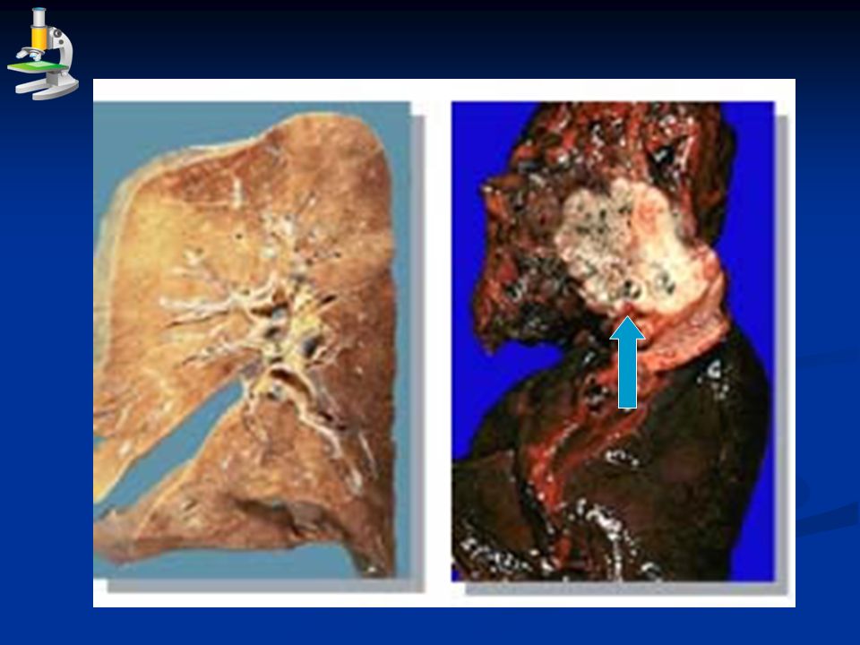
BRONCHOGENIC CARCINOMA BRONCHIAL CARCINOID 4. Cell of origin Epithelial cells of bronchial mucosa. Neuroendocrine cells “ kulchitsky cells ” of bronchial mucosa (APUD system). 5. Gross a. Central (hilar) type: 85%. - Arises from the main bronchus. - Forms a polypoid, ulcerative, or infiltrative growth. b. Peripheral type: 15%. - Arises from peripheral bronchi or bronchiole. - Forms a single or multiple masses. Site: near the hilum, arise from a main bronchus. N/E: a brownish yellow spherical mass projects into the bronchial lumen. May infiltrtae the lung tissue and appears as dumbell-shaped mass. The covering mucosa is usually intact.
. 5. Gross a. Central (hilar) type: 85%. - Arises from the main bronchus. - Forms a polypoid, ulcerative, or infiltrative growth. b. Peripheral type: 15%. - Arises from peripheral bronchi or bronchiole. - Forms a single or multiple masses. Site: near the hilum, arise from a main bronchus. N/E: a brownish yellow spherical mass projects into the bronchial lumen. May infiltrtae the lung tissue and appears as dumbell-shaped mass. The covering mucosa is usually intact..")
5
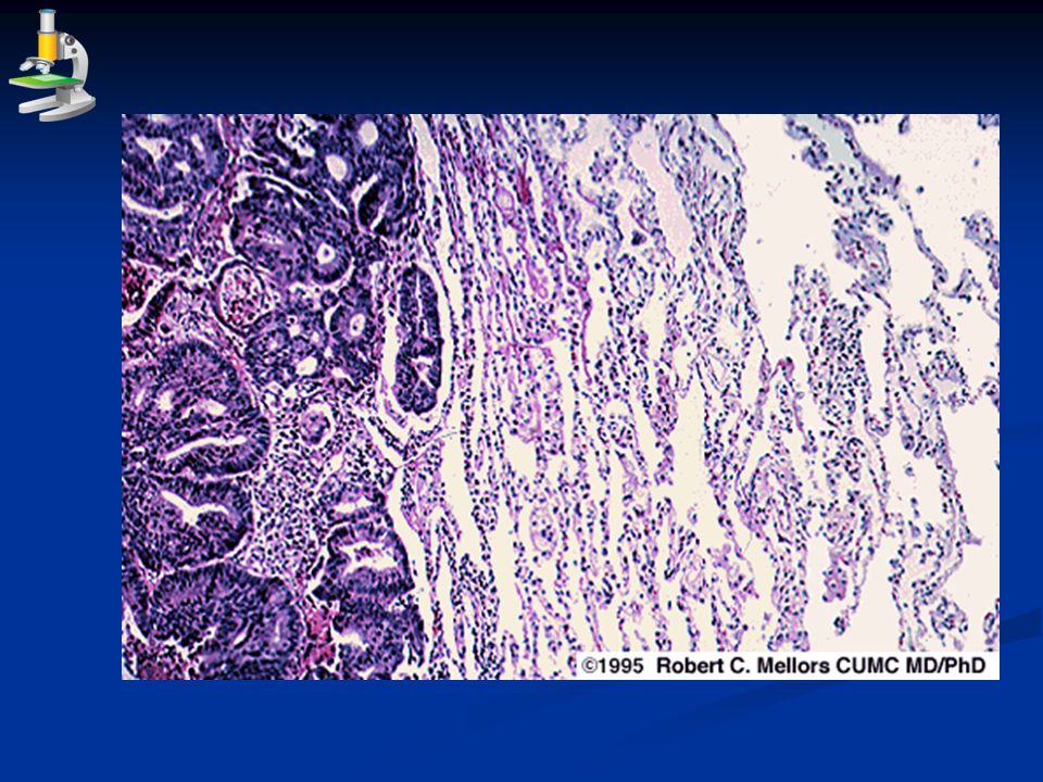
BRONCHOGENIC CARCINOMA BRONCHIAL CARCINOID 6. M/P * One of the following; 1. Squamous cell carcinoma (describe) 2. Adenocarcinoma (describe). 3. Small cell carcinoma (oat cell carcinoma): Sheets of malignant small cells, having increased nuclear/cytoplasmic ratio and frequent mitosis. The sheets are separated by fibrous tissue stroma. Wide areas of necrosis. 4. Large cell carcinoma: sheets of large sized malignant cells, having anaplastic features. The sheets are separated by scanty fibrous tissue stroma. - Nests, cords or masses of small cuboidal cells separated by delicate fibrous stroma. - The cells are uniform with rounded nuclei.the cytoplasm contain argyrophilic granules that stain with silver.
 2. Adenocarcinoma (describe). 3. Small cell carcinoma (oat cell carcinoma): Sheets of malignant small cells, having increased nuclear/cytoplasmic ratio and frequent mitosis. The sheets are separated by fibrous tissue stroma. Wide areas of necrosis. 4. Large cell carcinoma: sheets of large sized malignant cells, having anaplastic features. The sheets are separated by scanty fibrous tissue stroma. - Nests, cords or masses of small cuboidal cells separated by delicate fibrous stroma. - The cells are uniform with rounded nuclei.the cytoplasm contain argyrophilic granules that stain with silver..")
6
Small cell (oat cell) carcinoma
 carcinoma")
7
BRONCHOGENIC CARCINOMA BRONCHIAL CARCINOID 7. Effects and complications 1. Spread: a. Local: lung tissue, pleura, esophagus, phrenic nerve, recurrent laryngeal nerve, SVC & pericardium. b. Lymphatic: to hilar, mediastinal, supraclavicular L.Ns. c. Blood spread: through pulmonary artery to the lung and through pulmonary veins to systemic organs. 2. Hemoptysis. 3. Bronchial obstruction: leads to emphysema, atelectasis, retained secretion and suppuration, pneumonia. 4. Paraneoplastic syndrome. 1. Malignant transformation with metastasis. 2. Hemoptysis. 3. Bronchial obstruction. 4. Carcinoid syndrome.

8
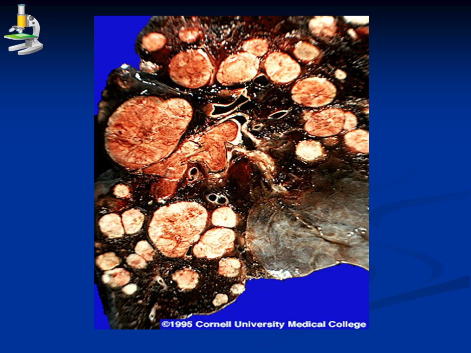
LUNG METASTASES (SECONDARIES) More common than the 1ry tumors. *Two forms I. Metastases reach through pulmonary artery: From malignant melanoma, hepatoma, encocrine carcinomas, urogenital carcinoma (renal cell carcinoma and testicular tumors), sarcomas and leukaemias. N/E: multiple nodules, variable in size scattered all over the lung lobes especially at the periphery. Metastases from RCC and seminoma are large in size and spherical in shape called “ cannon- ball secondaries ”. M/P: like its 1ry. II. Metastases reach through lymphatics: From cancer breast, abdominal carcinomas and lymphoma.
, sarcomas and leukaemias. N/E: multiple nodules, variable in size scattered all over the lung lobes especially at the periphery. Metastases from RCC and seminoma are large in size and spherical in shape called cannon- ball secondaries . M/P: like its 1ry. II. Metastases reach through lymphatics: From cancer breast, abdominal carcinomas and lymphoma..")
Similar presentations


 Environmental.>")


 have two basic components. Proliferating neoplastic cells that constitute.>")

 SHEN JIN The First Affiliated Hospital of Kunming Medical College.>")


>")
