Download presentation
Presentation is loading. Please wait.

1
Chapter 8 Lymphoid system Chapter 8 Lymphoid system Li Zhong Jie (李仲杰), Ph. D School of Medicine,Zhejiang University

2
The immune system: consists of the lymphatic organs, lymphocytes of the blood and lymph, and collections of different types of lymphocytes dispersed throughout the connective tissue. consists of the lymphatic organs, lymphocytes of the blood and lymph, and collections of different types of lymphocytes dispersed throughout the connective tissue. is essential to the body’s immunological defense against pathogens, foreign substences, infectious agents(bacteria and viruses) and abnormal cells. is essential to the body’s immunological defense against pathogens, foreign substences, infectious agents(bacteria and viruses) and abnormal cells.
 and abnormal cells. is essential to the body’s immunological defense against pathogens, foreign substences, infectious agents(bacteria and viruses) and abnormal cells..")
3
Immune response: Cellular immunity: involving mainly T-lymphocytesCellular immunity: involving mainly T-lymphocytes Humoral immunity: involving mainly B-lymphocytesHumoral immunity: involving mainly B-lymphocytes Mast cells, macrophages and otherMast cells, macrophages and other white blood cells are also involved in white blood cells are also involved in the immune response. Function: i. immunologic defense ii. immune surveillance iii. immune homeostasis

4
The cells of lymphatic system

5
1) Lymphocyte : a. T-lymphocytes: cytotoxic T cell: Tc kill the tumor cell, virus infective cell and foreign cell cytotoxic T cell: Tc kill the tumor cell, virus infective cell and foreign cell helper T cell: Th stimulate the B-lymphocyte differentiate into plasma cell helper T cell: Th stimulate the B-lymphocyte differentiate into plasma cell suppressor T cell: Ts regulate the function of B-lymphocyte and T-lymphocyte suppressor T cell: Ts regulate the function of B-lymphocyte and T-lymphocyte b. B-lymphocytes: become into plasma cell c. NK cell: attack virus infective cell and tumor cell without previous stimulation

6
Lymphocytes B-cells mature in bone marrow then concentrate in lymph nodes and spleen B-cells mature in bone marrow then concentrate in lymph nodes and spleen T-cells mature in thymus T-cells mature in thymus B and T cells mature then circulate in the blood and lymph B and T cells mature then circulate in the blood and lymph Circulation ensures they come into contact with pathogens and each other Circulation ensures they come into contact with pathogens and each other

7
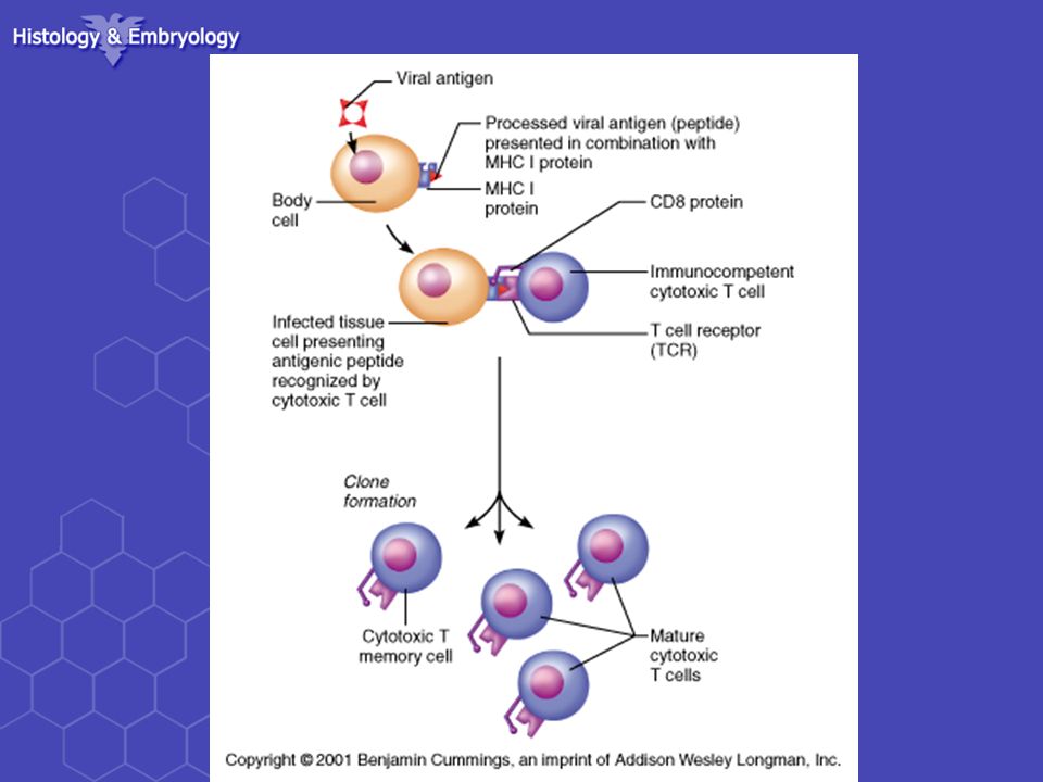
T-Lymphocytes Mature T-cells have T cell receptors which have a very similar structure to antibodies and are specific to 1 antigen. Mature T-cells have T cell receptors which have a very similar structure to antibodies and are specific to 1 antigen. They are activated when the receptor comes into contact with the antigen with another host cell (e.g. on a macrophage membrane or an invaded body cell) They are activated when the receptor comes into contact with the antigen with another host cell (e.g. on a macrophage membrane or an invaded body cell) Release perforin and granzyme Release perforin and granzyme
 They are activated when the receptor comes into contact with the antigen with another host cell (e.g. on a macrophage membrane or an invaded body cell) Release perforin and granzyme Release perforin and granzyme.")
9
B -Lymphocytes There are circa 10 million different B- lymphocytes, each of which make a different antibody. There are circa 10 million different B- lymphocytes, each of which make a different antibody. There are a small group of clones of each type of B-lymphocyte There are a small group of clones of each type of B-lymphocyte

10
B -Lymphocytes

11
Some activated B cells PLASMA CELLS these produce lots of antibodies Some activated B cells PLASMA CELLS these produce lots of antibodies The antibodies travel to the blood, lymph, lining of gut and lungs. The antibodies travel to the blood, lymph, lining of gut and lungs.

12
B -Lymphocytes Some activated B cells MEMORY CELLS. Some activated B cells MEMORY CELLS. Memory cells divide rapidly as soon as the antigen is reintroduced. Memory cells divide rapidly as soon as the antigen is reintroduced. There are many more memory cells than there were clone cells. There are many more memory cells than there were clone cells. When the pathogen/infection infects again it is destroyed before any symptoms show. When the pathogen/infection infects again it is destroyed before any symptoms show.

13
2) Plasma cell 3) Antigen presenting cell: a. dendritic cell b. macrophage ---mononuclear ---mononuclear ---phagocytic system ---phagocytic system Antigen processing is a necessary preliminary step for activation of T cell.

14
4) other cells: granulated cell granulated cell mast cell mast cell blood platelet blood platelet blood-borne stem cell blood-borne stem cell
 other cells: granulated cell granulated cell mast cell mast cell blood platelet blood platelet blood-borne stem cell blood-borne stem cell")
15
–2 types Diffuse lymphatic tissue Diffuse lymphatic tissue –No capsule present –Found in connective tissue of almost all organs Lymphatic nodules Lymphatic nodules –No capsule present –Oval-shaped masses –Found singly or in clusters Lymphoid tissue

16
General organization of lymphoid tissue: ---components: ---components: Reticular tissue: stellate-shaped with processes to form network Reticular tissue: stellate-shaped with processes to form network Cells: Cells: Lymphocyte (chiefly) Lymphocyte (chiefly) Plasma cell Plasma cell antigen presenting cell antigen presenting cell other cells other cells
 Lymphocyte (chiefly) Plasma cell Plasma cell antigen presenting cell antigen presenting cell other cells other cells")
17
a. Diffuse lymphoid tissue: no clear boundary no clear boundary mainly consists of T-lymphocyte mainly consists of T-lymphocyte postcapillary venules: postcapillary venules: -high endothelial venules, lining of tall cuboidal cells -high endothelial venules, lining of tall cuboidal cells -opening for lymphocyte enter lymphoid tissue from blood -opening for lymphocyte enter lymphoid tissue from blood Diffuse LT Lymphoid nodule

18
b. Lymphoid nodule: spherical or ovoid spherical or ovoid have clear boundaries have clear boundaries mainly composed of B- lymphocyte mainly composed of B- lymphocyte germinal center: lighter- stained central zone, these cells produce antibody- synthesizing plasma cells germinal center: lighter- stained central zone, these cells produce antibody- synthesizing plasma cells * primary LN → secondary LN

19
Lymph Nodules

20
Lymphatic organs: central lymphatic organs: ---produce the lymphocyte ---antigen-independent proliferation ---contain: thymus---T-lymphocyte bone marrow---B-lymphocyte peripheral lymphatic organs: ---receive the lymphocyte and perform the immune response ---antigen-dependent proliferation ----contain: lymph node spleen tonsils

21
Thymus – Location – behind the sternum in the mediastinum – Is a flat, capsule divides it into 2 lobes – Development Infant – conspicuous Infant – conspicuous Puberty – maximum size Puberty – maximum size Maturity – decreases in size Maturity – decreases in size – Function Differentiation and maturation Differentiation and maturation of T cells of T cells

22
Capsule ( C.T.) interlobular septa (trabecula) Cortex Cortex: Endothelial reticular cell (ERC) Lymphocyte: thymocyte Medulla: Medulla: ERC Lymphocyte: few Thymic corpuscles Structure:
 interlobular septa (trabecula) Cortex Cortex: Endothelial reticular cell (ERC) Lymphocyte: thymocyte Medulla: Medulla: ERC Lymphocyte: few Thymic corpuscles Structure:")
23
Thymus Cortex Medulla

24
Cortex medulla

25
Cortex thymocytes: the T cells in different developing stages. thymocytes: the T cells in different developing stages. epithelial reticular cells: provide a meshwork. Secret thymosin and thymopoietin epithelial reticular cells: provide a meshwork. Secret thymosin and thymopoietin A very active site of lymphocyte production A very active site of lymphocyte production

26
Thymic cortex epithelial reticular cells thymocytes

27
Blood-thymus barrier: endothelial cell of continuous capillary very thick basement membrane of EC perivascular space with macrophage in it basement membrane of ERC ERC or their processes with desmosome Consists of : Function: provide a stable environment for lymphocytes developing in the cortex---prevents circular antigens from reaching the thymic cortex

28
Medulla Thymocytes:few Thymocytes:few epithelial reticular cells: A large number epithelial reticular cells: A large number thymic corpuscles:A distinguishing feature of the thymic medulla. thymic corpuscles:A distinguishing feature of the thymic medulla.

29
thymic corpuscles

30
Hassall’s ( thymic) corpuscles a characteristic feature of the thymus a characteristic feature of the thymus their significance is as yet unknown their significance is as yet unknown
 corpuscles a characteristic feature of the thymus a characteristic feature of the thymus their significance is as yet unknown their significance is as yet unknown")
31
Hassall’s ( thymic) corpuscles spherical or ovoid body, 20-150um in diameter, composed of concentrically-arranged epithelial reticular cells, the central cells often show degeneration spherical or ovoid body, 20-150um in diameter, composed of concentrically-arranged epithelial reticular cells, the central cells often show degeneration /peripheral cell: inmature /near centra: mature /center cell: keratinased- eosinophilic
 corpuscles spherical or ovoid body, um in diameter, composed of concentrically-arranged epithelial reticular cells, the central cells often show degeneration spherical or ovoid body, um in diameter, composed of concentrically-arranged epithelial reticular cells, the central cells often show degeneration /peripheral cell: inmature /near centra: mature /center cell: keratinased- eosinophilic")
32
the thymic epithelial reticular cells can secrete thymic hormones (thymosin, thymolin, thymopoietin) the thymic epithelial reticular cells can secrete thymic hormones (thymosin, thymolin, thymopoietin) induce the stem cell to differentiate into T-lymphocyte More than 90% of the thymocyte degenerate in this area, only less than 10% of thymocyte will mature and leave the thymus as T-lymphocytes through the postcapillary venules. Thymus function:

33
Adult thymus has high infiltration of adipose tissue (A)
")
34
Older adult thymus has large amounts of adipose tissue

35
Lymph node A mass of lymphatic tissue enclosed in a capsule of connective tissue A mass of lymphatic tissue enclosed in a capsule of connective tissue Widely scattered along the course of lymph vessels Widely scattered along the course of lymph vessels Concentrated in area in neck, axilla (arm pits), and groin. Concentrated in area in neck, axilla (arm pits), and groin.
, and groin..")
36
Structure ---capsule: CT, trabeculae or septa ---cortex: outer densely-stained part ---medulla: inner paler-stained part hilum

37
Structure: capsule trabecula several afferent lymph vessels several afferent lymph vessels cortex cortex out cortex: (superficial cortex) lymphoid nodule (B-LC) internodule zone inner cortex: diffuse lymphoid tissue (T-LC) (paracortical zone) cortical lymphatic sinuses subcapsular sinus: peritrabecular sinus macrophage
 lymphoid nodule (B-LC) internodule zone inner cortex: diffuse lymphoid tissue (T-LC) (paracortical zone) cortical lymphatic sinuses subcapsular sinus: peritrabecular sinus macrophage")
38
Lymph node

39
Afferent lymph vessels GC

40
Superficial cortex(Nodular cortex) The outer cortex,situated under the capsule,consists of many Lymph nodules with or without germinal center. The outer cortex,situated under the capsule,consists of many Lymph nodules with or without germinal center.

41
Lymph nodule primary LN → secondary LN *Germinal center: central pale area /dark zone: large, immature BLC, Th /light zone: medium-sized BLC, Th C, macrophage, FDC /cap:small BLC:

42
paracortex zone (deep cortex) Composed of diffuse lymphatic tissue occupied by T cells. Also called deep cortex unit. Superficial cortex paracortex zone

43
outer cortex inner cortex capsule

44
SS: subcapsular sinus T: trabecular

45
subcapsular sinus peritrabecular sinus cortical lymphatic sinuses

46
Subcapsular sinuses Trabeculae Capsule peritrabec ular sinus

47
high endothelial venules ( venules-postcapillary ) ---opening for lymphocyte enter lymphoid tissue from blood
 ---opening for lymphocyte enter lymphoid tissue from blood")
48
high endothelial venules ( venules- postcapillary )
")
49
Medulla ---medullary cord: LT cord: LT cord: B-lymphocyte, plasma cell, macrophage, mast cell B-lymphocyte, plasma cell, macrophage, mast cell ---medullary sinus: similar to cortical sinus and connect with them similar to cortical sinus and connect with them more macrophagemore macrophage

50
Passage of lymph in lymph node Afferent lymph vessels Subcapsular sinus Cortical sinuses Medullary sinuses Efferent lymph vessels

51
Efferent lymph vessels in hilum

52
Recirculation of lymphocytes: Lymphoid tissue Efferent lymphatic vessel Postcapillary venules Blood circulation

53
Functions of lymph node Filter the lymph Filter the lymph Place to perform the immune response Place to perform the immune response Involve in the recirculation of lymphocyte Involve in the recirculation of lymphocyte

54
Spleen –Largest lymphatic organ –Located between the stomach & diaphragm –Structure is similar to a node Capsule present Capsule present But no afferent vessels or sinuses But no afferent vessels or sinuses –Histology Red pulp contains all the components of circulating blood Red pulp contains all the components of circulating blood White pulp is similar to lymphatic nodules White pulp is similar to lymphatic nodules –Functions Filters blood Filters blood Stores blood Stores blood

55
Spleen located in the passages of blood structure structure capsule parenchyma white pulp red pulp marginal zone

56
Structure: capsule: D.C.T. containing smooth muscle----trabecular; capsule is covered by mesothelium D.C.T. containing smooth muscle----trabecular; capsule is covered by mesothelium The splenic artery branch into trabecular arteries

57
White pulp : gray-white spots gray-white spots --- Isolated and randomly distributed. periarterial lymphatic sheath: periarterial lymphatic sheath: - diffuse lymphoid tissue : T-lymphocyte, macrophage - diffuse lymphoid tissue : T-lymphocyte, macrophage - central artery - central artery splenic corpuscle: splenic corpuscle: -lymphoid nodules: B-lymphocyte, macrophage -lymphoid nodules: B-lymphocyte, macrophage

58
Marginal zone: between White pulp and Red pulp between White pulp and Red pulp T-, B-lymphocyte, macrophage, less erythrocyte T-, B-lymphocyte, macrophage, less erythrocyte marginal sinus: central artery’s branch- channel for antigen and lymphocyte enter lymphoid tissue marginal sinus: central artery’s branch- channel for antigen and lymphocyte enter lymphoid tissue function: first capture, recognize antigen and induce immune reaction function: first capture, recognize antigen and induce immune reaction

59
Red pulp: splenic cord: Lymph tissue cord Lymph tissue cord B-,T-lymphocyte, macrophage, erythrocytes B-,T-lymphocyte, macrophage, erythrocytes function: filter blood function: filter blood splenic cord splenic sinus

60
splenic sinus: Blood sinus; 12-14um endothelial cell: rod-liked, gap Reticular fiber basal lamina: incomplete Abundant macrophage splenic sinus endothelial cell splenic cord

61
splenic sinus:

62
Blood supply of spleen Study by yourself splenic A→trabecular A→central A branches → marginal sinuses → splenic A→trabecular A→central A branches → marginal sinuses → penicillar arterioles (including: pulp arteriole→ sheathed capillary→ arterial capillary) → splenic sinus→ pulp venule→ trabecular vein→ splenic vein penicillar arterioles (including: pulp arteriole→ sheathed capillary→ arterial capillary) → splenic sinus→ pulp venule→ trabecular vein→ splenic vein
 → splenic sinus→ pulp venule→ trabecular vein→ splenic vein penicillar arterioles (including: pulp arteriole→ sheathed capillary→ arterial capillary) → splenic sinus→ pulp venule→ trabecular vein→ splenic vein")
63
Blood supply of spleen

64
Function: a. filter the blood a. filter the blood b. immunological defence b. immunological defence c. production of blood cells in fetus c. production of blood cells in fetus d. blood storage: 40 ml d. blood storage: 40 ml

65
5.Tonsil (Study by yourself) ---palatine tonsil ---palatine tonsil ---pharyngeal tonsil ---pharyngeal tonsil ---lingual tonsil ---lingual tonsil
 ---palatine tonsil ---palatine tonsil ---pharyngeal tonsil ---pharyngeal tonsil ---lingual tonsil ---lingual tonsil")
66
structure Mucosa Mucosa Epithelium stratified squamous epithelium form crypts lymphocyte infiltration Lamina propria lymphatic nodules diffuse lymphatic tissue Capsule: C.T. Capsule: C.T.

67
Tonsil

68
Mononuclear phagocyte system (Study by yourself) Bone marrow young monocyte Blood monocyte Tissues or organs CT: macrophage Liver: Kuffer cell Lung: dust cell Nerve tissue: microglia Osseous tissue: osteoclast Skin: Langerhans cell
 Bone marrow young monocyte Blood monocyte Tissues or organs CT: macrophage Liver: Kuffer cell Lung: dust cell Nerve tissue: microglia Osseous tissue: osteoclast Skin: Langerhans cell")
69
Function of MPS: phagocytosis phagocytosis participate in immune reaction secrete bioactive substances

70
Immunodeficiency Diseases Severe Combined Immunodeficienc y Disease Severe Combined Immunodeficienc y Disease –hereditary lack of T and B cells –vulnerability to opportunistic infection

71
Disorders of Immunity: Autoimmune Diseases Examples of autoimmune diseases Examples of autoimmune diseases –Multiple sclerosis – white matter of brain and spinal cord are destroyed –Myasthenia gravis – impairs communication between nerves and skeletal muscles –Juvenile diabetes – destroys pancreatic beta cells that produce insulin –Rheumatoid arthritis – destroys joints

72
Thanks for your attention!

Similar presentations

 System Color Textbook of Histology, 3rd ed. Gartner & Hiatt Copyright.>")


 -mast cells -interdigitating.>")


 from the lamina propria. Absorb.>")


