Download presentation
Presentation is loading. Please wait.

1
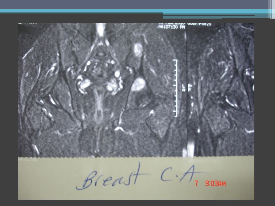
Jalal Jalal Shokouhi-MD Radiologist, Jam e jam and koorosh medical imaging center General secretary of Iranian society of radiology - President of Iranian radiologic cooperative Hasan Joorabchi-MD OB & GY, Tabriz medical university, Iran Mohammad Hossein Herischi – MD Orthopedist, Baku, Republic Azerbayjan Shahyar Pashaie – PhD Anatomist, Tehran, Iran Dariush Etemadi –MD Radiologist, Koorosh medical imaging center, Tehran, Iran jalaljalalshokouhi@hotmail.comjalaljalalshokouhi@hotmail.com www.medimage.irwww.medimage.ir Women's imaging Pelvic pain MRI in chronic pelvic pain (C.P.P.)
")
2
1/3 of women pelvic pain in lifetime Most common complain Most difficult to diagnosis and manage 60% (three of five) never find out the cause of their pelvic pain jalaljalalshokouhi@hotmail.comjalaljalalshokouhi@hotmail.com www.medimage.irwww.medimage.ir
 never find out the cause of their pelvic pain")
3
Women seek help from: Gynecologists Urologists Family practitioners jalaljalalshokouhi@hotmail.comjalaljalalshokouhi@hotmail.com www.medimage.irwww.medimage.ir

6
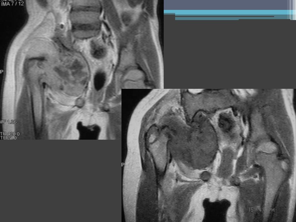
Pelvic masses

7
PET/CT technique & imaging issues

8
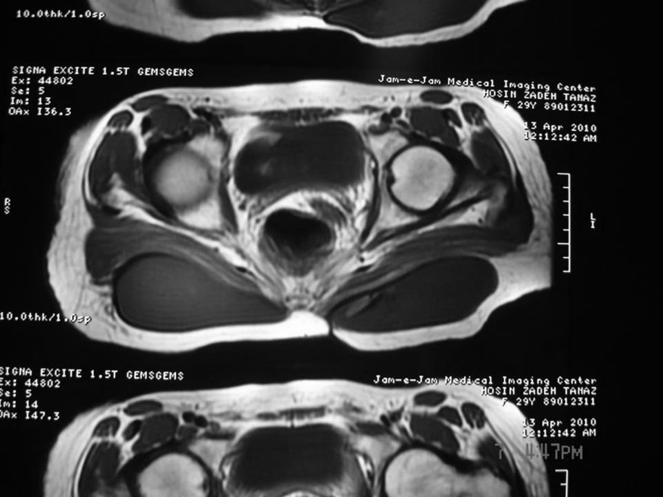
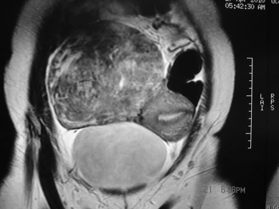
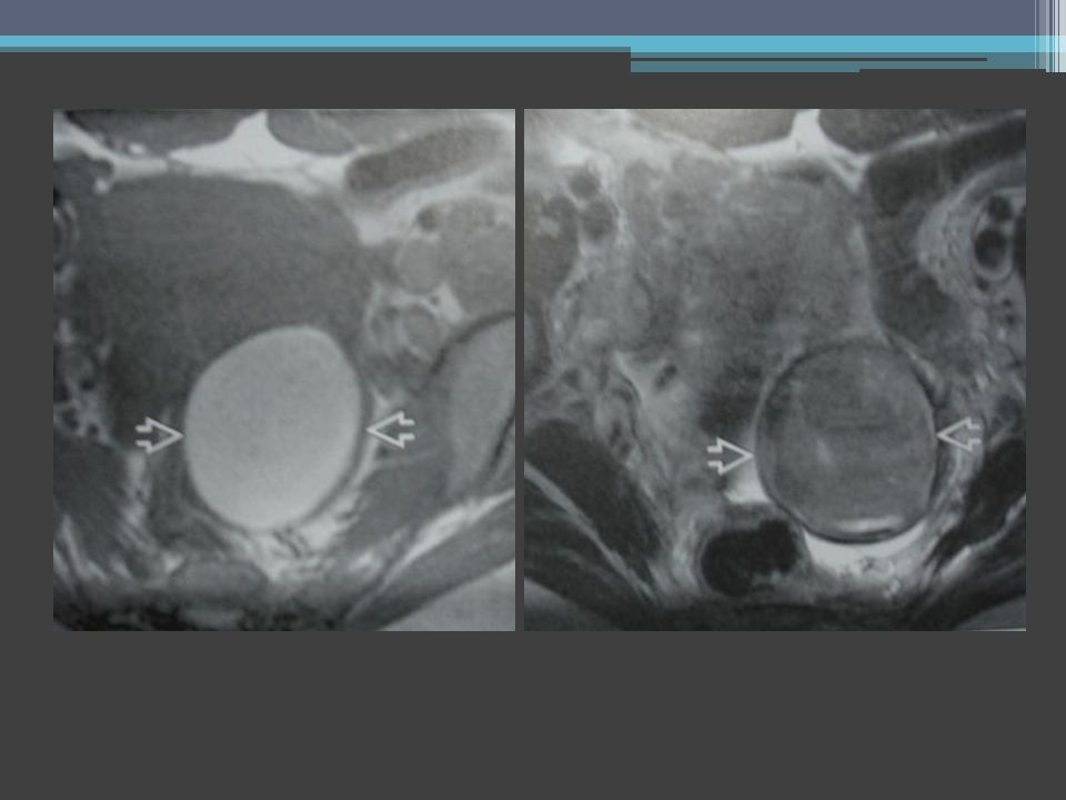
Low midline pelvic mass Prolapsing LeiomyomaMucinous Rectal Cancer

9
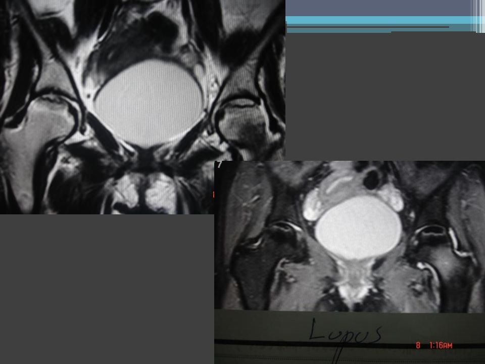
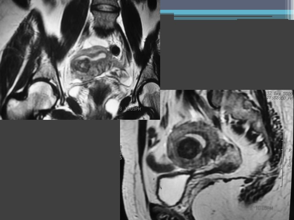
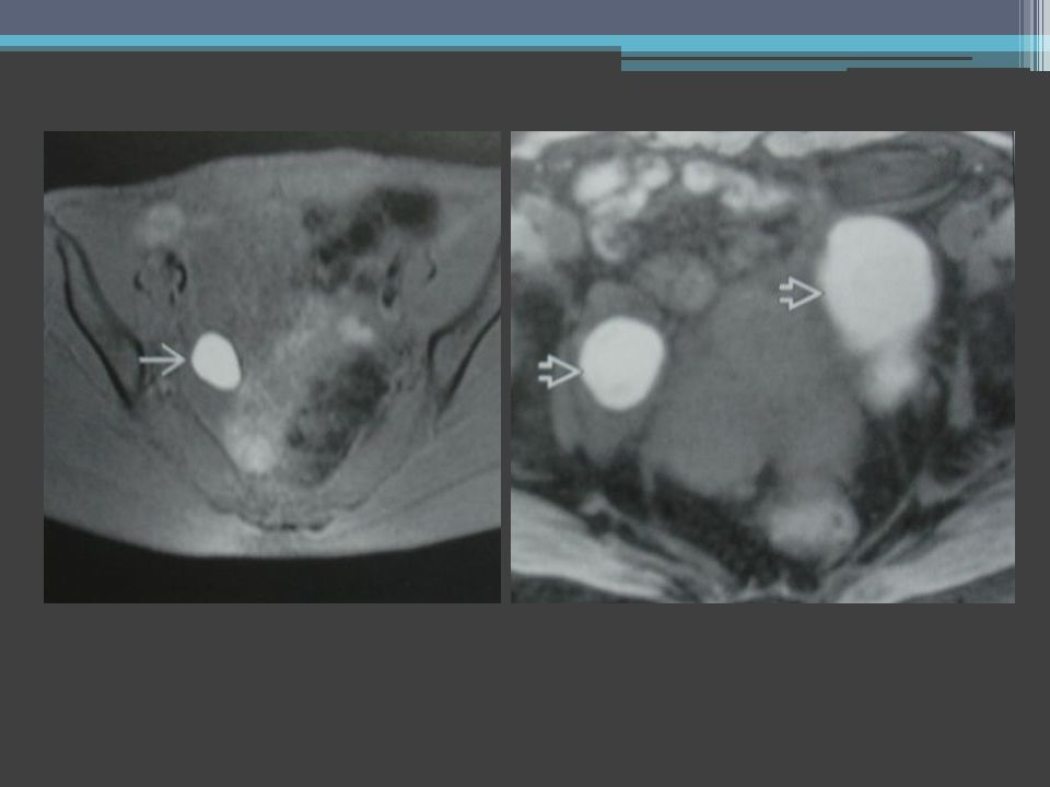
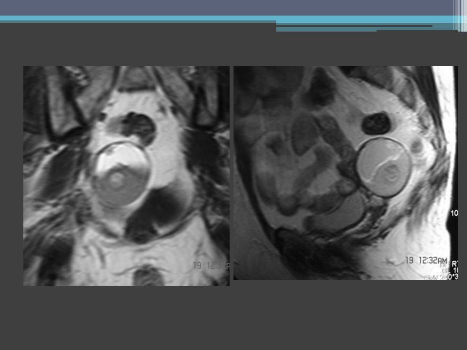
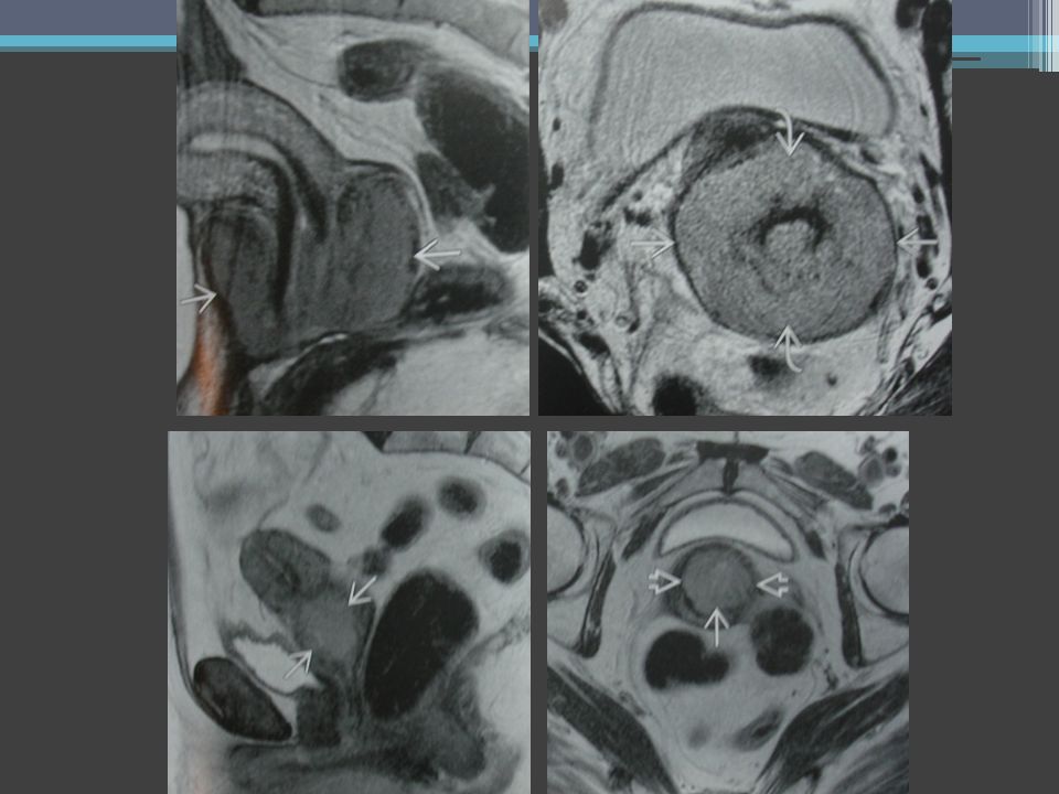
Anorectal Mass Prolapsing rectal cancerRectum duplication cyst

18
Chronic pelvic pain: Any pelvic pain lasting more than six months 1 woman of five women (20% incidence) It is a non specific pain (acute pain may indicate a specific injury) Cause: change in nervous system, tissues or muscles jalaljalalshokouhi@hotmail.comjalaljalalshokouhi@hotmail.com www.medimage.irwww.medimage.ir
 It is a non specific pain (acute pain may indicate a specific injury) Cause: change in nervous system, tissues or muscles")
19
Sacral teratoma

20
Sacral Masses Anterior Meningocele Sacrococcygeal chordoma

22
Signs and symptoms: 1. Severe and steady pain (dull to sharp, mild to severe) 2. Pain that comes and goes (intermittent) 3. Sharp pains or cramping 4. Dull aching 5. Limited physical activity 6. Pressure or heaviness deep within pelvis 7. Painful intercourse 8. Pain when urination and bowel movement 9. Severe cramping during periods 10. Depression and hopelessness and addiction
 3. Sharp pains or cramping 4. Dull aching 5. Limited physical activity 6. Pressure or heaviness deep within pelvis 7. Painful intercourse 8. Pain when urination and bowel movement 9. Severe cramping during periods 10. Depression and hopelessness and addiction.")
23
Accurate diagnosis: Pelvic exam Cultures of excretions Pelvic ultrasound and sonohisterography (TA, TV,TR) Laparascopy X-ray, X-ray CT, CT PET MRI (most accurate) jalaljalalshokouhi@hotmail.comjalaljalalshokouhi@hotmail.com www.medimage.irwww.medimage.ir
 Laparascopy X-ray, X-ray CT, CT PET MRI (most accurate)")
24
Causes (Gynecologic) : 1.Endometriosis (asymptomatic, marked pain, possible infertility) 2.Pelvic congestion syndrome 3.Adenomyosis 4.Chronic pelvic inflammatory disease *sexually transmitted organism *pelvic surgery *appendix perforation pain, bleeding, fever, chills if acute 5.Ovarian remnant 6.Fibroids 7.Irritable bowel syndrome 8.Interstitial cystitis (painful bladder syndrome) 9.Vaginal fistula 10.Vesicovaginal fistula 11.Uterovaginal fistula 12.Urethrovaginal fistula 13.Rectovaginal fistula 14.Tumors (benign and malignant) 15.Neuropathic pain 16.Diverticulitis 17.Pelvic floor pain (painful urination, constipation, painful intercourse) 18.Fibromyalgia connective tissue disease, pain and depression Non-gynecologic Bone and joint diseases Muscular Neurogenic
 : 1.Endometriosis (asymptomatic, marked pain, possible infertility) 2.Pelvic congestion syndrome 3.Adenomyosis 4.Chronic pelvic inflammatory disease *sexually transmitted organism *pelvic surgery *appendix perforation pain, bleeding, fever, chills if acute 5.Ovarian remnant 6.Fibroids 7.Irritable bowel syndrome 8.Interstitial cystitis (painful bladder syndrome) 9.Vaginal fistula 10.Vesicovaginal fistula 11.Uterovaginal fistula 12.Urethrovaginal fistula 13.Rectovaginal fistula 14.Tumors (benign and malignant) 15.Neuropathic pain 16.Diverticulitis 17.Pelvic floor pain (painful urination, constipation, painful intercourse) 18.Fibromyalgia connective tissue disease, pain and depression Non-gynecologic Bone and joint diseases Muscular Neurogenic")
39
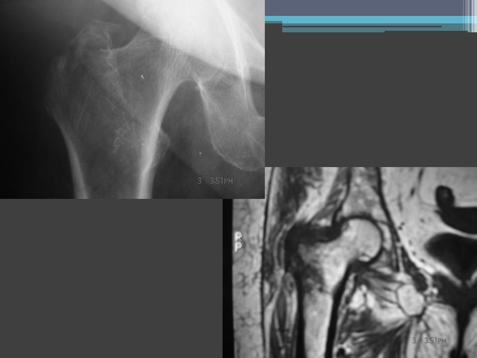
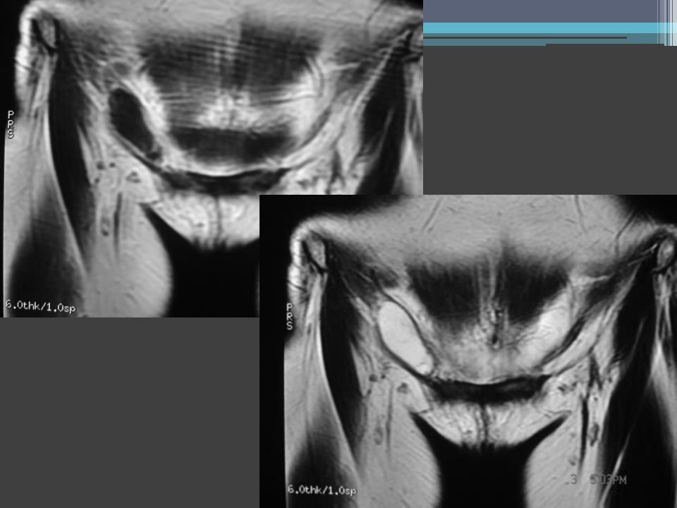
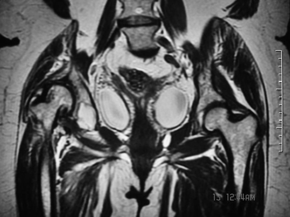
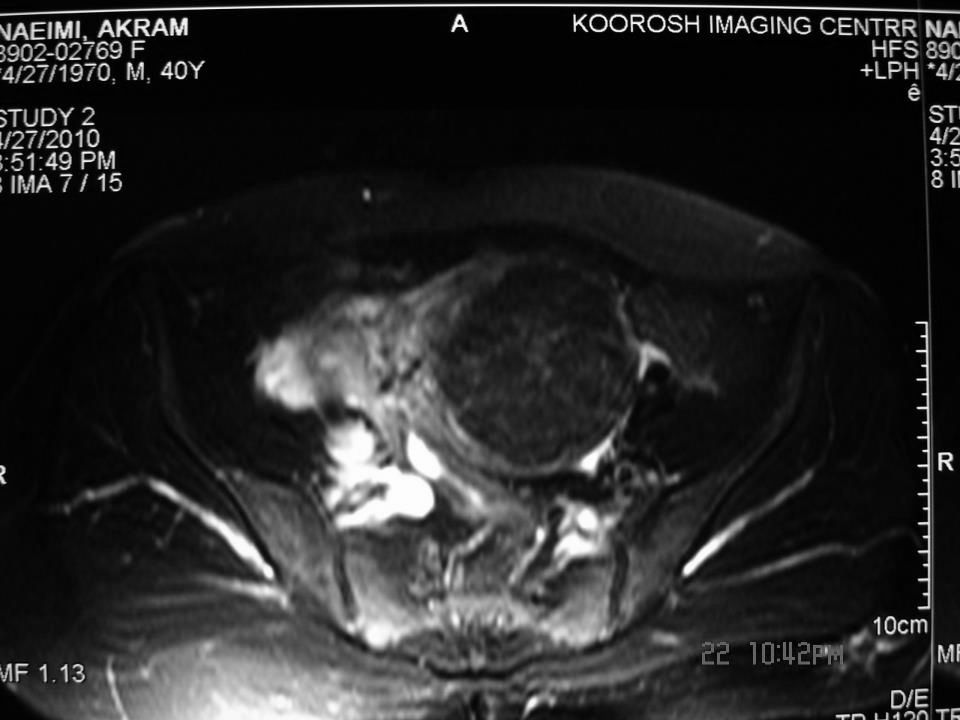
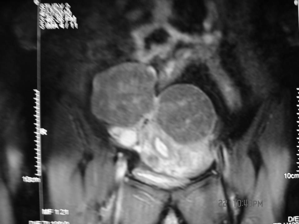
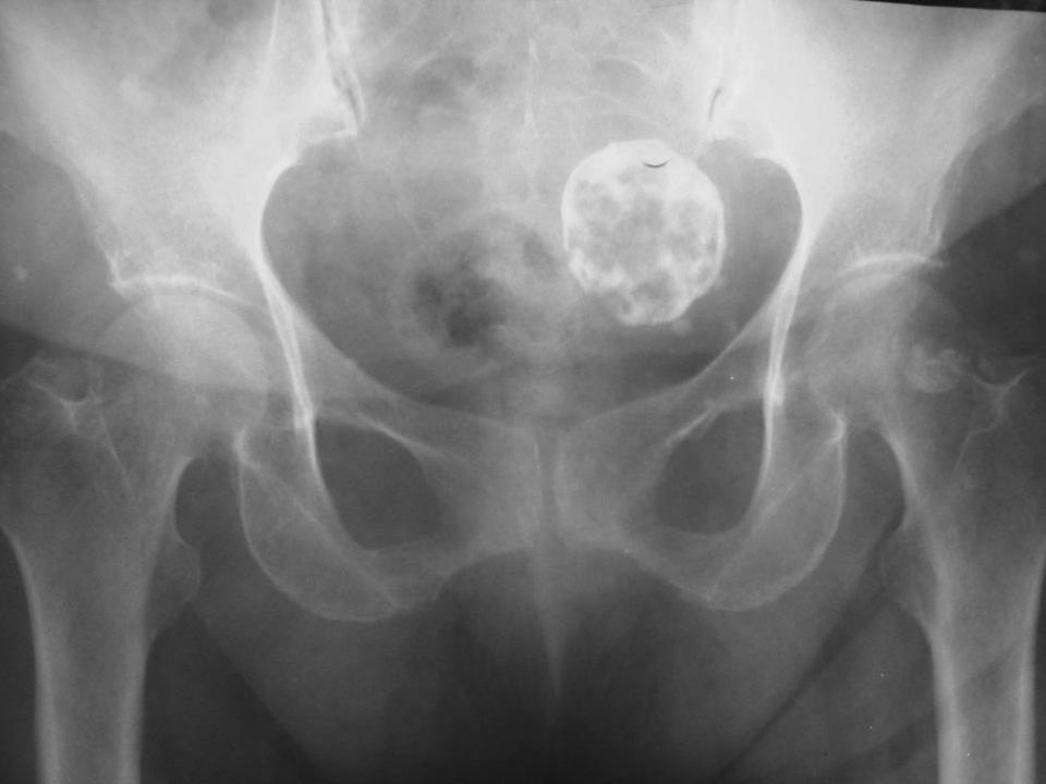
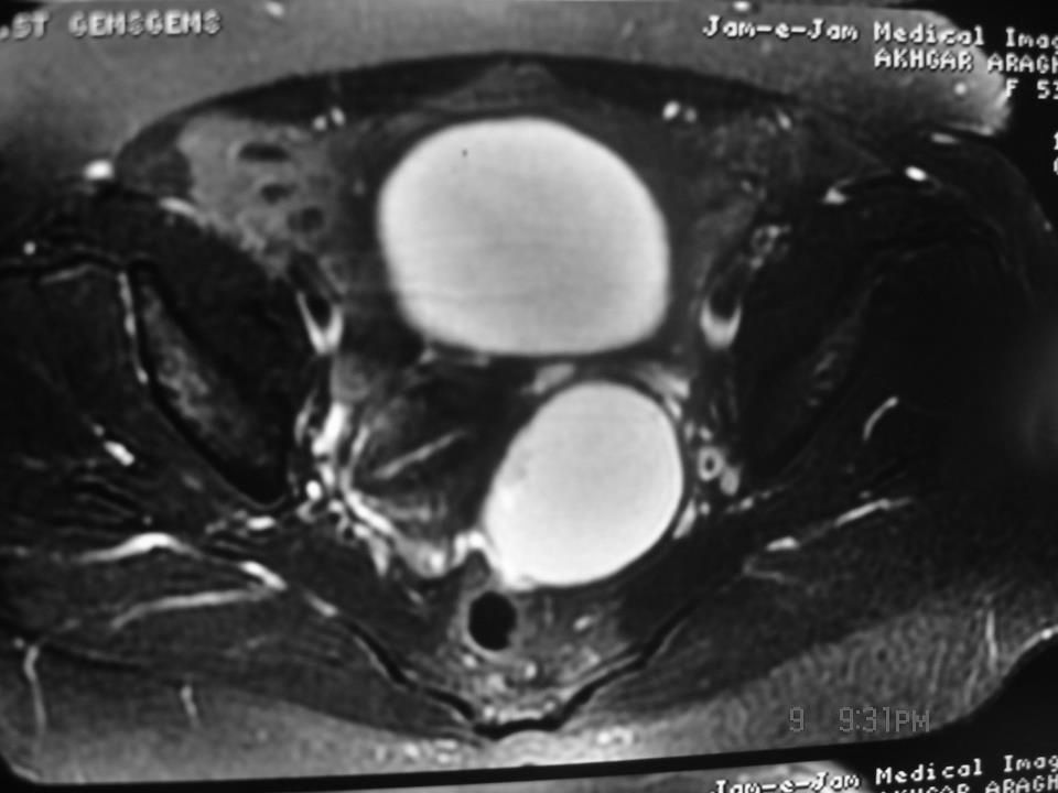
Hemangioma, Pelvis

40
Hemangiopericytoma Liposarcoma

41
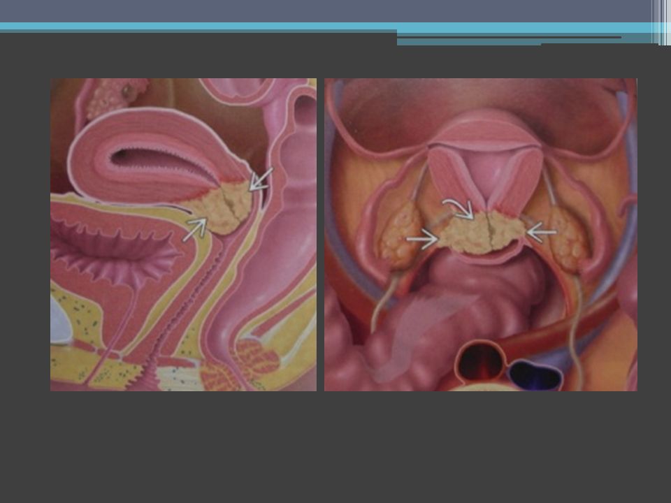
Pelvic congestion syndrome

42
Lymphadenopathy

43
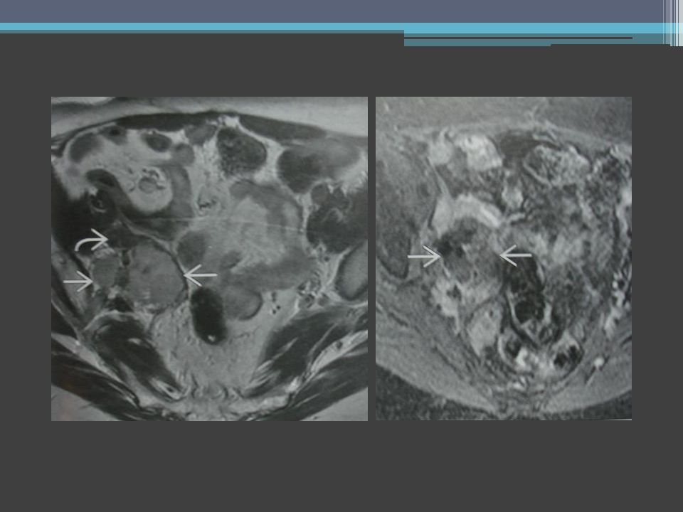
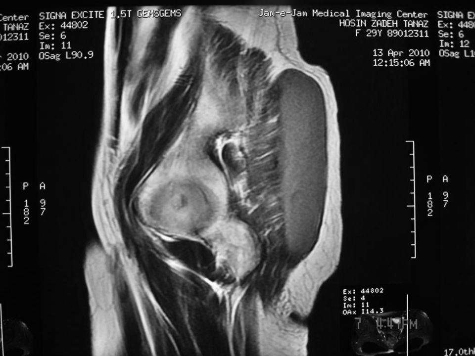
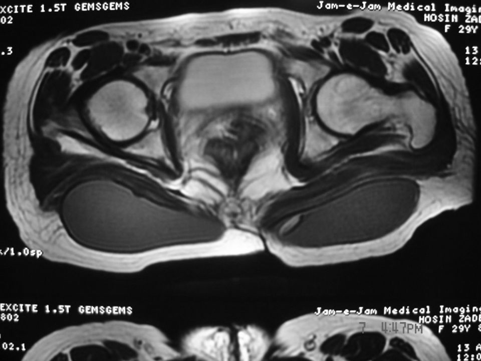
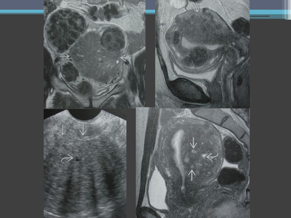
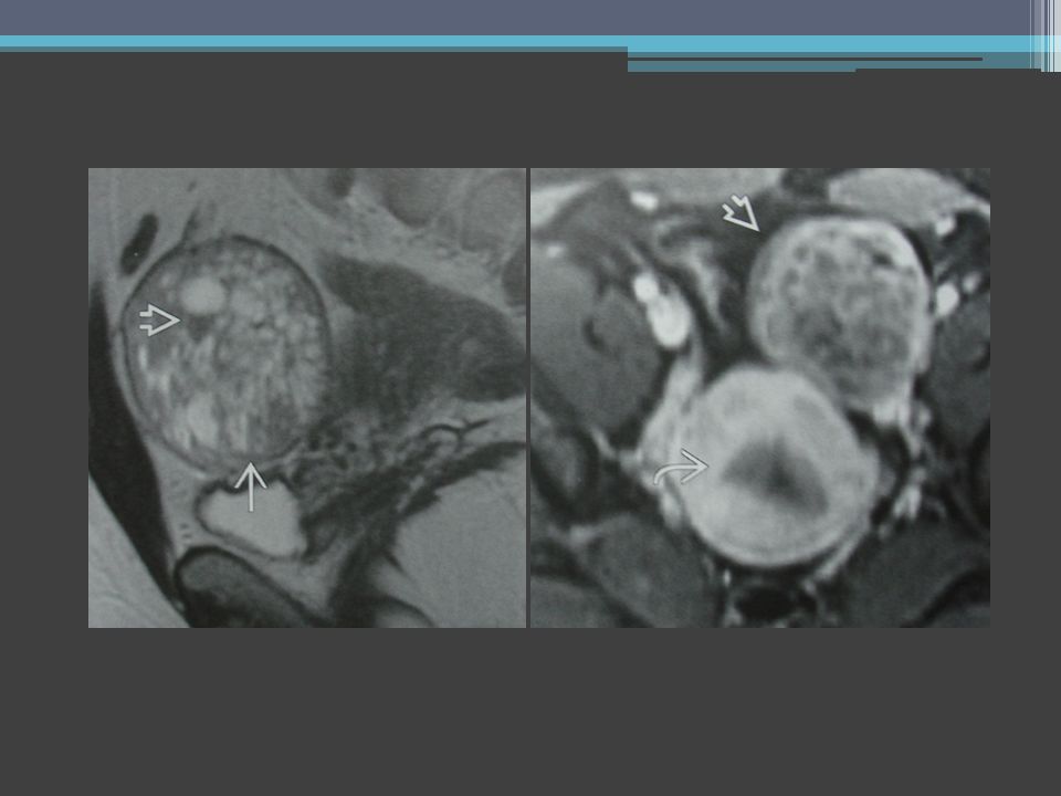
CYSTIC ADENOMYOSIS

44
ADENOMYOSIS

49
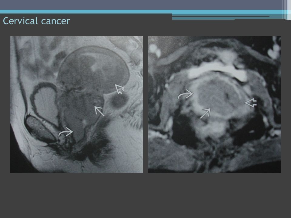
Cervical cancer

51
URETHRAL ANATOMY & IMAGING ISSUE

52
Vaginal fistula

54
Pelvic MRI: Non-invasive 3 Dimensional (x-y-z) Accurate Gives vital information Uncover, covered conditions Elegant imaging technology Diagnosis and staging Can diagnose pelvic floor lesions T1 - T2 - T1,F.S - T1,GD - 3D,GRE - T1 dynamic - MR lymphangiography MR PET jalaljalalshokouhi@hotmail.comjalaljalalshokouhi@hotmail.com www.medimage.irwww.medimage.ir
 Accurate Gives vital information Uncover, covered conditions Elegant imaging technology Diagnosis and staging Can diagnose pelvic floor lesions T1 - T2 - T1,F.S - T1,GD - 3D,GRE - T1 dynamic - MR lymphangiography MR PET")
57
MUCINOUS CYSTADENOMA

58
PELVIC FLOOD DESCENT

59
MR TECHNIQUE & ANATOMY

62
Interventional treatments: Acupuncture Biofeed back and relaxation therapies Nerve stimulation devices Local anesthesia injection in tender areas Help women who have become dependent on narcotics Fibroid embolization jalaljalalshokouhi@hotmail.comjalaljalalshokouhi@hotmail.com www.medimage.irwww.medimage.ir

Similar presentations


>")


