Download presentation
Presentation is loading. Please wait.

1
Tricia Pang November 25, 2008 Three-Dimensional Human Airway Segmentation for Sleep Apnea Diagnosis using Tubular Deformable Organisms

2
Motivation Approach Preliminary Investigation Deformable Organisms Preliminary Results Conclusion OVERVIEW

3
Motivation Approach Preliminary Investigation Deformable Organisms Preliminary Results Conclusion OVERVIEW

4
Motivation Obstructed sleep apnea (OSA) disorder Caused by collapse of soft tissue walls in the airway → model patient's airway to help diagnosis Hand-segmentation: laborious Goal: to develop automated tool for creating a patient-specific model of the airway Credit: Wikipedia
 disorder Caused by collapse of soft tissue walls in the airway → model patient s airway to help diagnosis Hand-segmentation: laborious Goal: to develop automated tool for creating a patient-specific model of the airway Credit: Wikipedia")
5
Motivation Artisynth [2] & OPAL Project (OPAL = Dynamic Modeling of the Oral, Pharyngeal and Laryngeal Complex for Biomedical Engineering) Import resulting airway into dynamic throat and mouth model for simulation
![Motivation Artisynth [2] & OPAL Project (OPAL = Dynamic Modeling of the Oral, Pharyngeal and Laryngeal Complex for Biomedical Engineering) Import resulting airway into dynamic throat and mouth model for simulation](http://images.slideplayer.com/26/8499393/slides/slide_5.jpg "Motivation Artisynth [2] & OPAL Project (OPAL = Dynamic Modeling of the Oral, Pharyngeal and Laryngeal Complex for Biomedical Engineering) Import resulting airway into dynamic throat and mouth model for simulation")
6
Motivation Approach Preliminary Investigation Deformable Organisms Preliminary Results Conclusion OVERVIEW

7
Data Source - MRI Normal subjects, OSA patients, various treatments Volumetric and cross-sectional measurements

8
Motivation Approach Preliminary Investigation Deformable Organisms Preliminary Results Conclusion OVERVIEW

9
Preliminary Investigation Combined 2D segmentation of axial slices in Matlab Procedure: User-indicated start point at base of airway Starting on axial slice at start point, grow ellipse outward Iterate on all axial slices moving upwards along airway, and use previous segmentation as starting contour “Active contours without edges” (Chan-Vese) [1]: Based on Mumford-Shah framework Evolve curve by minimizing energy from image (interior/exterior mean) and curvature
![Preliminary Investigation Combined 2D segmentation of axial slices in Matlab Procedure: User-indicated start point at base of airway Starting on axial slice at start point, grow ellipse outward Iterate on all axial slices moving upwards along airway, and use previous segmentation as starting contour Active contours without edges (Chan-Vese) [1]: Based on Mumford-Shah framework Evolve curve by minimizing energy from image (interior/exterior mean) and curvature](http://images.slideplayer.com/26/8499393/slides/slide_9.jpg "Preliminary Investigation Combined 2D segmentation of axial slices in Matlab Procedure: User-indicated start point at base of airway Starting on axial slice at start point, grow ellipse outward Iterate on all axial slices moving upwards along airway, and use previous segmentation as starting contour Active contours without edges (Chan-Vese) [1]: Based on Mumford-Shah framework Evolve curve by minimizing energy from image (interior/exterior mean) and curvature")
10
Motivation Approach Preliminary Investigation Deformable Organisms Preliminary Results Conclusion OVERVIEW

11
Deformable Organism I-DO: framework for ITK (McIntosh & Hamarneh) [4] Geometrical and physical layers of classical deformable models (data-driven) Behavioral and cognitive layers for intelligent deformation control (knowledge-driven) Related work: Spinal crawler [5] Vessel crawler [6]
![Deformable Organism I-DO: framework for ITK (McIntosh & Hamarneh) [4] Geometrical and physical layers of classical deformable models (data-driven) Behavioral and cognitive layers for intelligent deformation control (knowledge-driven) Related work: Spinal crawler [5] Vessel crawler [6]](http://images.slideplayer.com/26/8499393/slides/slide_11.jpg "Deformable Organism I-DO: framework for ITK (McIntosh & Hamarneh) [4] Geometrical and physical layers of classical deformable models (data-driven) Behavioral and cognitive layers for intelligent deformation control (knowledge-driven) Related work: Spinal crawler [5] Vessel crawler [6]")
12
Deformable Organism Goal: automatically segment airway by growing a tubular organism, guided by image data and a priori anatomical knowledge Advantages: Increased accuracy Analysis and labeling capabilities Ability to incorporate shape-based prior knowledge Modular framework

13
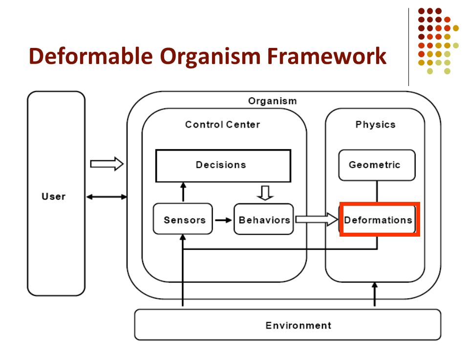
Deformable Organism Framework

15
Cognitive Centre class Ctrl_AirwayOrg decideNextBehavior() Grow: activates Beh_Growing with appropriate params for new layer (major/minor axis, location, radius, distance from previous layer) Terminate Update() Processing? DecideNextBehavior() IsFinished()? Yes No Yes CleanUp()
 IsFinished(). Yes No Yes CleanUp().")
16
Deformable Organism Framework

17
Sensor Modules class Sense_Hessian and Sense_SenseToGrow Compute gradient using itk::GradientRecursiveGaussianImageFilter

18
Deformable Organism Framework

19
Behavior Modules Decisions will result in the execution of behavior class Beh_Growing Calls physics module to create new layer (with params from Ctrl_AirwayOrg), connect to current front-most layer and activate springs class Beh_Fitting Calls gradient-driven deformation
, connect to current front-most layer and activate springs class Beh_Fitting Calls gradient-driven deformation")
20
Deformable Organism Framework

22
Deformation Module 3D Newtonian physics-based spring-mass system Based on sample code for Euler Physics Merged with Physics class: Phys_AirwayOrg When called from Beh_Growing, manipulate geometry by moving nodes, actuating springs, applying forces External forces from image gradient and drag force Internal forces from Hooke’s law and dampening spring forces

23
Deformable Organism Framework

24
Geometry Modules class Geom_AirwayOrg Parameterized by distance between medial nodes and circumferential boundary, major and minor axis length for each slice

25
Summary of Layers Control Center Grow, terminate, (branch) Behavior Grow, fit, (branch) Physics/Deformation Spring-mass system Medial and boundary nodes Radial, circumferential and sheer springs Geometric Medial-based shape representation Tubular with symmetric cross-section (often elliptical) Sensors ‘GrowSense’ ‘HessianSense’ (‘BranchSense’)
 Behavior Grow, fit, (branch) Physics/Deformation Spring-mass system Medial and boundary nodes Radial, circumferential and sheer springs Geometric Medial-based shape representation Tubular with symmetric cross-section (often elliptical) Sensors ‘GrowSense’ ‘HessianSense’ (‘BranchSense’)")
26
Viewer Adaptor Graphical interface for viewing geometry of DOs and their deformations in real time

27
Motivation Approach Preliminary Investigation Deformable Organisms Preliminary Results Conclusion OVERVIEW

28
Motivation Approach Preliminary Investigation Deformable Organisms Preliminary Results Conclusion OVERVIEW

29
Summary Model of a patient’s airway valuable to diagnosing the OSA disorder Tubular deformable organisms spring-mass system initiated at a user-indicated point grown along the airway boundary using a priori knowledge of upper airway anatomy

30
References [1] Chan, T. and Vese L. Active Contours Without Edges. IEEE Transactions on Image Processing, 10 (2001) [2] Fels, S., et al. Artisynth: A biomechanical simulation platform for the vocal tract and upper airway. International Seminar on Speech Production (2006) [3] Hamarneh, G. and McIntosh, C. Physics-Based Deformable Organisms for Medical Image Analysis. Proc of SPIE 5747 (2005) 326-335 [4] McIntosh, C. and Hamarneh, G. I-DO: A “Deformable Organisms” framework for ITK. Medical Image Analysis Lab, SFU. Release 0.50. [5] McIntosh, C. and Hamarneh, G. Spinal Crawlers: Deformable Organisms for Spinal Cord Segmentation and Analysis. MICCAI (2006) 808–815 [6] McIntosh, C. and Hamarneh, G. Vessel Crawlers: 3D Physically-based Deformable Organisms for Vasculature Segmentation and Analysis. Proceedings of IEEE CVPR (2006)
![References [1] Chan, T. and Vese L. Active Contours Without Edges.](http://images.slideplayer.com/26/8499393/slides/slide_30.jpg "IEEE Transactions on Image Processing, 10 (2001) [2] Fels, S., et al. Artisynth: A biomechanical simulation platform for the vocal tract and upper airway. International Seminar on Speech Production (2006) [3] Hamarneh, G. and McIntosh, C. Physics-Based Deformable Organisms for Medical Image Analysis. Proc of SPIE 5747 (2005) [4] McIntosh, C. and Hamarneh, G. I-DO: A Deformable Organisms framework for ITK. Medical Image Analysis Lab, SFU. Release [5] McIntosh, C. and Hamarneh, G. Spinal Crawlers: Deformable Organisms for Spinal Cord Segmentation and Analysis. MICCAI (2006) 808–815 [6] McIntosh, C. and Hamarneh, G. Vessel Crawlers: 3D Physically-based Deformable Organisms for Vasculature Segmentation and Analysis. Proceedings of IEEE CVPR (2006).")
31
Thank you! Questions?

Similar presentations


 1.>")


 Faculty of Mechanical Engineering, University of Kragujevac, Kragujevac, Serbia 2) Harvard School of Public Health, Boston,>")


