Download presentation
Presentation is loading. Please wait.

1
Skin (Integument) Heaviest single organ in the body (16% BW) Thin skin Thick skin (smooth and hairless hand and palms) Consists of three major regions – Epidermis – outermost superficial region – Dermis – middle region – Hypodermis – deepest region Basic Histology P360-370
 Heaviest single organ in the body (16% BW) Thin skin Thick skin (smooth and hairless hand and palms) Consists of three major regions – Epidermis – outermost superficial region – Dermis – middle region – Hypodermis – deepest region Basic Histology P")
2
Cells of the Epidermis Keratinocytes – produce the fibrous protein keratin Melanocytes – cells with cytoplasmic extensions produce the brown pigment melanin Langerhans’ cells –star shaped epidermal macrophages that help to activate the immune system Merkel cells – function as touch receptors in association with sensory nerve endings

3
EPIDERMIS: Cell types II Keratinocytes principal cell Langerhans APC cell migrates to node for immunity Melanocyte to make & transfer pigment Merkel cell sensory receptor dead alive Nerve cell represented by its axon

4
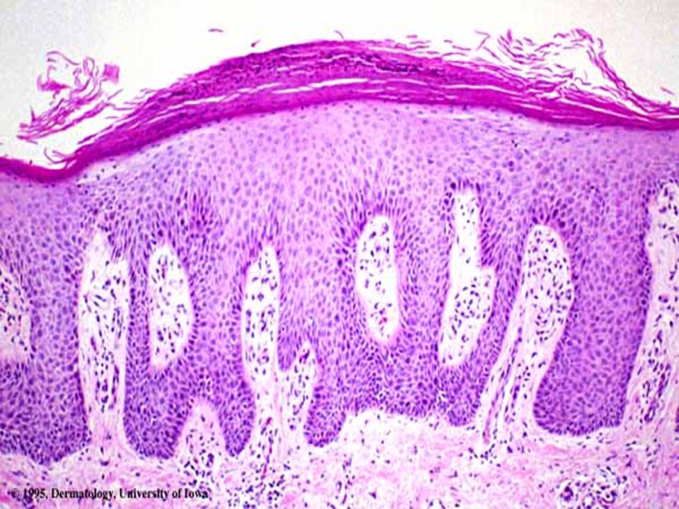
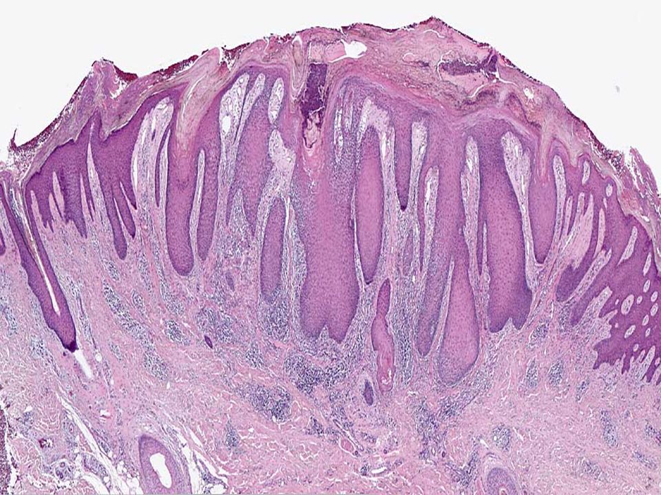
Layers of epidermis Stratum basale/germinativum (“basal or “forming” layer) – One layer thick mitotic cells – 10-25% melanocytes with processes into next layer – Merkel cells with sensory neurons Stratum spinosum (“prickly” layer) – Cells appear spiny due to numerous desmosomes – Many Langerhans cells Stratum granulosum (“grainy” layer) – Cells flatten – Organelles/nuclei begin to disintegrate – Keratin precursor granules begin to form – Lamellated granules with water-proof lipid form and will be spewed out between cells Stratum corneum (“horny” layer) – Cells are dead—too far from underlying capillaries to live – 20-30 cells thick up to ¾ of dermal thickness – Keratin, thickened membranes and glycolipids between cells provide “overcoat” for body to protect against water loss and other possible “assaults” on body
 – One layer thick mitotic cells – 10-25% melanocytes with processes into next layer – Merkel cells with sensory neurons Stratum spinosum ( prickly layer) – Cells appear spiny due to numerous desmosomes – Many Langerhans cells Stratum granulosum ( grainy layer) – Cells flatten – Organelles/nuclei begin to disintegrate – Keratin precursor granules begin to form – Lamellated granules with water-proof lipid form and will be spewed out between cells Stratum corneum ( horny layer) – Cells are dead—too far from underlying capillaries to live – cells thick up to ¾ of dermal thickness – Keratin, thickened membranes and glycolipids between cells provide overcoat for body to protect against water loss and other possible assaults on body")
6
Deepest epidermal layer firmly attached to the dermis Consists of a single row of columnar or cuboidal cells the youngest Keratinocytes Layers of the Epidermis: Stratum Basale (Basal Layer)
")
7
Layers of the Epidermis

8
Epidermis Stratified squamous epithelium Contains no blood vessels/avascular 4 types of cells 5 distinct strata (layers) of cells
 of cells")
9
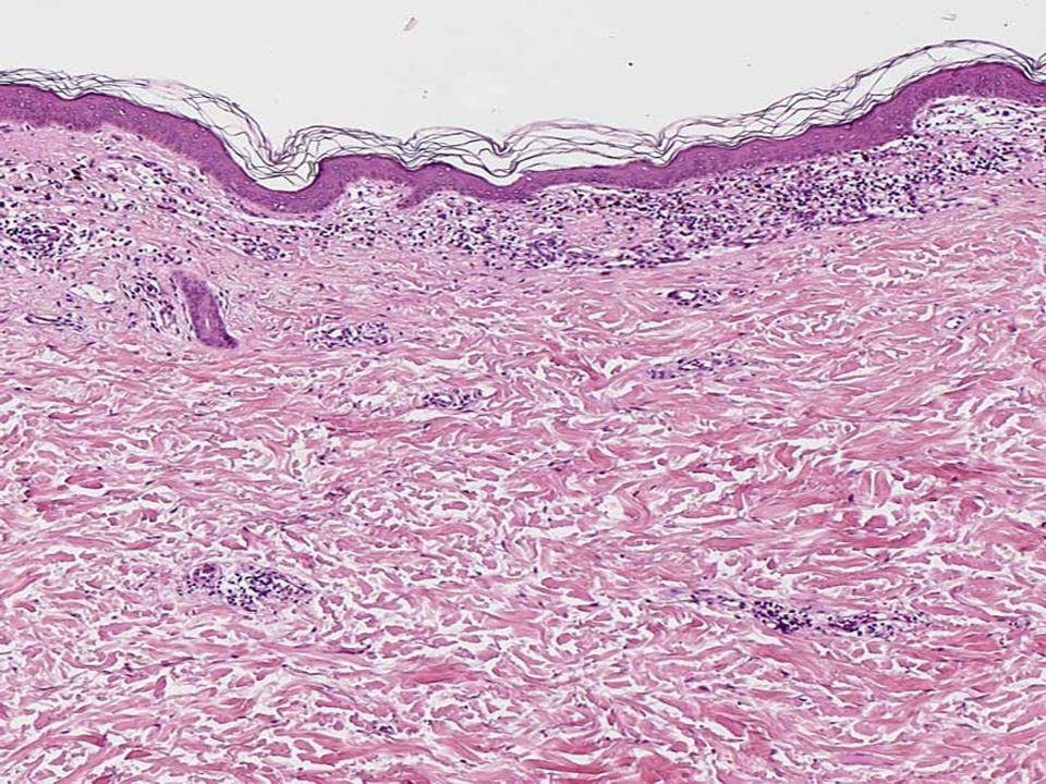
Layers of the Dermis Papillary Reticular

10
Layers of the Dermis: Papillary Layer Papillary layer – Thin areolar connective tissue with collagen and elastic fibers – Its superior surface contains peglike projections called dermal papillae. – Separated from the germinal layer by basal lamina – Dermal papillae contain capillary loops, Meissner’s corpuscles, and free nerve endings

11
Hypodermis Subcutaneous layer deep to the skin Composed of adipose and areolar connective tissue

12
Histopathologic terms for abnormalities of the epidermis Acanthosis- thickening of the spinous layer of the epidermis due to hyperplasia of keratinocytes Atrophy- thinning of the epidermis (also a clinical term)
")
16
Dyskeratosis- Premature keratinization of single cells within the epidermis and adnexal epithelium Necrosis

19
Acantholysis- loss of connection between keratinocytes

23
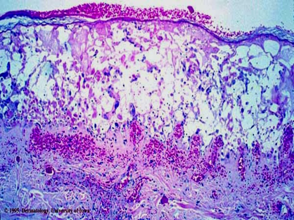
Spongiosis- intercellular edema of epidermis or adnexa, may culminate in intraepidermal vesicles

35
Scale, crust - keratinous material on the skin surface (scale), plasma with white and\or red blood cells (crust), or both (scale-crust)- also clinical Erosion- interference in the continuity of the epidermis, heals without a scar-also clinical Ulcer- interference in the continuity of the skin including epidermis and dermis. Following healing there is a residual scar- also clinical

43
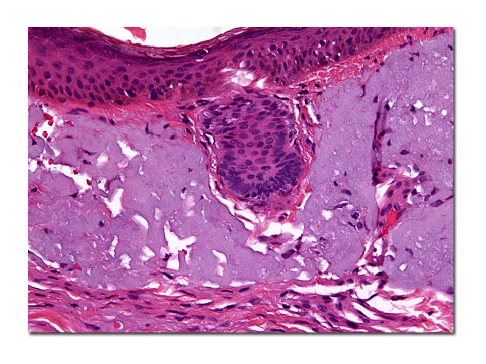
Premalignant lesions Actinic keratosis Bowen's disease

44
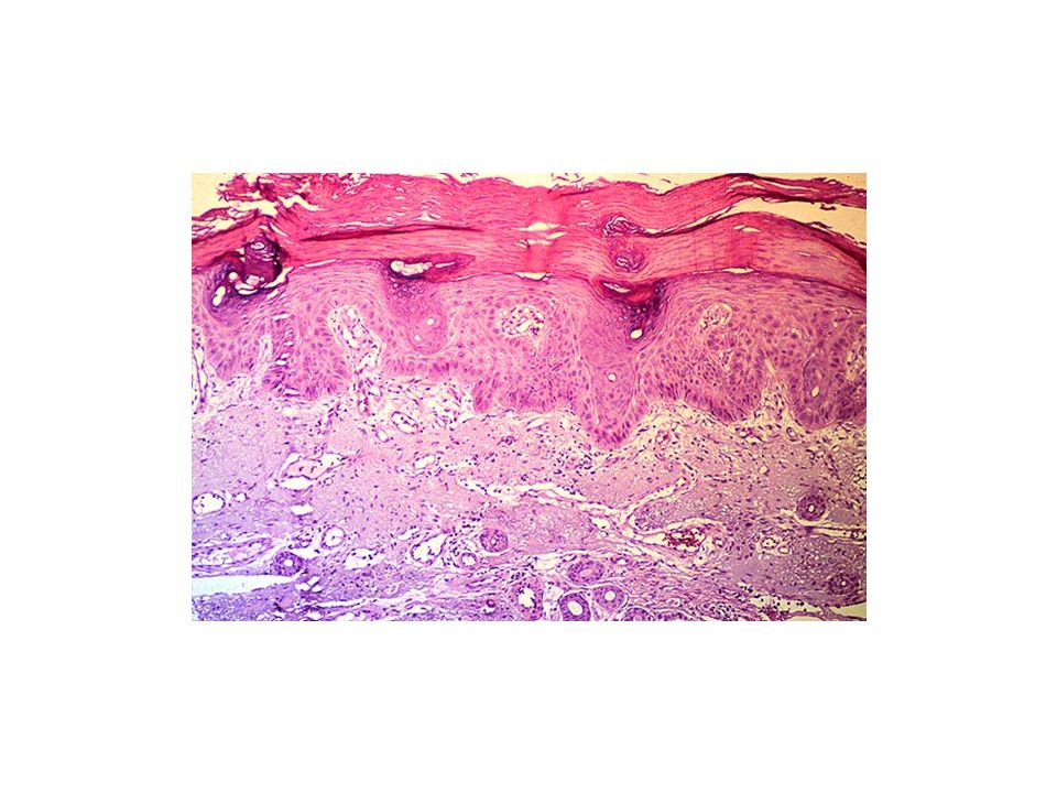
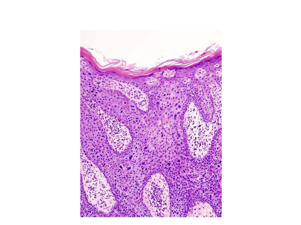
Actinic keratosis Aetiology : – Exposure to ionising radiation – Chronic exposure to sun – Hydrocarbons – Arsenicals Clinical features: – Usually less than 1 cm in diameter – Tan-brown or red coloured – Have a rough, sandpaper like consistency.

46
Microscopy: – Cytological atypia is seen in the lower-most layers of the epidermis – May be associated with hyperplasia of basal cells

49
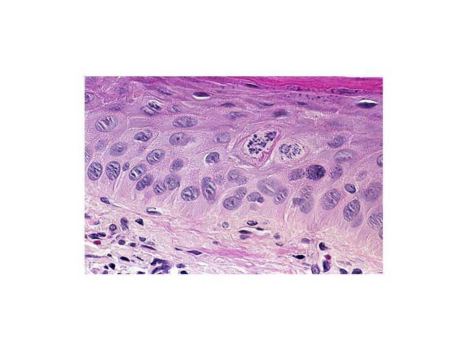
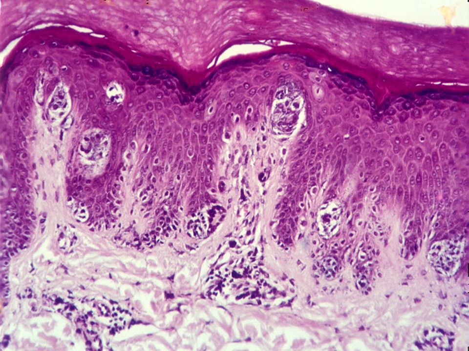
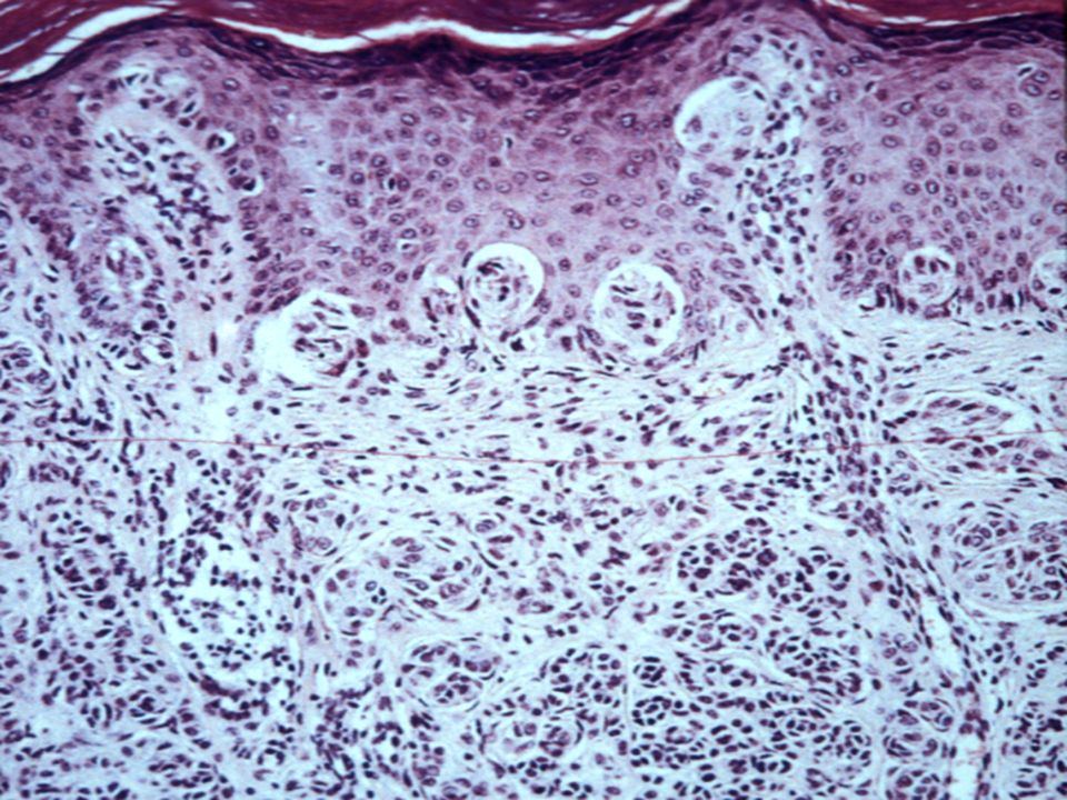
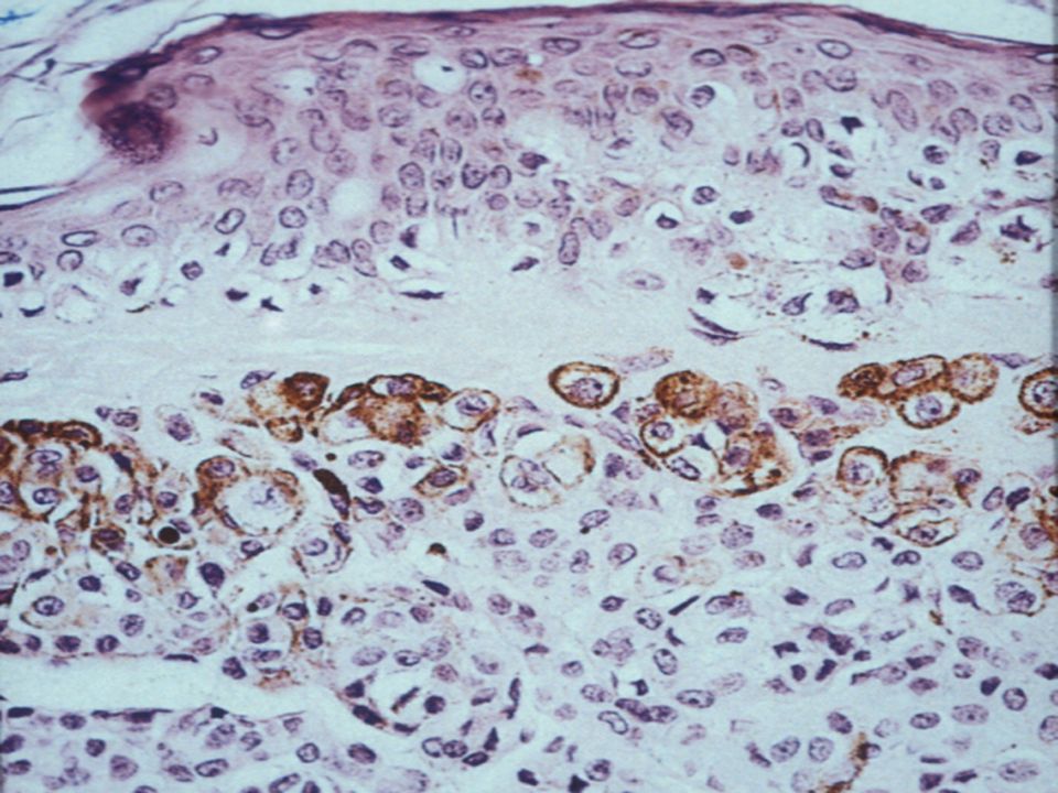
BOWEN DISEASE

50
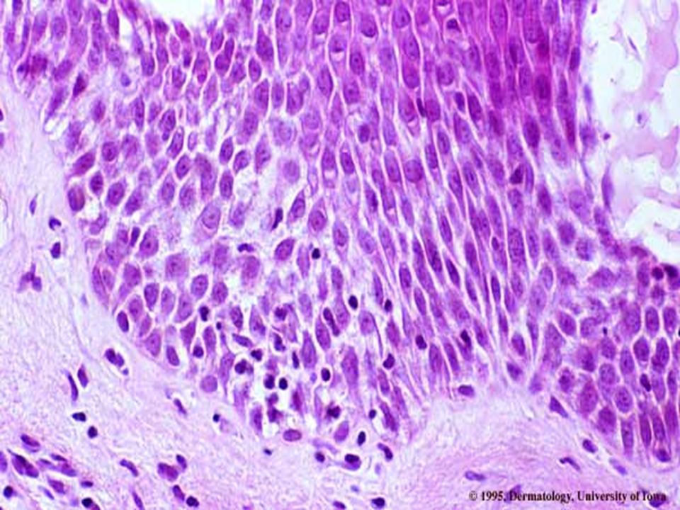
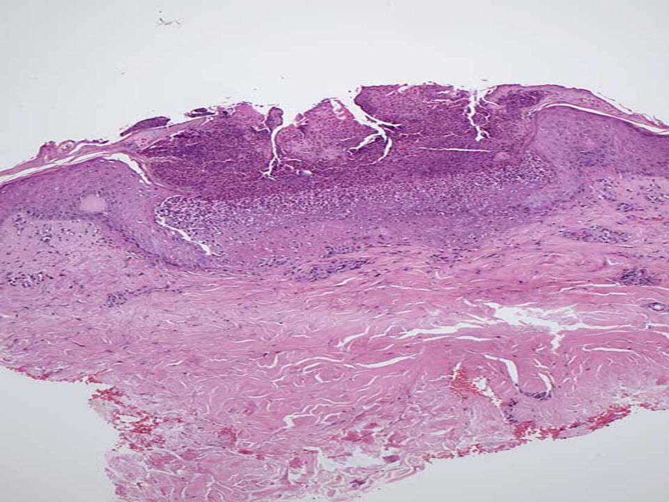
BOWEN DISEASE ( Carcinoma in situ) Squamous cell carcinoma that have not invaded through basement membrane of the dermoepidermal junction Microscopy: – Characterized by cells with atypical nuclei (enlarged and hyperchromatic) at all levels of the epidermis.
 Squamous cell carcinoma that have not invaded through basement membrane of the dermoepidermal junction Microscopy: – Characterized by cells with atypical nuclei (enlarged and hyperchromatic) at all levels of the epidermis.")
55
u ¿ Skin cancer ä basal cell carcinoma ä squamous cell carcinoma ä malignant melanoma types ä secondary skin cancers

56
Basal cell carcinoma – From stratum basale – Least malignant - 99% full cure Squamous cell carcinoma – From stratum spinosum – Prognosis is good if removed early Melanoma – Melanocyte cancer – Highly metastatic – Resistant to chemotherapy ABCD Rule – Asymmetry – Border irregularity – Color: several present – Diameter: greater than 6 mm

57
Squamous cell carcinoma Aetiology: – Exposure to sunlight – Industrial carcinogens (tars and oils) – Chronic ulcers and osteomyelitis – Old burn scars – Ingestion of arsenicals – Ionising radiation – Tobacco and betel nut chewing in oral cavity – Patients with xeroderma pigmentosum
 – Chronic ulcers and osteomyelitis – Old burn scars – Ingestion of arsenicals – Ionising radiation – Tobacco and betel nut chewing in oral cavity – Patients with xeroderma pigmentosum")
58
SQUAMOUS CELL CARCINOMA

62
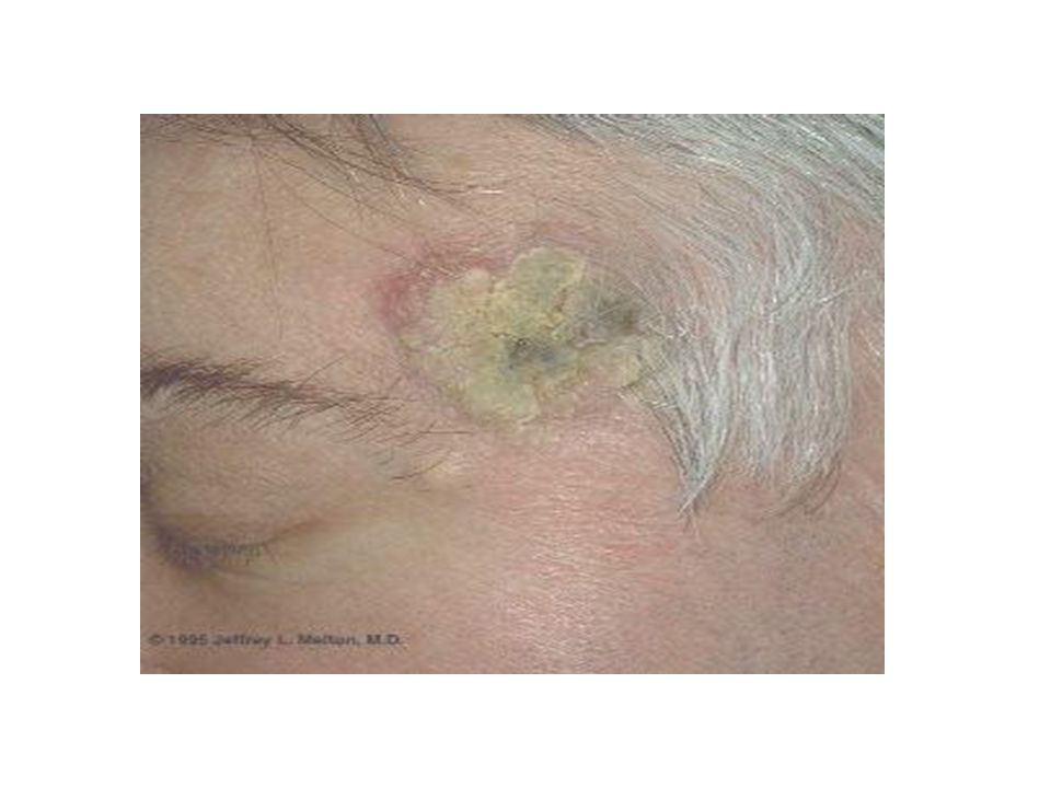
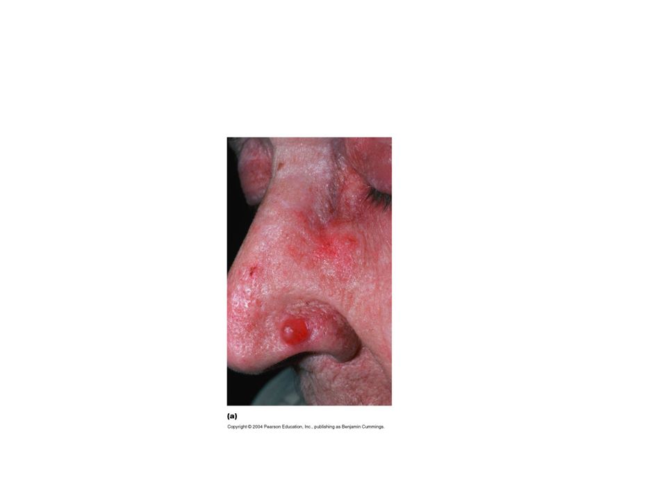
Basal cell carcinoma (Rodent ulcer) Slow growing tumors that rarely metastasize Aetiology: – Chronic sun exposure – Immunosuppression – Patients with inherited defects in DNA(xeroderma pigmentosum) Clinical features: – Lesions appear as pearly papules often containing prominent dilated subepidermal blood vessels (telangiectasias) – Advanced lesions may ulcerate,and extensive local invasion of bone or facial sinuses may occur (rodent ulcers). Site: occurs on hairy skin, the most common location being face, usually above a line from the lobe of ear to the corner of mouth. – The tumour enlarges in size by burrowing and by destroying the tissues locally like a rodent and hence the name ‘rodent ulcer’

64
What is this?

65
NODULAR BCC

66
SUPEFICIAL BCC

67
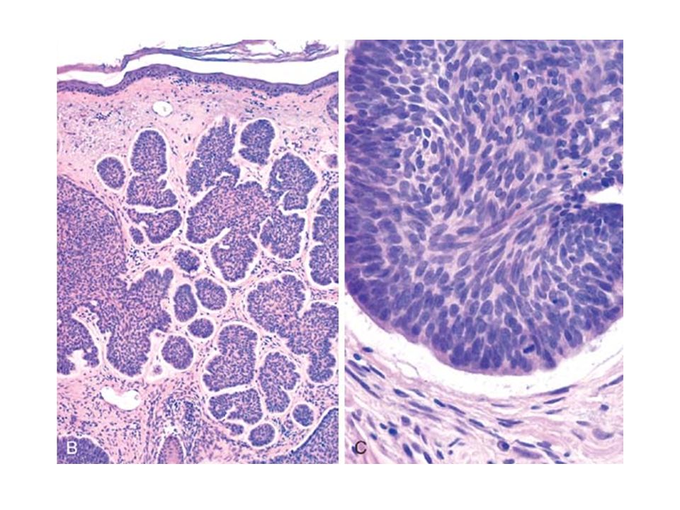
Microscopy : – Malignant Cells are seen arising from basal layer of epidermis and extending into dermis – Arranged as cords and islands – Composed of cells with basophilic hyperchromatic nuclei – Stroma is mucinous with many fibroblasts and lymphocytes – Malignant cells at the periphery of the islands are arranged radially with their long their long axis parallel to each other (palisading) – The islands of tumor cells are seperated from the surrounding stroma by clefts (due to shinkage of epithelial tumor nests).
 – The islands of tumor cells are seperated from the surrounding stroma by clefts (due to shinkage of epithelial tumor nests).")
72
Melanocytic lesions of the skin Benign and Malignant

75
Disorders of pigmentetion Vitiligo – Is a common diorder charecterised by partial or complete loss of pigment producing melanocytes. Melasma – Mask like zone of facial hyperpigmentatiom

76
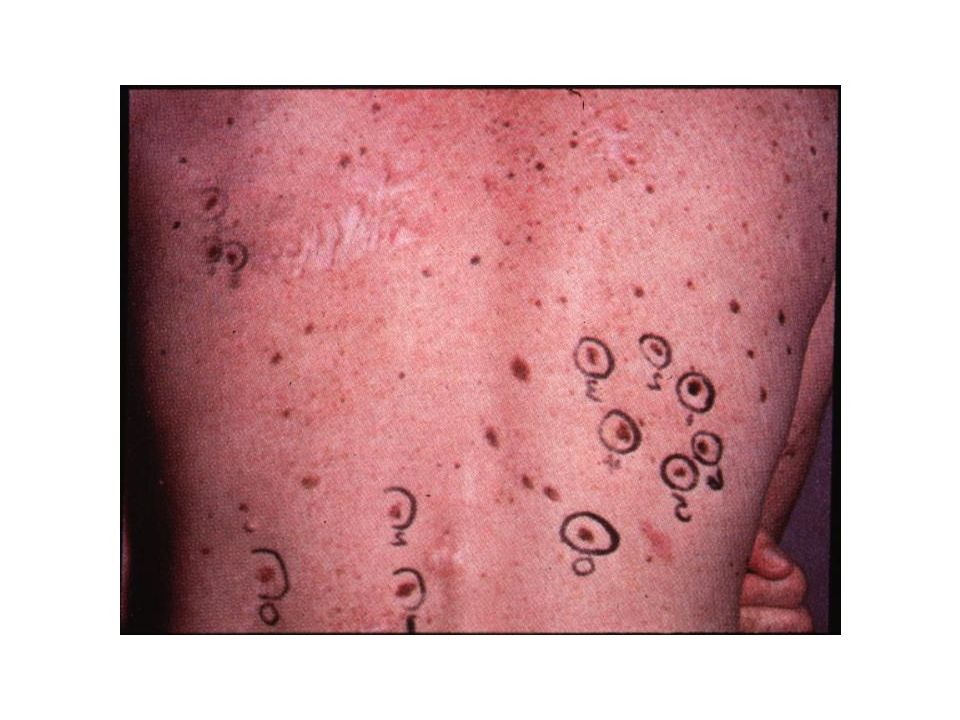
Melanocytic nevi Localized benign abnormality (malformation or neoplasia) of the melanocytic system Usually acquired Most nevi develop during the second and third decades Variable number- 20-30 Distribution- more common in skin of head, neck, trunk Exposure to UV light is an exacerbating factor for the development of nevi
 of the melanocytic system Usually acquired Most nevi develop during the second and third decades Variable number Distribution- more common in skin of head, neck, trunk Exposure to UV light is an exacerbating factor for the development of nevi")
77
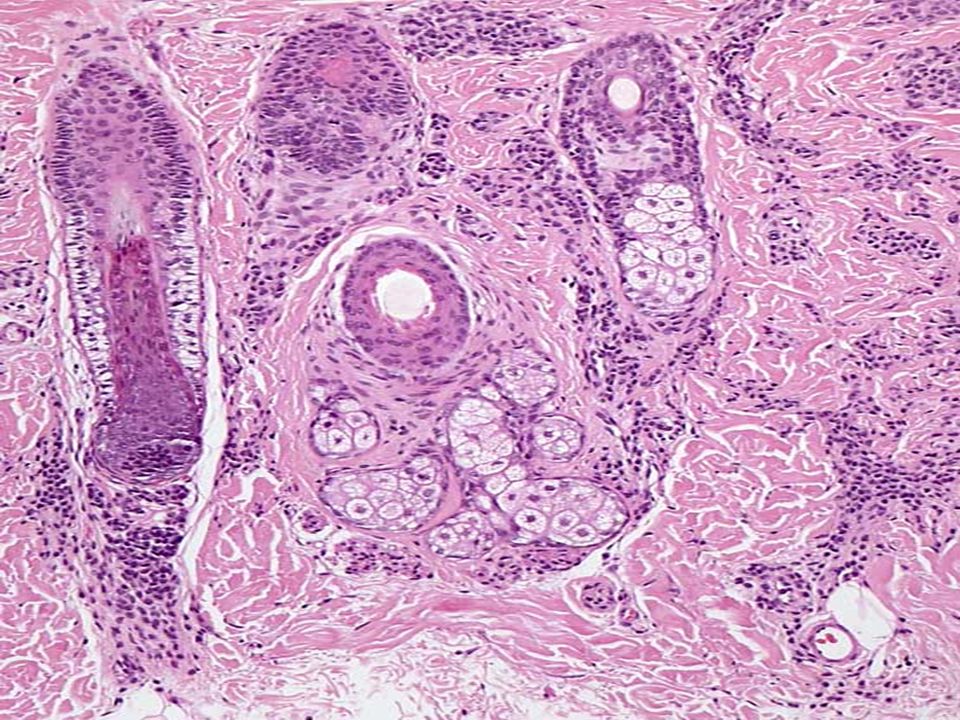
Common melanocytic nevi Junctional nevus- only epidermal component, flat or slightly elevated, non hairy, light brown Compound nevus- both epidermal and dermal components, slightly elevated or dome-shaped Intradermal nevus- only dermal component, flat or dome-shaped, often hairy

86
Congenital nevi

88
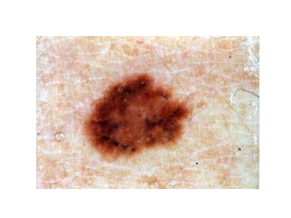
Dysplastic nevi Dysplastic nevus syndrome- genetically determined syndrome in families prone to develop melanomas Clinically- atypical, >5mm, irregular, variegated Appear in adolescence, continue to appear in adult life Architectural and cytologic atypia

94
Dysplastic nevi and melanoma Individuals with dysplastic nevi have an increased risk of melanoma Familial dysplastic nevi and history of melanoma- risk of melanoma approaching 100% by age 75y

95
Malignant melanoma Increasing incidence- 3-8% per year

96
Malignant melanoma- risk factors Solar radiation is the major cause of MM in light-pigmented populations Most melanomas arise in intermittently sun- exposed areas (males- trunk, upper back; females- lower legs, upper back) In whites- higher rates in the less pigmented
 In whites- higher rates in the less pigmented")
Similar presentations


 Largest organ of the body (15% of body weight) Skin thickness variable, normally 1-2 mm Protection –chemical barrier (waterproof)>")

 like an onion?>")
 Consists of three major regions.>")
