Download presentation
Presentation is loading. Please wait.

1
HISTOLOGY SPECIAL SENSES

3
DIVISION Functionally –Sensory (retina) –Dioptric cornea lens ant. & post. chambers vitreous body Anatomically - walls tunica ext / fibrosa tunica media / vasculosa tunica intima / retina - lens - ant./ post. Chambers - vitreous body

4
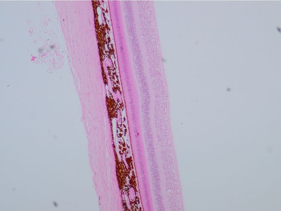
Wall of the eye

6
Cornea: It is colourless and transparent. It consists of five layers. 1.Corneal Epithelium –It is stratified, squamous, and non-keratinized. 2. Bowman’s Membrane-contributes greatly to the strength and stability of the cornea. 3. Stroma- It is made up of many layers of parallel collagen bundles. 4. Descmet’s membrane-It is homogenous structure composed of fine collagenous filament. 5. Endothelium-It is simple squamous epithelium.

7
Cornea:

8
Choroid It is a highly vascularised coat, with loose connective tissue between its blood vessels. Melanocytes are abundant in this layer and give it its characteristic black colour. The inner layer of the choroid called is the choriocapillary layer and it has an important function in the nutrition of retina. This layer is separated from the retina by a hyaline membrane called as Bruch’s membrane.

9
Ciliary body: It is a continuous thickened ring. Histologically the ciliary body is made up of loose connective tissue surrounding ciliary muscle. Ciliary muscle plays an important role in visual accommodation.

10
Ciliary Processes: The ciliary processes are ridge like extensions of the ciliary body. From the Ciliary processes emerged zonule fibres {suspensory ligament of the lens} that insert into the capsule of the lens and anchor it in place. Another important function of the ciliary process is to produce aqueous humor.

12
Iris The iris is the extension of the choroid that partially covers the lens, leaving a round opening in the centre called the pupil. The anterior surface is formed of a discontinuous layer of pigment cells and fibroblasts. Beneath this layer is a poorly vascularised connective tissue with few fibres and many fibroblasts and melanocytes. The function of the melanocytes or pigment cells containing melanin in several regions of the eye is to keep stray light rays from interfering with image formation. The melanocytes of the stroma of the iris are responsible for the colour of the eyes.

14
The smooth posterior surface of the iris is covered by two layers of epithelium, which also cover the ciliary body and its processes. The inner epithelium, in contact with the posterior chamber, is heavily pigmented with melanin granules. The outer epithelial have radially directed tongue like extensions of the basal region, they are filled with overlapping myofilaments, creating the dilator papillae muscle of the iris.

15
The constrictor muscle of the pupil (constrictor pupillae) CP consists of a band of circumferentially oriented smooth muscle fibres situated in the stroma near to the free edge of the iris.
 CP consists of a band of circumferentially oriented smooth muscle fibres situated in the stroma near to the free edge of the iris.")
17
Canal of Schlemm trabeculae Glaucoma

18
Pupillary constriction - sphincter pupillae muscle miosis - III cranial nerve- Parasympathetic. Pupillary dilation - dilator pupillae muscle mydriasis – sympathetic

19
The lens has three components: 1. Lens Capsule: It is very thick basement membrane and consists mainly of collagen type IV and glycoprotein.

20
2. Subcapsular Epithelium: It consists of a single layer of cuboidal epithelial cells that is present only on the anterior surface of the lens. 3. Lens Fibres: Lens fibres are elongated and appear as thin, flattened structures. These cells are filled with a group of proteins called crytstallins. They are produced throughout life, at an ever- decreasing rate.

23
Bipolar cells Ganglion cells Photoreceptor cells Pigmented epith. Cells Muller cell Optic nerve fibre Amacrine Horizontal cell

24
sclera choroid 1 2 3 4 5 6 7 8 9 10 RETINA

25
sclera choroid 1 2 3 4 5 6 7 8 9 10 RETINA

27
Anatomy Summary:Inner ear

28
Anatomy Summary: The Cochlea

29
Perilymph in vestibular and tympanic duct Similar to plasma Endolymph in cochlear duct Secreted by epithelial cells, Similar to intracellular fluid(high in potassium) Fluid Compartments in the Inner Ear
 Fluid Compartments in the Inner Ear")
38
1.The structure at the pointers: A.Synthesize Vitamin D B.Lack melanin granules C.Forms part of the Blood-retina barrier D.Is the Ganglion cell layer

39
1.The structure at the pointer: A.Is scala vestibuli B.Contains perilymph C.Is scala tympani D.Contains endolymph

40
1.The circumscribed structure : A.Makes the vitreous body B.Is part of the fibrous tunic C.Has numerous fenestrated capillaries D.Attached to the lens via collagen fibres

41
1.The structure enclosed by the rectangle: A.Rests on the vestibular (Reissner’s) membrane B.Is the helicotrema C.Is the spiral ganglion D.Is the organ of Corti
 membrane B.Is the helicotrema C.Is the spiral ganglion D.Is the organ of Corti")
42
Those who wish to sing always find a song !

43
The End

Similar presentations


 1. Cornea 2. Sclera Middle Tunic (pg. 470-474) 3. Choroid Coat 4. Ciliary Body 5. Lens & Accommodation 6. Aqueous.>")


>")


