Download presentation
Presentation is loading. Please wait.

1
Apoptosis is a highly regulated process that plays critical role in many cellular functions: 1: Cell differentiation and development (deletion of caspases leads to organ malformations) 2: T cell Homeostasis: (AICD) which is a process by which unwanted activated T cells Are eliminated. This is mainly mediated by Fas, TNFR and in some cases perforin and granzymes 3: killing virally infected cells and tumors: CTL mediated apoptosis.

2
IL-2-Driven Expansion/ Survival/ Differentiation Ag-responsive T cells expand, die, or become memory cells Death by Cytokine Deprivation Continued IL-2 driven expansion Effector Cell (short-lived) Memory Cell (long lived) Ag+APC IL-2 Survival Factor Stromal Activation-Induced Death (FasL/Fas) Naive T cell Ag’+APC’ ? Survivin

4
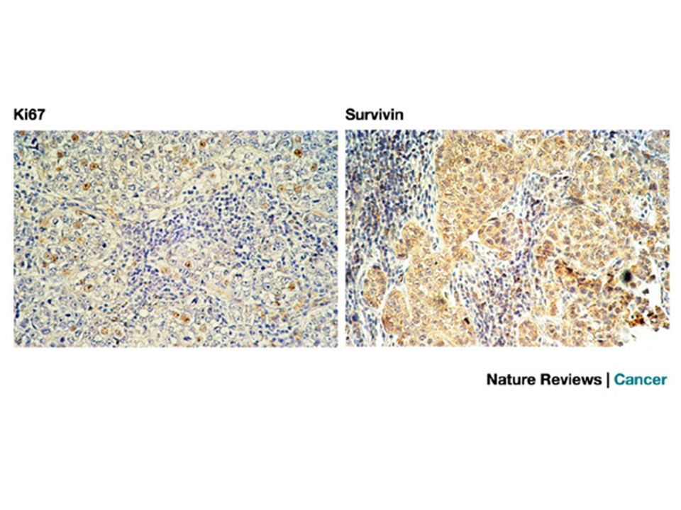
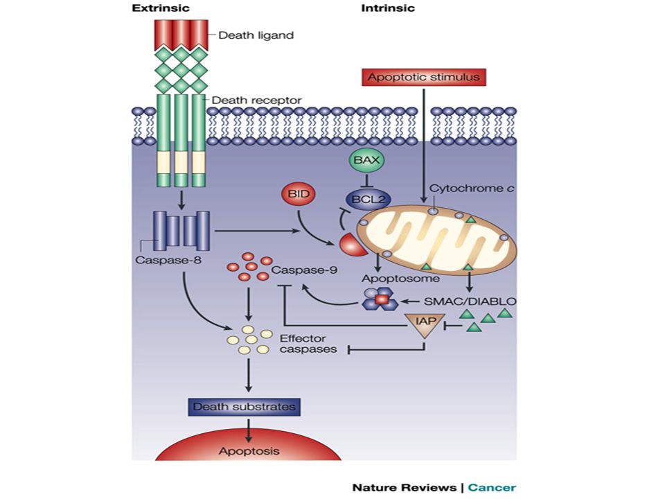
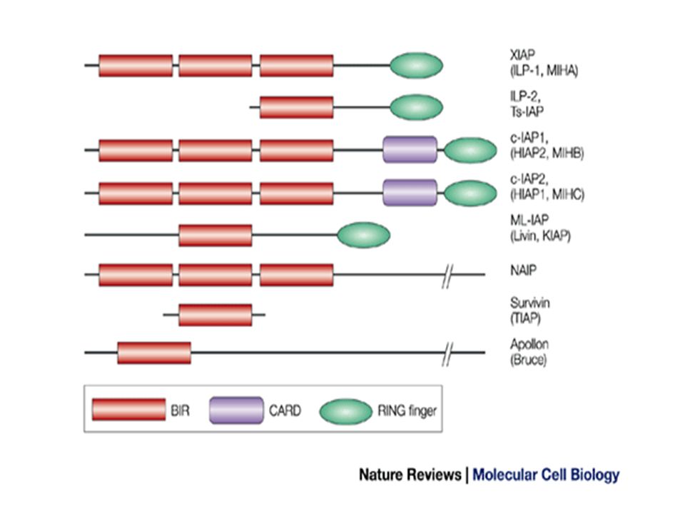
In mammalian cells, apoptosis is modulated by two protein families — the BCL2 and inhibitor of apoptosis (IAP) families. Survivin is a unique member of the IAP family. It is associated with several subcellular compartments and its expression is regulated by many signalling pathways. The survivin pathway interfaces with both the cell-death machinery and mechanisms of cell-cycle progression and microtubule stability. Survivin expression is undetectable in most normal adult tissues, but is overexpressed in virtually every human tumour that has been studied. Several mechanisms have been proposed to account for this overexpression, one of which is loss of wild-type p53. Indispensable (proliferation and apoptosis)
.")
8
IL-2-Driven Expansion/ Survival/ Differentiation Ag-responsive T cells expand, die, or become memory cells Death by Cytokine Deprivation Continued IL-2 driven expansion Effector Cell (short-lived) Memory Cell (long lived) Ag+APC IL-2 Survival Factor Stromal Activation-Induced Death (FasL/Fas) Naive T cell Ag’+APC’ ? Survivin

9
Structures of the B7-1/B7-2–CD28/CTLA-4 superfamily members. CD28 family members are IMMUNOGLOBULIN SUPERFAMILY members with a single immunoglobulin V-like domain. CD28 and CTLA-4 have a MYPPPY motif that is essential for binding B7-1 and B7-2, whereas ICOS has a FDPPPF motif and binds ICOSL but not B7-1 and B7-2. PD-1 is a receptor for both PD-L1 and PD-L2, which might also bind to other, as yet unidentified, receptors on T cells (indicated by the dotted arrows and the question mark).
..")
10
ICOS engagement promotes T-helper-cell differentiation and effector function, and is particularly important for interleukin 10 (IL-10) production but has a modest role in regulating T-cell expansion and IL-2 production. ICOS engagement can upregulate CD40L, and this pathway has a key role in promoting immunoglobulin isotype switching. Little is known about signalling pathways downstream of ICOS. ICOS has a phosphatidylinositol 3- kinase motif in its cytoplasmic tail.

11
PD-1 contains two tyrosines in its cytoplasmic tail, forming ITIM and ITSM motifs. Mutation of the ITSM tyrosine but not the ITIM tyrosine abolishes PD-1-mediated inhibitory activity. The signalling pathways by which PD-1 exerts its effects are just beginning to be understood. Ligation of TCR and PD-1 can lead to tyrosine phosphorylation (P) and activation of SHP-2, resulting in dephosphorylation of signalling molecules and reduced cytokine mRNA synthesis. Ligation of both PD-1 and the BCR can inhibit tyrosine phosphorylation of effector signalling molecules.
 and activation of SHP-2, resulting in dephosphorylation of signalling molecules and reduced cytokine mRNA synthesis. Ligation of both PD-1 and the BCR can inhibit tyrosine phosphorylation of effector signalling molecules..")
12
Hypothetical model of TNFR control of primary T-cell responses. As herpes-virus entry mediator (HVEM) and CD28 are constitutively expressed by naive T cells, and their ligands are expressed by resting antigen-presenting cells (APCs) or rapidly induced, they might control initial activation and cell division. CD27 might also function to regulate clonal expansion. Expression of OX40, 4-1BB and CD30 and their ligands is induced by T cells and APCs with delayed kinetics and might function later. OX40 and 4-1BB, and possibly CD30, prevent apoptosis and regulate the ability of effector cells to persist at the peak of the response. The timeline depicted is based on both in vitro and in vivo data from several studies. Most, but not all, data fit this model. A great deal of overlap in the timing of action of tumour-necrosis factor receptor (TNFR) molecules is probable and will vary depending on the nature of the antigen, its dose and pro-inflammatory factors.
 and CD28 are constitutively expressed by naive T cells, and their ligands are expressed by resting antigen-presenting cells (APCs) or rapidly induced, they might control initial activation and cell division. CD27 might also function to regulate clonal expansion. Expression of OX40, 4-1BB and CD30 and their ligands is induced by T cells and APCs with delayed kinetics and might function later. OX40 and 4-1BB, and possibly CD30, prevent apoptosis and regulate the ability of effector cells to persist at the peak of the response. The timeline depicted is based on both in vitro and in vivo data from several studies. Most, but not all, data fit this model. A great deal of overlap in the timing of action of tumour-necrosis factor receptor (TNFR) molecules is probable and will vary depending on the nature of the antigen, its dose and pro-inflammatory factors..")
13
Structural organization of the co-stimulatory TNFR–TNF-family members. The tumour-necrosis factor (TNF) ligands (top) are shown as homotrimeric type II transmembrane proteins. The TNF receptor (TNFR)-family molecules (bottom) are depicted as type I transmembrane monomers that are thought to associate in trimers when interacting with their ligands. The commonly used names are in bold, with alternate names and TNF/TNFR superfamily (SF) designations in parentheses. The total amino-acid length and number of intracellular amino acids (parentheses) are also indicated. Web access to TNF/TNFR nomenclature and sequences can be found at
 ligands (top) are shown as homotrimeric type II transmembrane proteins. The TNF receptor (TNFR)-family molecules (bottom) are depicted as type I transmembrane monomers that are thought to associate in trimers when interacting with their ligands. The commonly used names are in bold, with alternate names and TNF/TNFR superfamily (SF) designations in parentheses. The total amino-acid length and number of intracellular amino acids (parentheses) are also indicated. Web access to TNF/TNFR nomenclature and sequences can be found at.")
14
Generalized time course of expression of co-stimulatory TNFR-family members. The transition of naive T cells after activation to effector and memory stages is accompanied by the upregulation or downregulation of expression of tumour-necrosis factor receptor (TNFR)-family members. Few molecules have been studied at the same time or in similar model systems, therefore this generalized model is based on separate, mostly in vitro, studies, some of which represent different situations. So, expression levels are relative and kinetics depicted might vary depending on the system. Herpes-virus entry mediator (HVEM) and CD27 are constitutively expressed by naive T cells and some memory T cells. OX40, 4-1BB and CD30 are not expressed by naive T cells and the peak level of expression occurs after encounter with antigen, before and at the height of the effector response in both primary and secondary immune reactions.
-family members. Few molecules have been studied at the same time or in similar model systems, therefore this generalized model is based on separate, mostly in vitro, studies, some of which represent different situations. So, expression levels are relative and kinetics depicted might vary depending on the system. Herpes-virus entry mediator (HVEM) and CD27 are constitutively expressed by naive T cells and some memory T cells. OX40, 4-1BB and CD30 are not expressed by naive T cells and the peak level of expression occurs after encounter with antigen, before and at the height of the effector response in both primary and secondary immune reactions..")
15
Signalling intermediates in TNFR-family co-stimulation. Each co-stimulatory tumour-necrosis factor receptor (TNFR) binds TNFR-associated factors (TRAFs) that function as adaptor proteins for kinases. TRAF2 might be used by all TNFR molecules and link to several common signalling pathways, including those that involve JUN N-terminal kinase (JNK) and the AP1 (FOS/JUN) transcriptional complex; nuclear factor- B (NF- B); and phosphatidylinositol 3-kinase (PI3K) and protein kinase B (PKB, also known as AKT). Signalling through OX40 and 4-1BB upregulate the expression of anti- apoptotic members of the BCL2 family, and might negatively regulate pro- apoptotic members, such as BAD (BCL2- antagonist of cell death) and BIM (BCL2- interacting mediator of cell death). Although most co-stimulatory TNFR molecules can activate AP1 and NF- B, direct links to T-cell effector function, cell division or survival await further investigation (indicated by dashed arrows).
 binds TNFR-associated factors (TRAFs) that function as adaptor proteins for kinases. TRAF2 might be used by all TNFR molecules and link to several common signalling pathways, including those that involve JUN N-terminal kinase (JNK) and the AP1 (FOS/JUN) transcriptional complex; nuclear factor- B (NF- B); and phosphatidylinositol 3-kinase (PI3K) and protein kinase B (PKB, also known as AKT). Signalling through OX40 and 4-1BB upregulate the expression of anti- apoptotic members of the BCL2 family, and might negatively regulate pro- apoptotic members, such as BAD (BCL2- antagonist of cell death) and BIM (BCL2- interacting mediator of cell death). Although most co-stimulatory TNFR molecules can activate AP1 and NF- B, direct links to T-cell effector function, cell division or survival await further investigation (indicated by dashed arrows)..")
16
Control of antigen-specific T-cell numbers by TNFR-family co-signals. According to this model, in the absence of tumour-necrosis factor receptor (TNFR) co-signals, little expansion in the number of antigen-reactive T cells will occur in the primary response, and the frequency of effector T cells will be low. The number of antigen-specific memory T cells that develop is small. If memory T cells do not receive clonal expansion and survival signals from TNFR molecules during secondary responses to recall antigen, the frequency of secondary effector T cells will be markedly reduced. Preventing a single TNFR–TNF interaction might have a less pronounced effect, although suppressive effects have been seen in several primary and secondary responses. It is probable that blocking several interactions at once inhibits either stage of a response more markedly. Hypothesis: Co-stimulation and Signaling Thru OX40 (TNFRSF) leads to activation of survivin (survival and proliferation)
 co-signals, little expansion in the number of antigen-reactive T cells will occur in the primary response, and the frequency of effector T cells will be low. The number of antigen-specific memory T cells that develop is small. If memory T cells do not receive clonal expansion and survival signals from TNFR molecules during secondary responses to recall antigen, the frequency of secondary effector T cells will be markedly reduced. Preventing a single TNFR–TNF interaction might have a less pronounced effect, although suppressive effects have been seen in several primary and secondary responses. It is probable that blocking several interactions at once inhibits either stage of a response more markedly. Hypothesis: Co-stimulation and Signaling Thru OX40 (TNFRSF) leads to activation of survivin (survival and proliferation).")
17
Sustained proliferation of recently activated T cells is dependent on OX40 Survivin expression is lower OX40 KO and is specific (cell cycle proteins and aurora) Survivin expression is dependent on OX40 CD4 T cells from wt or OX40 KO AND TCR transgenic mice were stimulated in vitro with APCs and peptide. (A) Naive T cell proliferation over 7 days. Data are mean thymidine incorporation ±SD in triplicate cultures and are representative of three experiments. (B and C) Naive T cell Survivin expression after stimulation of wt or KO T cells (B) or wt T cells in the presence of agonist anti-OX40 (C). Relative levels of Survivin over time are shown compared to peak levels in wt T cells (value of 1) based on densitometric scans. Similar data were obtained in two to four experiments. Figure 1. Defective Survivin Expression in the Absence of OX40
 Naive T cell proliferation over 7 days. Data are mean thymidine incorporation ±SD in triplicate cultures and are representative of three experiments. (B and C) Naive T cell Survivin expression after stimulation of wt or KO T cells (B) or wt T cells in the presence of agonist anti-OX40 (C). Relative levels of Survivin over time are shown compared to peak levels in wt T cells (value of 1) based on densitometric scans. Similar data were obtained in two to four experiments. Figure 1. Defective Survivin Expression in the Absence of OX40.")
18
Re-expression of survivin in effector T cells was also impaired The number of survivin positive dividing cells was reduced especially at d3 CD28KO mice show deficiency in early proliferation of T cells. However, this was accompanied by defects in genes involved in proliferation (Cyclin E, Cyclin D, CDK2, and Aurora). Naive wt or OX40 KO T cells were stimulated with anti-CD3 and CD28 for 7 days. The primed T cells were restimulated with Ag and APCs, and Survivin, Cyclin E, Cyclin D, CDK2, and Aurora B visualized. Similar data were obtained in three experiments. (E and F) Survivin intracellular staining, after labeling naive wt or OX40 KO T cells with CFSE and stimulating with Ag/APCs (E). Quantification of percent Survivin-positive cells in each division on day 5 (F). Data are representative of three experiments. Data are mean ± SD from triplicate cultures.
. Naive wt or OX40 KO T cells were stimulated with anti-CD3 and CD28 for 7 days. The primed T cells were restimulated with Ag and APCs, and Survivin, Cyclin E, Cyclin D, CDK2, and Aurora B visualized. Similar data were obtained in three experiments. (E and F) Survivin intracellular staining, after labeling naive wt or OX40 KO T cells with CFSE and stimulating with Ag/APCs (E). Quantification of percent Survivin-positive cells in each division on day 5 (F). Data are representative of three experiments. Data are mean ± SD from triplicate cultures..")
19
Active PKB fully restore survivin expression in the absence of OX40 signaling IP experiments showed that survivin is phorsphorylated Thr34 as seen in cancer cells Dominant negative mutant of PKB inhibited survivin expression. Figure 2. PKB Regulates Survivin Naive CD4 cells from wt or OX40 KO AND mice were stimulated with peptide and APCs. On day 2 and 3, T cells were transduced with retroviral vectors expressing GFP alone (Mig), GFP with myristoylated PKB (Mig-myr-PKB) (A and B, wt and OX40 KO), or GFP with dominant-negative PKB (Mig-dn-PKB) (C, wt). (A and C) On day 6 of primary culture, GFP+ CD4 cells were sorted, and lysates were analyzed for Survivin and PKB. (B) On day 6 of primary culture, GFP+ CD4 cells were sorted, lysates were immunoprecipitated with anti-Survivin and analyzed for phosphorylated threonine. What signaling pathway link OX40 to up-regulation of apoptosis?
, GFP with myristoylated PKB (Mig-myr-PKB) (A and B, wt and OX40 KO), or GFP with dominant-negative PKB (Mig-dn-PKB) (C, wt). (A and C) On day 6 of primary culture, GFP+ CD4 cells were sorted, and lysates were analyzed for Survivin and PKB. (B) On day 6 of primary culture, GFP+ CD4 cells were sorted, lysates were immunoprecipitated with anti-Survivin and analyzed for phosphorylated threonine. What signaling pathway link OX40 to up-regulation of apoptosis .")
20
Blocking PI3K with specific inhibitor inhibited survivin Blocking PI3Kinase in effector T cells after re-expression suppressed survivin expression Sustained or periodic PKB activation is nceccessary to maintain survivin. (D) Naive wt T cells were stimulated with peptide and APCs, and LY294002 was added for the last 24 hr of culture (days 2–3 or 3–4). Survivin expression was determined at the end of the 24 hr period. (E) Activated wt T cells, taken after 7 days of primary culture, were restimulated with peptide/APCs in the presence of DMSO, LY294002, or Wortmannin added at time 0
 Naive wt T cells were stimulated with peptide and APCs, and LY was added for the last 24 hr of culture (days 2–3 or 3–4). Survivin expression was determined at the end of the 24 hr period. (E) Activated wt T cells, taken after 7 days of primary culture, were restimulated with peptide/APCs in the presence of DMSO, LY294002, or Wortmannin added at time 0.")
21
Figure 3. Survivin Is Expressed in G1 and Is Regulated by OX40 Signals (A) Naive CD4 T cells from wt AND mice were stimulated with Ag and APCs in the presence of DMSO (control), Hydroxyurea, Aphidicolin, or Nocodazole added at time 0 hr. After 2 days, Survivin expression was analyzed in various phases of the cell cycle after BrdU pulsing and 7-AAD staining. Left: cell cycle analysis shown in red with Survivin expression overlaid in blue, after gating on CD4 cells. Right: total percent Survivin expression in CD4 cells. Rationale: report suggested that survivin is only expressed in mitosis and is predominantly involved in the induction of proliferation. Survivin is expressed in all phases Inhibitors of S phase progression did not influence survivin expression
 Naive CD4 T cells from wt AND mice were stimulated with Ag and APCs in the presence of DMSO (control), Hydroxyurea, Aphidicolin, or Nocodazole added at time 0 hr. After 2 days, Survivin expression was analyzed in various phases of the cell cycle after BrdU pulsing and 7-AAD staining. Left: cell cycle analysis shown in red with Survivin expression overlaid in blue, after gating on CD4 cells. Right: total percent Survivin expression in CD4 cells. Rationale: report suggested that survivin is only expressed in mitosis and is predominantly involved in the induction of proliferation. Survivin is expressed in all phases Inhibitors of S phase progression did not influence survivin expression.")
22
Figure 3. Survivin Is Expressed in G1 and Is Regulated by OX40 Signals (B–D) Survivin induction in effector T cells. Wt effector T cells taken after 7–11 days of primary culture were stimulated for 24 hr with agonist anti-OX40 alone or control IgG in the presence of DMSO, Hydroxyurea, Aphidicolin, or Nocodazole (B and C); or DMSO, ethanol, cyclohexamide (CHX), or LY294002 (D), added at time 0 hr. Survivin expression by intracellular staining (B) and RT-PCR (C and D). Similar data were obtained in one repeat experiment. Errors are mean ± SD from triplicate PCR wells. OX40 had direct effect on survivin expressoion OX40 regulate survivin expression thru AKT signaling pathway.
 Survivin induction in effector T cells. Wt effector T cells taken after 7–11 days of primary culture were stimulated for 24 hr with agonist anti-OX40 alone or control IgG in the presence of DMSO, Hydroxyurea, Aphidicolin, or Nocodazole (B and C); or DMSO, ethanol, cyclohexamide (CHX), or LY (D), added at time 0 hr. Survivin expression by intracellular staining (B) and RT-PCR (C and D). Similar data were obtained in one repeat experiment. Errors are mean ± SD from triplicate PCR wells. OX40 had direct effect on survivin expressoion OX40 regulate survivin expression thru AKT signaling pathway..")
23
Figure 4. Survivin Rescues Proliferation and Death of OX40-Deficient T Cells In Vitro CD4 T cells from wt or OX40 KO AND mice were stimulated with peptide and APCs, and transduced on days 2 and 3 with retroviral vectors expressing GFP (Mig) or GFP with wt Survivin (Mig-Survivin). (A) Primary T cell survival on day 8 after reculturing unsorted T cells without further stimulation on day 3. Data show percent recovery calculated based on assigning the input number of GFP+ cells in each culture as 100%. Data are mean ± SD from three experiments. Cell lysates were analyzed by immunoblotting for Survivin (top). (B) Primary proliferation on day 4 and day 8 measured in unseparated cultures by pulsing with thymidine for the last 20 hr. Data are mean ± SD from triplicate cultures and are representative of three experiments. (C) Primary apoptosis on day 6 measured in unseparated cultures by gating on CD4+ cells (GFP+ and GFP−) and staining with annexin V. Open histograms are unstained cells. Data are representative of three experiments. Data showed that in primary T cell culture, introducing survivin leads 1.Recovery of T cells in OX40 KO 2.Sustained division of OX40 deficient T cells 3.Reduced apoptosis
 or GFP with wt Survivin (Mig-Survivin). (A) Primary T cell survival on day 8 after reculturing unsorted T cells without further stimulation on day 3. Data show percent recovery calculated based on assigning the input number of GFP+ cells in each culture as 100%. Data are mean ± SD from three experiments. Cell lysates were analyzed by immunoblotting for Survivin (top). (B) Primary proliferation on day 4 and day 8 measured in unseparated cultures by pulsing with thymidine for the last 20 hr. Data are mean ± SD from triplicate cultures and are representative of three experiments. (C) Primary apoptosis on day 6 measured in unseparated cultures by gating on CD4+ cells (GFP+ and GFP−) and staining with annexin V. Open histograms are unstained cells. Data are representative of three experiments. Data showed that in primary T cell culture, introducing survivin leads 1.Recovery of T cells in OX40 KO 2.Sustained division of OX40 deficient T cells 3.Reduced apoptosis.")
24
Recall T cell survival after sorting GFP+ T cells on day 6 and re-stimulating with peptide and APC in secondary response. Results: OX40 cells transduced with wt survivin showed enhanced recover. Increase accumulation of T cells in the S phase

25
Figure 5. Survivin Restores Defective Expansion of OX40-Deficient T Cells In Vivo CD4 T cells from wt or OX40 KO OT-II mice were stimulated with peptide in vitro and transduced on days 2/3 with retroviral vectors expressing GFP (Mig) or GFP with wt Survivin (Mig-Survivin). After 6 days, GFP+ CD4 cells were sorted, labeled with PKH26, and adoptively transferred to syngeneic recipients. Mice were challenged with OVA in PBS (closed histograms) or PBS without antigen (open histograms). (A) T cell expansion after 3 and 7 days, with the number of GFP+Vβ5+CD4+ cells enumerated in the spleen and lymph nodes. Data are mean response ± SD from three individual mice per group and are representative of three experiments. OX40 T cells expanded less after 3 days and it was rescued by survivin The defect was more evident at day 7 PBS antigen
 or GFP with wt Survivin (Mig-Survivin). After 6 days, GFP+ CD4 cells were sorted, labeled with PKH26, and adoptively transferred to syngeneic recipients. Mice were challenged with OVA in PBS (closed histograms) or PBS without antigen (open histograms). (A) T cell expansion after 3 and 7 days, with the number of GFP+Vβ5+CD4+ cells enumerated in the spleen and lymph nodes. Data are mean response ± SD from three individual mice per group and are representative of three experiments. OX40 T cells expanded less after 3 days and it was rescued by survivin The defect was more evident at day 7 PBS antigen.")
26
Figure 5. Survivin Restores Defective Expansion of OX40-Deficient T Cells In Vivo (B) Division of transferred T cells was monitored for 3 days after challenge by determining either the extent of dilution of PKH26 (left) or the incorporation of BrdU after an in vivo pulse for 2 days (middle). Apoptosis was measured by annexin V staining after gating on Vβ5+CD4+ (GFP+ and GFP−) cells (right). Bold line, Mig-Survivin group; dashed line, Mig-control group. Shaded histograms are recipients of Mig-Survivin T cells injected with PBS. Data are representative of three experiments. Increase in cell division and less apoptosis in wt transgenic mice. Overall, Sustained survivin expression thru OX40:OX40L interaction is required to maintain T cell division overtime.
 Division of transferred T cells was monitored for 3 days after challenge by determining either the extent of dilution of PKH26 (left) or the incorporation of BrdU after an in vivo pulse for 2 days (middle). Apoptosis was measured by annexin V staining after gating on Vβ5+CD4+ (GFP+ and GFP−) cells (right). Bold line, Mig-Survivin group; dashed line, Mig-control group. Shaded histograms are recipients of Mig-Survivin T cells injected with PBS. Data are representative of three experiments. Increase in cell division and less apoptosis in wt transgenic mice. Overall, Sustained survivin expression thru OX40:OX40L interaction is required to maintain T cell division overtime..")
27
Figure 6. Survivin Does Not Sustain Long-Term CD4 T Cell Survival CD4 T cells from wt or OX40 KO AND mice were stimulated with peptide and transduced with retroviral vectors on day 2/3. (A) Expression of total (human/mouse) and endogenous (mouse) Survivin and Bcl-2-like proteins on day 6 in lysates of GFP+CD4+-sorted cells. (B) In vitro recall T cell recovery after 12 days of secondary culture after sorting GFP+CD4 cells and restimulating with peptide. Data show mean percent recovery ± SD calculated based on the input number of cells. (C) In vivo T cell recovery 14 days after immunization, after sorting GFP+CD4 cells, adoptively transferring to syngeneic recipients, and challenging with PCC in PBS (closed histograms) or PBS alone (open histograms). Data are mean number of GFP+Vβ3+CD4+ cells ± SD enumerated in the spleen and lymph nodes from three individual mice per group. Results representative of two or three experiments. A: Survivin did induce expression of BCl-2 B: In vitro 12 day secondary immune response, survivin was not able to recover specific T cells C: In vivo secondary immune response gave the same resuts
 Expression of total (human/mouse) and endogenous (mouse) Survivin and Bcl-2-like proteins on day 6 in lysates of GFP+CD4+-sorted cells. (B) In vitro recall T cell recovery after 12 days of secondary culture after sorting GFP+CD4 cells and restimulating with peptide. Data show mean percent recovery ± SD calculated based on the input number of cells. (C) In vivo T cell recovery 14 days after immunization, after sorting GFP+CD4 cells, adoptively transferring to syngeneic recipients, and challenging with PCC in PBS (closed histograms) or PBS alone (open histograms). Data are mean number of GFP+Vβ3+CD4+ cells ± SD enumerated in the spleen and lymph nodes from three individual mice per group. Results representative of two or three experiments. A: Survivin did induce expression of BCl-2 B: In vitro 12 day secondary immune response, survivin was not able to recover specific T cells C: In vivo secondary immune response gave the same resuts.")
28
Figure 7. Dominant-Negative Survivin Inhibits T Cell Expansion CD4 T cells from wt OT-II TCR transgenic mice were stimulated with peptide/APCs and transduced with retroviral vectors expressing GFP (Mig) or GFP with dominant-negative Survivin (Mig-T34A- Survivin). (A) Expression of total (human and mouse) and endogenous (mouse) Survivin, Bcl-xL, and Bcl-2, in GFP/CD4-sorted cells on day 6. (B–D) In vitro response. T cell expansion (B) based on enumerating GFP+ cells and expressed as percent of input number, proliferation (C) based on incorporation of thymidine, and cell cycle progression (D) based on BrdU/7-AAD staining, all after in vitro restimulation of GFP/CD4-sorted cells. Data are representative of two or three experiments. Errors indicate mean ± SD of triplicate cultures.
 or GFP with dominant-negative Survivin (Mig-T34A- Survivin). (A) Expression of total (human and mouse) and endogenous (mouse) Survivin, Bcl-xL, and Bcl-2, in GFP/CD4-sorted cells on day 6. (B–D) In vitro response. T cell expansion (B) based on enumerating GFP+ cells and expressed as percent of input number, proliferation (C) based on incorporation of thymidine, and cell cycle progression (D) based on BrdU/7-AAD staining, all after in vitro restimulation of GFP/CD4-sorted cells. Data are representative of two or three experiments. Errors indicate mean ± SD of triplicate cultures..")
29
Figure 7. Dominant-Negative Survivin Inhibits T Cell Expansion (E and F) In vivo response. GFP+CD4+ T cells were sorted, labeled with PKH26, and adoptively transferred into naive syngeneic recipients. Mice were challenged with OVA in PBS (closed histograms) or PBS alone (open histograms). (E) T cell expansion after 3 or 7 days to assess the number of GFP+/Vβ5+/CD4+ cells in combined spleen and lymph nodes. Data are mean ± SD from three individual mice per group and are representative of three experiments. (F) Cell division or apoptosis after 3 days by determining the extent of dilution of PKH26 (top) and incorporation of BrdU after a 2 day pulse (middle) after gating on Vβ5+GFP+ cells; and staining for annexin V after gating on Vβ5+CD4+ (GFP+ and GFP−) cells. Bold line, Mig control; dashed line, Mig-T34A-Survivin. Shaded histograms are profiles from recipients of Mig-T34A-Survivin T cells injected with PBS. Data are representative of three experiments.
 or PBS alone (open histograms). (E) T cell expansion after 3 or 7 days to assess the number of GFP+/Vβ5+/CD4+ cells in combined spleen and lymph nodes. Data are mean ± SD from three individual mice per group and are representative of three experiments. (F) Cell division or apoptosis after 3 days by determining the extent of dilution of PKH26 (top) and incorporation of BrdU after a 2 day pulse (middle) after gating on Vβ5+GFP+ cells; and staining for annexin V after gating on Vβ5+CD4+ (GFP+ and GFP−) cells. Bold line, Mig control; dashed line, Mig-T34A-Survivin. Shaded histograms are profiles from recipients of Mig-T34A-Survivin T cells injected with PBS. Data are representative of three experiments..")
30
Hypothetical model of TNFR control of primary T-cell responses. As herpes-virus entry mediator (HVEM) and CD28 are constitutively expressed by naive T cells, and their ligands are expressed by resting antigen-presenting cells (APCs) or rapidly induced, they might control initial activation and cell division. CD27 might also function to regulate clonal expansion. Expression of OX40, 4-1BB and CD30 and their ligands is induced by T cells and APCs with delayed kinetics and might function later. OX40 and 4-1BB, and possibly CD30, prevent apoptosis and regulate the ability of effector cells to persist at the peak of the response. The timeline depicted is based on both in vitro and in vivo data from several studies. Most, but not all, data fit this model. A great deal of overlap in the timing of action of tumour-necrosis factor receptor (TNFR) molecules is probable and will vary depending on the nature of the antigen, its dose and pro-inflammatory factors.
 and CD28 are constitutively expressed by naive T cells, and their ligands are expressed by resting antigen-presenting cells (APCs) or rapidly induced, they might control initial activation and cell division. CD27 might also function to regulate clonal expansion. Expression of OX40, 4-1BB and CD30 and their ligands is induced by T cells and APCs with delayed kinetics and might function later. OX40 and 4-1BB, and possibly CD30, prevent apoptosis and regulate the ability of effector cells to persist at the peak of the response. The timeline depicted is based on both in vitro and in vivo data from several studies. Most, but not all, data fit this model. A great deal of overlap in the timing of action of tumour-necrosis factor receptor (TNFR) molecules is probable and will vary depending on the nature of the antigen, its dose and pro-inflammatory factors..")
31
Current approaches for survivin targeting in cancer therapy. a | Generation of antigen-specific cytolytic T cells against survivin peptides is being considered for vaccination strategies. b | Molecular antagonists of survivin, including antisense, ribozymes and expression of dominant-negative mutants, have been consistently associated with induction of apoptosis, enhancement of cell-death stimuli and inhibition of tumour growth in vivo. c | Interference of mitotic phosphorylation of survivin by CDC2 by sequential administration of cyclin-dependent kinase inhibitors flavopiridol or Purvalanol A after chemotherapy-induced mitotic arrest has resulted in Strong anticancer activity in vitro and in vivo. A similar response has been observed by adenoviral over expression of a phosphorylation-deficient survivin Thr34→Ala mutant (pAd-T34A).
..")
Similar presentations
 Prepared by Cai Chunhui.>")


 immune.>")


>")
