Download presentation
Presentation is loading. Please wait.

1
Apoptosis Yasir Waheed

2
The cells of a multicellular organism are members of a highly organized community. The number of cells in this community is tightly regulated not simply by controlling the rate of cell division, but also by controlling the rate of cell death. If cells are no longer needed, they commit suicide by activating an intracellular death program. This process is therefore called programmed cell death, although it is more commonly called apoptosis (from a Greek word meaning "falling off," as leaves from a tree). The amount of apoptosis that occurs in developing and adult animal tissues can be astonishing. In the developing vertebrate nervous system, for example, up to half or more of the nerve cells normally die soon after they are formed. In a healthy adult human, billions of cells die in the bone marrow and intestine every hour.
. The amount of apoptosis that occurs in developing and adult animal tissues can be astonishing. In the developing vertebrate nervous system, for example, up to half or more of the nerve cells normally die soon after they are formed. In a healthy adult human, billions of cells die in the bone marrow and intestine every hour..")
3
Figure 17-46. The function of cell death in matching the number of developing nerve cells to the number of target cells they contact. More nerve cells are produced than can be supported by the limited amount of survival factors released by the target cells. Therefore, some cells receive an insufficient amount of survival factors to keep their suicide program suppressed and, as a consequence, undergo apoptosis.

4
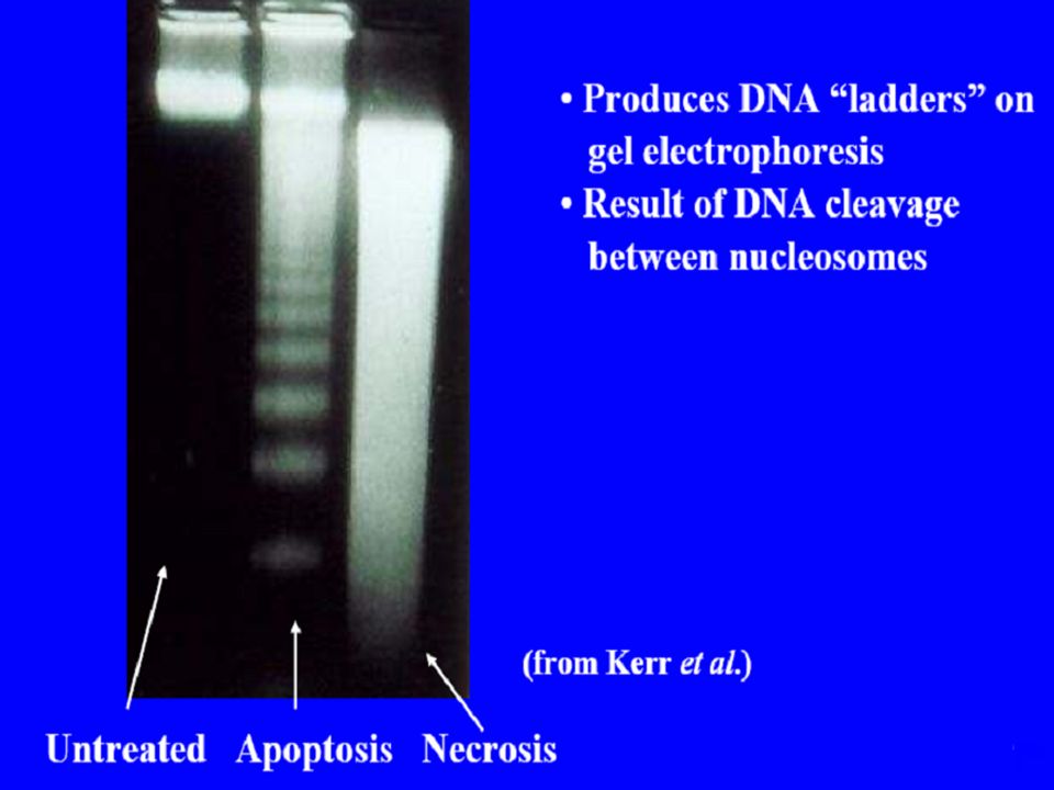
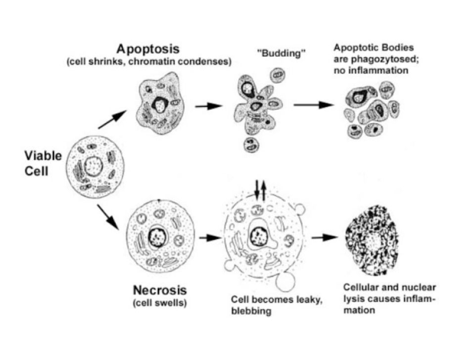
Necrosis Apoptosis

5
Figure 17-35. Sculpting the digits in the developing mouse paw by apoptosis. (A) The paw in this mouse embryo has been stained with a dye that specifically labels cells that have undergone apoptosis. The apoptotic cells appear as bright green dots between the developing digits. (B) This interdigital cell death eliminates the tissue between the developing digits, as seen one day later, when few, if any, apoptotic cells can be seen.
 The paw in this mouse embryo has been stained with a dye that specifically labels cells that have undergone apoptosis. The apoptotic cells appear as bright green dots between the developing digits. (B) This interdigital cell death eliminates the tissue between the developing digits, as seen one day later, when few, if any, apoptotic cells can be seen..")
6
Figure 17-36. Apoptosis during the metamorphosis of a tadpole into a frog. As a tadpole changes into a frog, the cells in the tadpole tail are induced to undergo apoptosis; as a consequence, the tail is lost.

7
The intracellular machinery responsible for apoptosis seems to be similar in all animal cells. This machinery depends on a family of proteases that have a cysteine at their active site and cleave their target proteins at specific aspartic acids. They are therefore called caspases. Caspases are synthesized in the cell as inactive precursors, or procaspases, which are usually activated by cleavage at aspartic acids by other caspases Apoptosis Is Mediated by an Intracellular Proteolytic Cascade

8
Figure 17-38. The caspase cascade involved in apoptosis. (A) Each suicide protease is made as an inactive proenzyme (procaspase), which is usually activated by proteolytic cleavage by another member of the caspase family. As indicated, two of the cleaved fragments associate to form the active site of the caspase. The active enzyme is thought to be a tetramer of two of these units (not shown). (B) Each activated caspase molecule can cleave many procaspase molecules, thereby activating them, and these can then activate even more procaspase molecules. In this way, an initial activation of a small number of procaspase molecules (called initiator caspases) can lead, via an amplifying chain reaction (a cascade), to the explosive activation of a large number of procaspase molecules. Some of the activated caspases (called effector caspases) then cleave a number of key proteins in the cell, including specific cytosolic proteins and nuclear lamins, leading to the controlled death of the cell.
 Each suicide protease is made as an inactive proenzyme (procaspase), which is usually activated by proteolytic cleavage by another member of the caspase family. As indicated, two of the cleaved fragments associate to form the active site of the caspase. The active enzyme is thought to be a tetramer of two of these units (not shown). (B) Each activated caspase molecule can cleave many procaspase molecules, thereby activating them, and these can then activate even more procaspase molecules. In this way, an initial activation of a small number of procaspase molecules (called initiator caspases) can lead, via an amplifying chain reaction (a cascade), to the explosive activation of a large number of procaspase molecules. Some of the activated caspases (called effector caspases) then cleave a number of key proteins in the cell, including specific cytosolic proteins and nuclear lamins, leading to the controlled death of the cell..")
9
Procaspase activation can be triggered from outside the cell by the activation of death receptors on the cell surface. Killer lymphocytes, for example, can induce apoptosis by producing a protein called Fas ligand, which binds to the death receptor protein Fas on the surface of the target cell. The clustered Fas proteins then recruit intracellular adaptor proteins that bind and aggregate procaspase-8 molecules, which cleave and activate one another. The activated caspase-8 molecules then activate downstream procaspases to induce apoptosis (Figure 17- 39A). Some stressed or damaged cells kill themselves by producing both the Fas ligand and the Fas protein, thereby triggering an intracellular caspase cascade. Extracellular activation of apoptosis
. Some stressed or damaged cells kill themselves by producing both the Fas ligand and the Fas protein, thereby triggering an intracellular caspase cascade. Extracellular activation of apoptosis.")
10
Figure 17-39. Induction of apoptosis by either extracellular or intracellular stimuli. (A) Extracellular activation. A killer lymphocyte carrying the Fas ligand binds and activates Fas proteins on the surface of the target cell. Adaptor proteins bind to the intracellular region of aggregated Fas proteins, causing the aggregation of procaspase-8 molecules. These then cleave one another to initiate the caspase cascade.
 Extracellular activation. A killer lymphocyte carrying the Fas ligand binds and activates Fas proteins on the surface of the target cell. Adaptor proteins bind to the intracellular region of aggregated Fas proteins, causing the aggregation of procaspase-8 molecules. These then cleave one another to initiate the caspase cascade..")
11
(B) Intracellular activation. Mitochondria release cytochrome c, which binds to and causes the aggregation of the adaptor protein Apaf-1. Apaf-1 binds and aggregates procaspase-9 molecules, which leads to the cleavage of these molecules and the triggering of a caspase cascade. Other proteins that contribute to apoptosis are also released from the mitochondrial intermembrane space (not shown).
..")
12
Figure 17-47. Two ways in which survival factors suppress apoptosis. (A) In mammalian cells, the binding of some survival factors to cell-surface receptors leads to the activation of various protein kinases, including protein kinase B (PKB), that phosphorylate and inactivate the Bcl-2 family member Bad. When not phosphorylated, Bad promotes apoptosis by binding and inhibiting Bcl-2. Once phosphorylated, Bad dissociates, freeing Bcl-2 to suppress apoptosis. As indicated, PKB also suppresses death by phosphorylating and thereby inhibiting gene regulatory proteins of that stimulate the transcription of genes that encode proteins that promote apoptosis.
 In mammalian cells, the binding of some survival factors to cell-surface receptors leads to the activation of various protein kinases, including protein kinase B (PKB), that phosphorylate and inactivate the Bcl-2 family member Bad. When not phosphorylated, Bad promotes apoptosis by binding and inhibiting Bcl-2. Once phosphorylated, Bad dissociates, freeing Bcl-2 to suppress apoptosis. As indicated, PKB also suppresses death by phosphorylating and thereby inhibiting gene regulatory proteins of that stimulate the transcription of genes that encode proteins that promote apoptosis..")
13
Figure 17-42. Cell-cycle arrest or apoptosis induced by excessive stimulation of mitogenic pathways. Abnormally high levels of Myc cause the activation of p19ARF, which binds and inhibits Mdm2 and thereby causes increased p53 levels. Depending on the cell type and extracellular conditions, p53 then causes either cell-cycle arrest or apoptosis.

14
Apoptosis Cell death Fas FasL TNF-R1 TNF TRADD FADD Cas8 Cas eff Mit Cyt c Protein degradation DNase

16
P53 & Rb One of the most frequently mutated tumor suppressor gene in sporadic human cancer is p53 P53 inhibits proliferation Stimulated Apoptosis The first tumor suppressor gene cloned in 1986 was RB Loss of both alleles of RB leads to the development of Retinoblastoma in humans RB is seldom mutated in sporadic human cancers RB inhibits proliferation, promotes differentiation but it also inhibits apoptosis.

19
THANKS

Similar presentations


. B.Mutant embryo.>")

, death domain, cytochrome.>")
 Apoptosis is a cell mechanism used to eliminate cells that are unnecessary to or that contain mutations that.>")


 In adult tissues cell death exactly balances cell division In apoptosis the cell destroys itself from within.>")


