Download presentation
Presentation is loading. Please wait.

1
Histology Techniques CLS 322
Tissue Processing Histology Techniques CLS 322 Dr. Samah Kotb 2015

2
Different histological methods
There are 3 main techniques which are used in preparing microscopical sections from tissues: The paraffin technique (It is the most common method) The celloidin technique (It is the most perfect method) The freezing technique (It is the most rapid method)
 The celloidin technique (It is the most perfect method) The freezing technique (It is the most rapid method)")
3
Different histological methods
Tissues from the body taken for diagnosis of disease processes must be processed in the histology laboratory to produce microscopic slides that are viewed under the microscope by pathologists. The techniques for processing the tissues, whether Biopsies, Larger specimens removed at surgery, Or Tissues from autopsy are described below. The persons who do the tissue processing and make the glass microscopic slides are HISTOTECHNOLOGISTS.

4
Specimen Accessioning
Tissue specimens received in the surgical pathology laboratory have a request form that lists the patient information and history along with a description of the site of origin. The specimens are accessioned by giving them a number that will identify each specimen for each patient.

5
Surgical Specimen

6
Gross examination Tissues removed from the body for diagnosis arrive in the Pathology Department and are examined by a pathologist, pathology assistant, or pathology resident. Gross examination consists of describing the specimen and placing all or parts of it into a small plastic cassette which holds the tissue while it is being processed to a paraffin block. Initially, the cassettes are placed into a fixative.

9
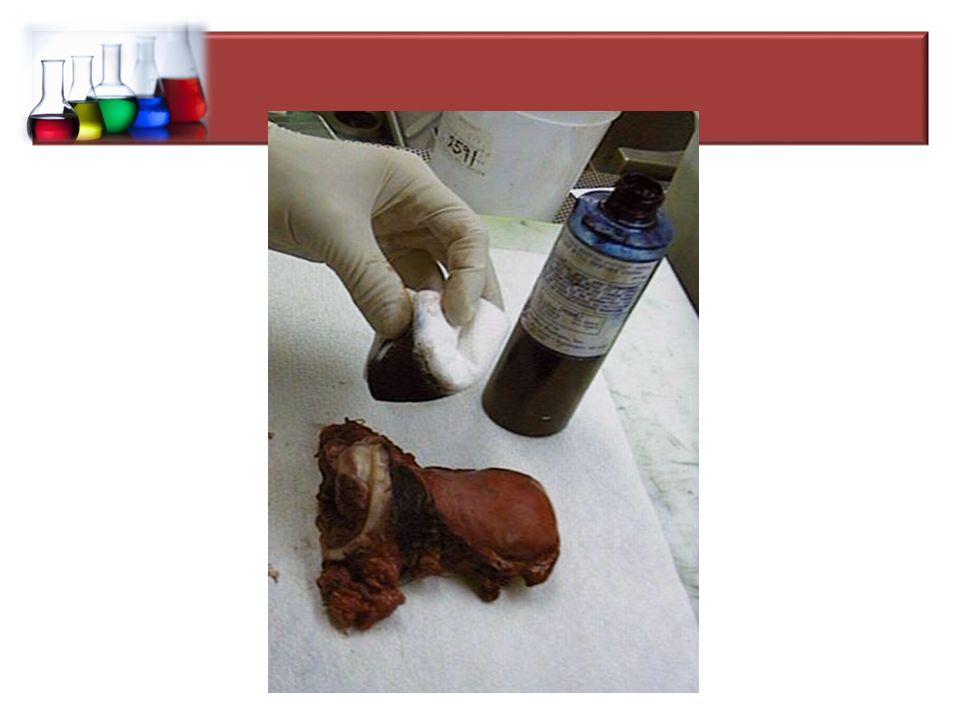
…Gross examination Note:
When a malignancy is suspected, then the specimen is often covered with ink in order to mark the margins of the specimen. Different colored inks can be used to identify different areas if needed. When sections are made and processed, the ink will mark the actual margin on the slide.

11
Tissue Processing steps
Biological tissues are generally rather soft, making it quite difficult to cut acceptably thin sections directly from the fresh or fixed tissues. Methods must be used to hold the tissues firm, which facilitates cutting thin sections with a sharp knife. Firmness can be achieved either by embedding the tissues in a suitable embedment or by freezing the tissue. Once the tissue has been fixed, it must be processed into a form in which it can be made into thin microscopic sections.

12
…Tissue Processing steps
The usual way this is done is with paraffin. Tissues embedded in paraffin, which is similar in density to tissue, can be sectioned at anywhere from 3 to 10 microns, usually 5-8 routinely. The technique of getting fixed tissue into paraffin is called tissue processing. The main steps in this process are dehydration and clearing.

13
…The paraffin technique
Tissues that come off the tissue processor are still in the cassettes and must be manually put into the blocks by a technician who must pick the tissues out of the cassette and pour molten paraffin over them. This "embedding" process is very important, because the tissues must be aligned, or oriented, properly in the block of paraffin.

15
The paraffin Technique
Washing Following fixation, the tissues should be washed from 15 to 30 minutes. The fixed tissues are washed in running tap water to remove the fixative from them.

16
...The paraffin Technique
Dehydration Wet fixed tissues (in aqueous solutions) cannot be directly infiltrated with paraffin. First, the water from the tissues must be removed by dehydration. This is usually done with a series of alcohols; say 70% to 95% to 100%. The organic solvent must replace the water gradually to prevent turbulence at the interface between water and pure ethanol. Turbulence could cause damage or distortion to cellular components. Sometimes the first step is a mixture of formalin and alcohol.
 cannot be directly infiltrated with paraffin. First, the water from the tissues must be removed by dehydration. This is usually done with a series of alcohols; say 70% to 95% to 100%. The organic solvent must replace the water gradually to prevent turbulence at the interface between water and pure ethanol. Turbulence could cause damage or distortion to cellular components. Sometimes the first step is a mixture of formalin and alcohol.")
17
…The paraffin technique
Clearing The next step is called "clearing" and consists of removal of the dehydrant with a substance that will be miscible with the embedding medium (paraffin). The commonest clearing agent is xylene. Toluene works well, and is more tolerant of small amounts of water left in the tissues, but is 3 times more expensive than xylene. Chloroform used to be used, but is a health hazard, and is slow. Methyl salicylate is rarely used because it is expensive, but it smells nice (it is oil of wintergreen). Excessive exposure to clearing reagents may cause excessive hardness or shrinkage.
. The commonest clearing agent is xylene. Toluene works well, and is more tolerant of small amounts of water left in the tissues, but is 3 times more expensive than xylene. Chloroform used to be used, but is a health hazard, and is slow. Methyl salicylate is rarely used because it is expensive, but it smells nice (it is oil of wintergreen). Excessive exposure to clearing reagents may cause excessive hardness or shrinkage.")
18
…The paraffin technique
Embedding The tissue is infiltrated with the embedding agent, almost always paraffin. Nearly 100 years ago, the method of embedding tissues in paraffin was developed. Paraffin is a derivative of crude petroleum. It is a group of variable length, long-chain hydrocarbons of the methane series. Most paraffins suitable as embedding media melt between 52° and 58°C. Since most paraffin have a melting point between 52-58°C, it must infiltrate the cells while it is hot.

19
The freezing Technique
In this method, the fresh or fixed tissues are frozen hardened and cut with a freezing microtome in the cryostat apparatus within few minutes. It is a quick and simple method which is commonly used during operations for rapid diagnosis of tumors e.g. carcinoma. The chemistry of tissues is preserved because we use no heat and no chemical solvents. Can be used in Histochemistry to demonstrate enzymes and chemical components of tissues. Disadvantage: It gives not-serial thick sections which may fragment into small pieces, so they are very difficult to be stained and to be stored.

Similar presentations


 Tissue processing.>")


