Download presentation
Presentation is loading. Please wait.

1
Histology and Embryology 组织学与胚胎学 Department of Histology and embryology Three Gorges University, Yichang, China

2
Introduction

3
I. What’s histology? II. Why we study it ? III. How to study it ?-Histological methods.

4
I. What’s histology? Histology (Greek words): /histo-tissue /logia-study of,or knowledge of So, histology means the knowledge of tissue, is a branch of Anatomy. Anatomy: ---gross anatomy ---microscopic anatomy Structures related to function. So, exactly, Histology is a science which study the microstructure and the relationship between the structure and function of human being.
: /histo-tissue /logia-study of,or knowledge of So, histology means the knowledge of tissue, is a branch of Anatomy. Anatomy: ---gross anatomy ---microscopic anatomy Structures related to function. So, exactly, Histology is a science which study the microstructure and the relationship between the structure and function of human being..")
5
Cell: smallest unit of structure and function of body ↓ tissue: group of cell+extracellular ground substance four basic tissue: ---epithelium ↓ ---connective tissue ---muscular tissue ---nervous tissue organ: made up of tissue, have special shape, structure and function ↓ system: organs Which have related function get together.

6
It is the bases of other subject in medicine. It intertwines the disciplines of cell biology, biochemistry, physiology, and as appropriate, pathology. Students will recognize the importance of this subject as they refer to the text later in your careers.

7
II.What’s Embryology? Embryology is a kind of science which study the processes and the regulations of the development of human fetus.

8
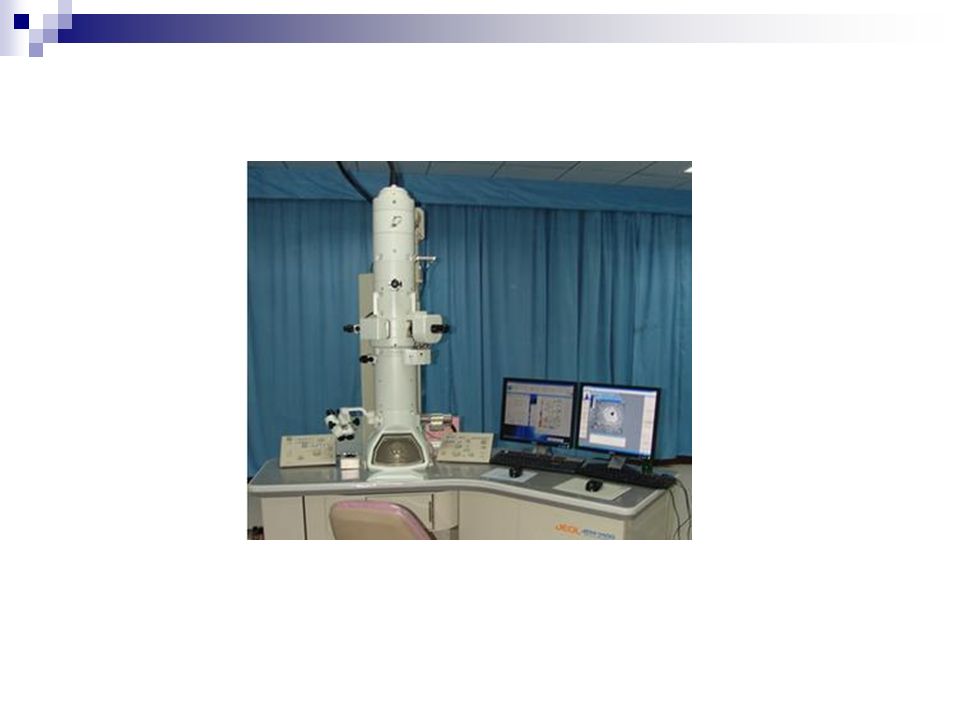
III. How to study it- histological methods ---Development of histology deponds on the development of technique. ---Histology studies the microstructures. So, we should have the aid of microscope to study. Several types of microscopes are available. According to the light source used, microscopes can be basally classified as: light microscope(LM) electron microscope(EM)
 electron microscope(EM).")
11
1. structure of Microscope LM EM ---useful magnification: 1500X 800,000X ---resolution: 0.2um 0.2nm

12
2. Preparation of tissue for LM The most routine one is paraffin section stained with hematoxylin and eosin(H&E) The steps: a. Obtaining th specimen: fresh, small pieces (less than 5 cubic milimeter ( mm 3 ) )-tissue block b. Fixation: fixatives: use formalin or Bouin’s to preserve structural organisation c. Dehydration: use ethyl alcohol to get rid of water of tissue and cell
 The steps: a. Obtaining th specimen: fresh, small pieces (less than 5 cubic milimeter ( mm 3 ) )-tissue block b. Fixation: fixatives: use formalin or Bouin’s to preserve structural organisation c. Dehydration: use ethyl alcohol to get rid of water of tissue and cell.")
13
d. Clearing: use xylene to get rid of alcohol alcohol and xylene are embedding mediums e. Embedding: firstly, heat the paraffin, make it melt, then put tissue block into melted paraffin, allow paraffin harden, the tissue block is embedded in.

14
f. Sectioning: use microtome to cut the tissue into 3-8um thick sections, then mounted them on glass slides g. H&E staining ---Hematoxylin: basic stain, combines with acidic components, make them appear blue color- we call such components as basophilic ---Eosin: acidic stain, combines with basic components, make them appear pink color- we call such components as acidophilic(eosinophilic)
.")
16
observing

18
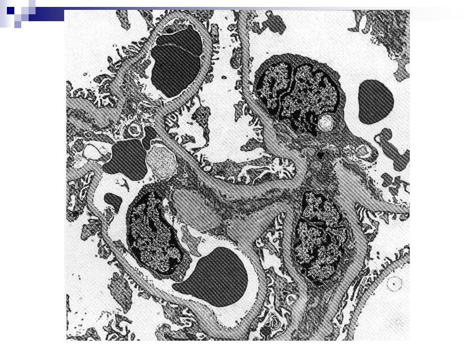
3. Preparation of tissue for EM The steps are same to preparation for LM a. tissue block: more small, less than 1mm 3 b. plastic materials for embedding c. ultra-thin sections is about 30-50nm thick( use ultramicrotome) d. heavy metal salts- increase staining contrast ---lead citrate ---uranyl acetate
 d. heavy metal salts- increase staining contrast ---lead citrate ---uranyl acetate.")
19
e. the beam of electron replace the light to illuminate the tissue sections Beam of electron illuminate the tissue section, we use a screen to receive the electron. In some areas, the beam of electron is impeded by those tissue element which are stained with heavy metal salts, so very few electrons penetrate to excite the screen, such areas appear dark, are described as electron- dense areas. Unstained areas, by contrast, appear light, we call them as electron-lucent areas.

21
4. Histochemistry 1) General Histochemistry: Combine histological methods with chemical or biochemical methods, make some compositions of tissue or cell become insoluble, colored or electron-dense,, to show those chemical compositions of tissue or cell in situ, such compositions includes protein,amino acid, nucleic acid, lipid and enzymes.
 General Histochemistry: Combine histological methods with chemical or biochemical methods, make some compositions of tissue or cell become insoluble, colored or electron-dense,, to show those chemical compositions of tissue or cell in situ, such compositions includes protein,amino acid, nucleic acid, lipid and enzymes..")
22
*Periodic acid schiff reaction(PAS reaction): schiff’s reagent (colorless) Polysaccharides → aldehydes → magenta complexes Glycogen oxidize combine (purple red colored)
: schiff’s reagent (colorless) Polysaccharides → aldehydes → magenta complexes Glycogen oxidize combine (purple red colored)")
23
2) immnohistochemistry: antigen- antibody
 immnohistochemistry: antigen- antibody")
26
3) in situ hybridization: nucleic acid: DNA(desoxyribose nucleic acid) RNA(ribose nucleic acid)
 in situ hybridization: nucleic acid: DNA(desoxyribose nucleic acid) RNA(ribose nucleic acid)")
Similar presentations


 histology is a lost art, especially if one is interested.>")


