Download presentation
Presentation is loading. Please wait.

1
Reproductive System Pathologies
FINAL

2
Female Reproductive System

4
Congenital Anomolies (FEMALE)
")
5
Bicornate Uterus

6
Bicornate Uterus

7
Unicornuate Uterus

8
Uterus Didelphys Uterus didelphys with an obstructed hemivagina. (a) Axial T2-weighted image shows two separate uteri and two cervices (arrows), all of which have normal zonal anatomy. Arrowheads = ovaries. (b) Coronal T2-weighted image shows a hematocele (H) due to obstruction of the right hemivagina. (c) Contrast-enhanced computed tomographic (CT) scan shows agenesis of the right kidney. Uterus didelphys with an obstructed hemivagina is termed Wunderlich syndrome and is usually associated with ipsilateral renal agenesis. (Reprinted, with permission, from reference 50.)
 Axial T2-weighted image shows two separate uteri and two cervices (arrows), all of which have normal zonal anatomy. Arrowheads = ovaries. (b) Coronal T2-weighted image shows a hematocele (H) due to obstruction of the right hemivagina. (c) Contrast-enhanced computed tomographic (CT) scan shows agenesis of the right kidney. Uterus didelphys with an obstructed hemivagina is termed Wunderlich syndrome and is usually associated with ipsilateral renal agenesis. (Reprinted, with permission, from reference 50.)")
9
Uterus Didelphys

10
Inflammatory Diseases

11
Pelvic Inflammatory Disease

12
Neoplastic Diseases (FEMALE)
")
13
Cystic Masses

14
Polycystic Ovaries

15
Endometriosis

16
Teratoma Dermoid Cyst

17
Dermoid Cyst

18
Dermoid Cyst

19
Cystadenocarcinoma

20
Ovarian Cancer

22
Cervical Cancer

23
Uterine Masses

24
Uterine Fibroid They are benign growths, arising from the muscular wall of the uterus. Their origin is thought to be the muscle in the walls of uterine blood vessels. Fibroids vary greatly in size, and can remain for years with little change. Others can grow much larger and reach the size of a 5 month pregnancy or more. In pregnancy, pre-existing fibroids can increase 3-5 times in size. This is thought to be due in part to the very high estrogen level in pregnancy, as well as to other factors stimulating the pregnancy changes. Quite remarkably after pregnancy, these same fibroids can shrink to their pre-pregnancy size. Menopausal patients who take estrogen show a varying response. Some who start with significant size fibroids may notice a slow increase in size, while others experience very little change at all. Fibroids are extremely common. They are estimated to reach significant size in 25-30% of all Caucasians, and in 50% of women of African background. If very tiny fibroids are included, some studies suggest that by the menopause virtually every woman has them. In most cases, there is more than one fibroid present. Sometimes there are many - 50 or more have been counted. A Solitary fibroid can occur, but is much less frequent. Cancer in a fibroid is very uncommon (perhaps 1:750 to 1000). There is some data that suggests this cancer (called a sarcoma) may not arise from a pre-existing fibroid at all, but develop in an area of the uterus not a fibroid. Fibroids are also called by other names such as: Myoma, Leiomyoma, Leiomyomata and Fibromyoma Serosal Fibroids (or those which develop in the outer portion of the uterus and expand giving the uterus a "knobby" appearance.) A serosal fibroid develops below the capsule of the uterus, and slowly expands outwards. (Observe the animated drawing to the left.) Probably because they are not trapped below the surface of the uterus, they can expand to large size. They produce no change in menstrual flow, and no increase in the miscarriage rate. They are compatible with pregnancy (though because of their size they can become uncomfortable by causing increasing pressure). Serosal fibroids produce a problem in pregnancy only if they are in the lower part of the uterus. There they can block the outlet of the pelvis making a C-Section the only way to deliver the baby. Intra-Mural Fibroids (or those which develop within the wall of the uterus and expand making the uterus feel larger than normal during a pelvic exam.) An Intra-mural fibroid develops below the capsule of the uterus, and slowly expands, increasing the bulk of the uterus. (Observe the animated drawing to the left.) When there are many fibroids within the wall, the uterine cavity also expands. This can result in heavier menstrual flows. Should the combined bulk of the fibroids (all types) be large enough to fill the pelvis tightly, a blockage of flow of urine from the kidneys may result. Though this is uncommon, it can damage the kidneys if left untreated. Hence once this blockage is discovered, these fibroids must be removed
. There is some data that suggests this cancer (called a sarcoma) may not arise from a pre-existing fibroid at all, but develop in an area of the uterus not a fibroid. Fibroids are also called by other names such as: Myoma, Leiomyoma, Leiomyomata and Fibromyoma. Serosal Fibroids (or those which develop in the outer portion of the uterus and expand giving the uterus a knobby appearance.) A serosal fibroid develops below the capsule of the uterus, and slowly expands outwards. (Observe the animated drawing to the left.) Probably because they are not trapped below the surface of the uterus, they can expand to large size. They produce no change in menstrual flow, and no increase in the miscarriage rate. They are compatible with pregnancy (though because of their size they can become uncomfortable by causing increasing pressure). Serosal fibroids produce a problem in pregnancy only if they are in the lower part of the uterus. There they can block the outlet of the pelvis making a C-Section the only way to deliver the baby. Intra-Mural Fibroids (or those which develop within the wall of the uterus and expand making the uterus feel larger than normal during a pelvic exam.) An Intra-mural fibroid develops below the capsule of the uterus, and slowly expands, increasing the bulk of the uterus. (Observe the animated drawing to the left.) When there are many fibroids within the wall, the uterine cavity also expands. This can result in heavier menstrual flows. Should the combined bulk of the fibroids (all types) be large enough to fill the pelvis tightly, a blockage of flow of urine from the kidneys may result. Though this is uncommon, it can damage the kidneys if left untreated. Hence once this blockage is discovered, these fibroids must be removed.")
25
LEIOMYOMA (FIBROID TUMOR)
STOOL LEIOMYOMA Due to the presence of needles, and possibly also due to her long-term disability, the patient has chronic ileus, causing severe constipation and stool impaction. By the way, the lesion projecting over the right iliac wing is a calcified leiomyoma uteri. The uterus is displaced to the right by the stool-distended sigmoid colon.

26
Endometrial Cancer

27
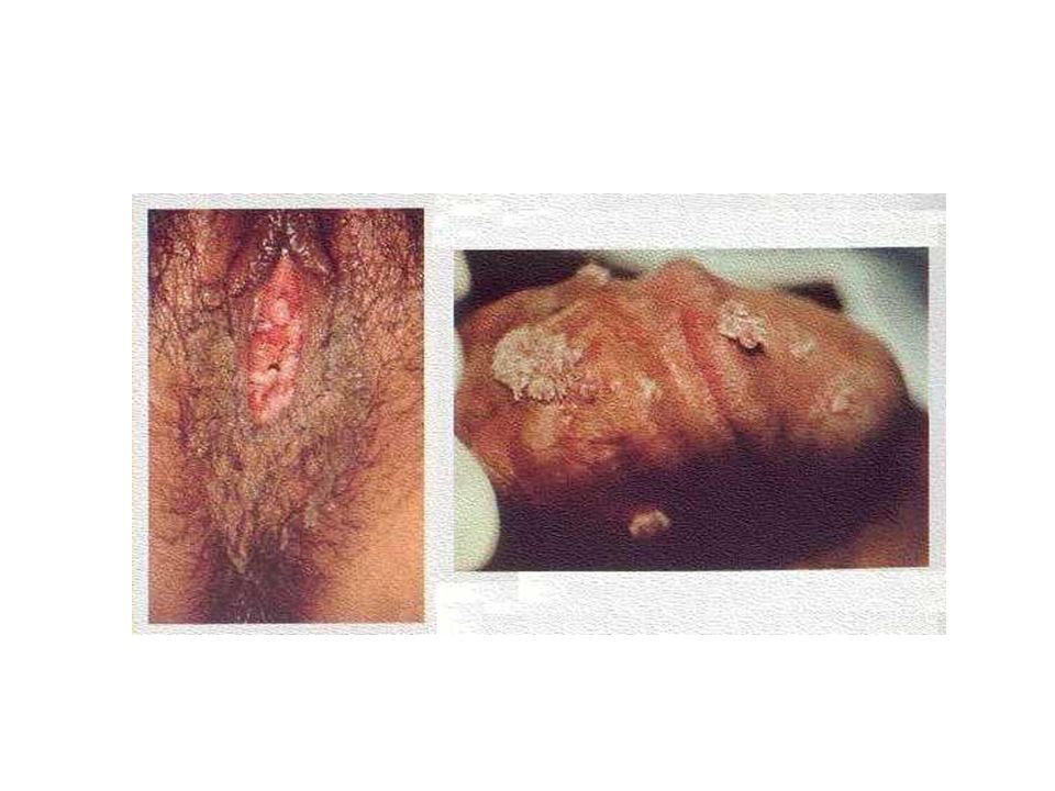
HPV Cervical Cancer -Caused by HPV types16 & 18 (high-risk HPV strains) -The virus enters the body through a cut, abrasion or tear in the outer layer of skin. It invades the cervical tissue cells and transfers genetic material. This leads to abnormal changes in the cells.

29
DES

30
Bladder Prolapse

31
Prolapsed Uterus

32
Breast Patholgies FEMALES

34
Fibroadenomas

35
Fibrocystic Breasts

36
Breast Carcinoma

37
Breast Cancer

38
Disorders During Pregnancy

40
Oligohydramnios

41
Polyhydramnios

42
Ectopic Pregnancy

43
Ectopic Pregnancy

44
Hydatidiform Mole

45
Congenital Anomolies (MALES)
")
46
Male Reproductive System

47
Cryptorchidism

48
Cryptorchidism

49
TSE

50
PSA Test

51
Inflammatory diseases

52
Epididymo-orchitis

53
Benign Prostatic Hyperplasia

54
Benign Prostatic Hyperplasia

55
Prostate Cancer

56
Testicular Masses

57
Hydroceles

58
Spermatoceles

59
Seminomas

60
Embryonal Carcinomas

61
Teratomas

62
Choriocarcinomas

63
Diabetes and Impotence
Impotence or erectile dysfunction is a very common problem that affects 20 million (1 out of 5) American Men. Erectile dysfunction is the result of a single, or more commonly a combination of multiple factors. At one time impotence was thought to be the result of psychological problems, but we now know that 90% of the cases are organic in nature. Some of the many causes of impotence include, diabetes, high blood pressure, heart and vascular disease, stress, hormone problems, pelvic surgery, trauma, venous leak, and side effects of frequently prescribed medications. No matter what the cause, most men have a secondary psychological reaction that can worsen the situation. Feelings of performance anxiety, guilt, and low self-esteem are common. The penis contains two chambers called the corpora cavernosa, which run the length of the organ (see figure 1). A spongy tissue fills the chambers. The corpora cavernosa are surrounded by a membrane, called the tunica albuginea. The spongy tissue contains smooth muscles, fibrous tissues, spaces, veins, and arteries. The urethra, which is the channel for urine and ejaculate, runs along the underside of the corpora cavernosa and is surrounded by the corpus spongiosum. Erection begins with sensory or mental stimulation, or both. Impulses from the brain and local nerves cause the muscles of the corpora cavernosa to relax, allowing blood to flow in and fill the spaces. The blood creates pressure in the corpora cavernosa, making the penis expand. The tunica albuginea helps trap the blood in the corpora cavernosa, thereby sustaining erection. When muscles in the penis contract to stop the inflow of blood and open outflow channels, erection is reversed
 American Men. Erectile dysfunction is the result of a single, or more commonly a combination of multiple factors. At one time impotence was thought to be the result of psychological problems, but we now know that 90% of the cases are organic in nature. Some of the many causes of impotence include, diabetes, high blood pressure, heart and vascular disease, stress, hormone problems, pelvic surgery, trauma, venous leak, and side effects of frequently prescribed medications. No matter what the cause, most men have a secondary psychological reaction that can worsen the situation. Feelings of performance anxiety, guilt, and low self-esteem are common. The penis contains two chambers called the corpora cavernosa, which run the length of the organ (see figure 1). A spongy tissue fills the chambers. The corpora cavernosa are surrounded by a membrane, called the tunica albuginea. The spongy tissue contains smooth muscles, fibrous tissues, spaces, veins, and arteries. The urethra, which is the channel for urine and ejaculate, runs along the underside of the corpora cavernosa and is surrounded by the corpus spongiosum. Erection begins with sensory or mental stimulation, or both. Impulses from the brain and local nerves cause the muscles of the corpora cavernosa to relax, allowing blood to flow in and fill the spaces. The blood creates pressure in the corpora cavernosa, making the penis expand. The tunica albuginea helps trap the blood in the corpora cavernosa, thereby sustaining erection. When muscles in the penis contract to stop the inflow of blood and open outflow channels, erection is reversed.")
64
Prostatitis

65
Prostatitis

66
Testicular Torsion

67
Testicular Torsion

68
Scrotal Hernia

Similar presentations


 have two basic components. Proliferating neoplastic cells that constitute.>")


 Moscow Regional Research Center of Obstetrics & Gynecology,>")