Download presentation
Presentation is loading. Please wait.

1
FOUNDATIONS OF BEHAVIORAL NEUROSCIENCE Copyright © 2014 Pearson Education, Inc. All Rights Reserved

2
Chapter 6 Vision Copyright © 2014 Pearson Education, Inc. All Rights Reserved

3
Vision Learning Objectives 1. Describe the characteristics of light and color, outline the anatomy of the eye and its connections with the brain, and describe the process of transduction of visual information. 2. Describe the coding of visual information by photoreceptors and ganglion cells in the retina. 3. Describe the striate cortex and discuss how its neurons respond to orientation, movement, spatial frequency, retinal disparity, and color. 4. Describe the anatomy of the visual association cortex and discuss the location and functions of the two streams of visual analysis that take place there. 5. Discuss the perception of color and the analysis of form by neurons in the ventral stream. 6. Describe the role of the visual association cortex in the perception of objects, faces, body parts, and places. 7. Describe the role of the visual association cortex in the perception of movement. 8. Describe the role of the visual association cortex in the perception of spatial location. Copyright © 2014 Pearson Education, Inc. All Rights Reserved

4
The Stimulus Anatomy of the Visual System The Eyes Photoreceptors Connections Between Eye and Brain Coding of Visual Information in the Retina Coding of Light and Dark Coding of Color Analysis of Visual Information: Role of the Striate Cortex Anatomy of the Striate Cortex Orientation and Movement Spatial Frequency Retinal Disparity Color Modular Organization of the Striate Cortex Copyright © 2014 Pearson Education, Inc. All Rights Reserved

5
The Stimulus Analysis of Visual Information: Role of the Visual Association Cortex Two Streams of Visual Analysis Perception of Color Perception of Form Perception of Movement Perception of Spatial Location Copyright © 2014 Pearson Education, Inc. All Rights Reserved

6
Prologue sensory receptor A specialized neuron that detects a particular category of physical events. sensory transduction The process by which sensory stimuli are transduced into slow, graded receptor potentials. receptor potential A slow, graded electrical potential produced by a receptor cell in response to a physical stimulus. Copyright © 2014 Pearson Education, Inc. All Rights Reserved

7
The Stimulus sensory receptor A specialized neuron that detects a particular category of physical events. sensory transduction The process by which sensory stimuli are transduced into slow, graded receptor potentials. receptor potential A slow, graded electrical potential produced by a receptor cell in response to a physical stimulus. Copyright © 2014 Pearson Education, Inc. All Rights Reserved

8
The Stimulus Copyright © 2014 Pearson Education, Inc. All Rights Reserved Our eyes detect the presence of light. For humans light is a narrow band of the spectrum of electromagnetic radiation. Electromagnetic radiation with a wavelength of between 380 and 760 nm (a nanometer, nm, is one-billionth of a meter) is visible to us. (See Figure 6.1.) Other animals can detect different ranges of electromagnetic radiation.
 is visible to us. (See Figure 6.1.) Other animals can detect different ranges of electromagnetic radiation..")
9
Copyright © 2014 Pearson Education, Inc. All Rights Reserved

10
The Stimulus hue One of the perceptual dimensions of color; the dominant wavelength. brightness One of the perceptual dimensions of color; intensity. saturation One of the perceptual dimensions of color; purity. Copyright © 2014 Pearson Education, Inc. All Rights Reserved

12
Anatomy of the Visual System Copyright © 2014 Pearson Education, Inc. All Rights Reserved The Eyes The eyes are suspended in the orbits, bony pockets in the front of the skull. They are held in place and moved by six extraocular muscles attached to the tough, white outer coat of the eye called the sclera. Normally, we cannot look behind our eyeballs and see these muscles, because their attachments to the eyes are hidden by the conjunctiva.

13
Anatomy of the Visual System Copyright © 2014 Pearson Education, Inc. All Rights Reserved The Eyes These mucous membranes line the eyelid and fold back to attach to the eye (thus preventing a contact lens that has slipped off the cornea from “falling behind the eye”). Figure 6.3 illustrates the anatomy of the eye. (See Figure 6.3.)
. Figure 6.3 illustrates the anatomy of the eye. (See Figure 6.3.).")
14
Anatomy of the Visual System Copyright © 2014 Pearson Education, Inc. All Rights Reserved The Eyes saccadic movement (suh kad ik) The rapid, jerky movement of the eyes used in scanning a visual scene. pursuit movement The movement that the eyes make to maintain an image of a moving object on the fovea.
 The rapid, jerky movement of the eyes used in scanning a visual scene. pursuit movement The movement that the eyes make to maintain an image of a moving object on the fovea..")
15
Copyright © 2014 Pearson Education, Inc. All Rights Reserved

16
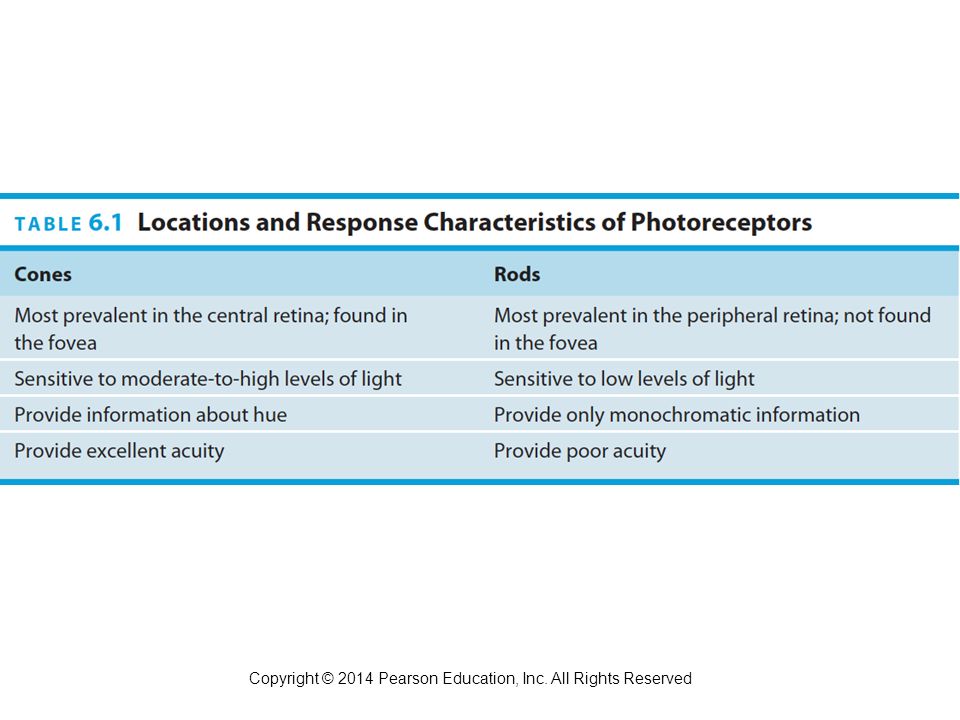
Anatomy of the Visual System Copyright © 2014 Pearson Education, Inc. All Rights Reserved The Eyes retina The neural tissue and photoreceptive cells located on the inner surface of the posterior portion of the eye. rod One of the receptor cells of the retina; sensitive to light of low intensity. cone One of the receptor cells of the retina; maximally sensitive to one of three different wavelengths of light and hence encodes color vision.

17
Anatomy of the Visual System The Eyes photoreceptor One of the receptor cells of the retina; transduces photic energy into electrical potentials. fovea (foe vee a) The region of the retina that mediates the most acute vision of birds and higher mammals. Color-sensitive cones constitute the only type of photoreceptor found in the fovea. Copyright © 2014 Pearson Education, Inc. All Rights Reserved
 The region of the retina that mediates the most acute vision of birds and higher mammals. Color-sensitive cones constitute the only type of photoreceptor found in the fovea. Copyright © 2014 Pearson Education, Inc. All Rights Reserved.")
19
Anatomy of the Visual System The Eyes optic disk The location of the exit point from the retina of the fibers of the ganglion cells that form the optic nerve; responsible for the blind spot. Copyright © 2014 Pearson Education, Inc. All Rights Reserved

21
Anatomy of the Visual System The Eyes bipolar cell A bipolar neuron located in the middle layer of the retina, conveying information from the photoreceptors to the ganglion cells. ganglion cell A neuron located in the retina that receives visual information from bipolar cells; its axons give rise to the optic nerve. Copyright © 2014 Pearson Education, Inc. All Rights Reserved

22
Anatomy of the Visual System The Eyes horizontal cell A neuron in the retina that interconnects adjacent photoreceptors and the outer processes of the bipolar cells. amacrine cell (amm a krin) A neuron in the retina that interconnects adjacent ganglion cells and the inner processes of the bipolar cells. Copyright © 2014 Pearson Education, Inc. All Rights Reserved
 A neuron in the retina that interconnects adjacent ganglion cells and the inner processes of the bipolar cells. Copyright © 2014 Pearson Education, Inc. All Rights Reserved.")
24
Anatomy of the Visual System Photoreceptors lamella A layer of membrane containing photopigments; found in rods and cones of the retina. photopigment A protein dye bonded to retinal, a substance derived from vitamin A; responsible for transduction of visual information. Copyright © 2014 Pearson Education, Inc. All Rights Reserved

25
Anatomy of the Visual System Photoreceptors opsin (opp sin) A class of protein that, together with retinal, constitutes the photopigments. retinal (rett i nahl) A chemical synthesized from vitamin A; joins with an opsin to form a photopigment. rhodopsin (roh dopp sin) A particular opsin found in rods. Copyright © 2014 Pearson Education, Inc. All Rights Reserved
 A chemical synthesized from vitamin A; joins with an opsin to form a photopigment. rhodopsin (roh dopp sin) A particular opsin found in rods. Copyright © 2014 Pearson Education, Inc. All Rights Reserved.")
26
Anatomy of the Visual System Connections Between Eye and Brain dorsal lateral geniculate nucleus (LGN) A group of cell bodies within the lateral geniculate body of the thalamus; receives inputs from the retina and projects to the primary visual cortex. magnocellular layer One of the inner two layers of neurons in the dorsal lateral geniculate nucleus; transmits information necessary for the perception of form, movement, depth, and small differences in brightness to the primary visual cortex. Copyright © 2014 Pearson Education, Inc. All Rights Reserved

27
Anatomy of the Visual System Connections Between Eye and Brain calcarine fissure (kal ka rine) A horizontal fissure on the inner surface of the posterior cerebral cortex; the location of the primary visual cortex. striate cortex (stry ate) The primary visual cortex. Copyright © 2014 Pearson Education, Inc. All Rights Reserved
 The primary visual cortex. Copyright © 2014 Pearson Education, Inc. All Rights Reserved.")
29
Anatomy of the Visual System Connections Between Eye and Brain optic chiasm A cross-shaped connection between the optic nerves, located below the base of the brain, just anterior to the pituitary gland. Copyright © 2014 Pearson Education, Inc. All Rights Reserved

31
Coding of Visual Information in the Brain Coding of Light and dark receptive field That portion of the visual field in which the presentation of visual stimuli will produce an alteration in the firing rate of a particular neuron. Copyright © 2014 Pearson Education, Inc. All Rights Reserved

33
Coding of Visual Information in the Brain Coding of Light and dark Kuffler (1952, 1953), recording from ganglion cells in the retina of the cat, discovered that their receptive field consists of a roughly circular center, surrounded by a ring. Stimulation of the center or surrounding fields had contrary effects: ON cells were excited by light falling in the central field (center) and were inhibited by light falling in the surrounding field (surround), whereas OFF cells responded in the opposite manner. Copyright © 2014 Pearson Education, Inc. All Rights Reserved
 and were inhibited by light falling in the surrounding field (surround), whereas OFF cells responded in the opposite manner. Copyright © 2014 Pearson Education, Inc. All Rights Reserved.")
34
Coding of Visual Information in the Brain Coding of Light and dark ON/OFF ganglion cells were briefly excited when light was turned on or off. In primates these ON/OFF cells project to the superior colliculus, which is primarily involved in visual reflexes in response to moving or suddenly- appearing stimuli (Schiller and Malpeli, 1977), which suggests that they do not play a direct role in form perception. (See Figure 6.9.) Copyright © 2014 Pearson Education, Inc. All Rights Reserved
, which suggests that they do not play a direct role in form perception. (See Figure 6.9.) Copyright © 2014 Pearson Education, Inc. All Rights Reserved.")
36
Coding of Visual Information in the Brain Coding of Color So far, we have been examining the monochromatic properties of ganglion cells—that is, their responses to light and dark. But, of course, objects in our environment selectively absorb some wavelengths of light and reflect others, which, to our eyes, gives them different colors. The retinas of humans and many species of nonhuman primates contain three different types of cones, which provides them (and us) with the most elaborate form of color vision (Jacobs, 1996; Hunt et al., 1998). Copyright © 2014 Pearson Education, Inc. All Rights Reserved
 with the most elaborate form of color vision (Jacobs, 1996; Hunt et al., 1998). Copyright © 2014 Pearson Education, Inc. All Rights Reserved.")
37
Coding of Visual Information in the Brain Photoreceptors: Trichromatic Coding Various theories of color vision have been proposed for many years—long before it was possible to disprove or validate them by physiological means. In 1802, Thomas Young, a British physicist and physician, proposed that the eye detected different colors because it contained three types of receptors, each sensitive to a single hue. Copyright © 2014 Pearson Education, Inc. All Rights Reserved

38
Coding of Visual Information in the Brain Photoreceptors: Trichromatic Coding His theory was referred to as the trichromatic (three-color) theory. It was suggested by the fact that for a human observer any color can be reproduced by mixing various quantities of three colors judiciously selected from different points along the spectrum. Copyright © 2014 Pearson Education, Inc. All Rights Reserved

39
Coding of Visual Information in the Brain Photoreceptors: Trichromatic Coding Three different types of photoreceptors (three different types of cones) are responsible for color vision. Investigators have studied the absorption characteristics of individual photoreceptors, determining the amount of light of different wavelengths that is absorbed by the photopigments. Copyright © 2014 Pearson Education, Inc. All Rights Reserved

40
Coding of Visual Information in the Brain Photoreceptors: Trichromatic Coding These characteristics are controlled by the particular opsin a photoreceptor contains; different opsins absorb particular wavelengths more readily. The peak sensitivities of the three types of cones are approximately 420 nm (blue-violet), 530 nm (green), and 560 nm (yellow-green). The peak sensitivity of the short-wavelength cone is actually 440 nm in the intact eye because the lens absorbs some short- wavelength light. For convenience the short-, medium-, and long- wavelength cones are traditionally called “blue,” “green,” and “red” cones, respectively. Copyright © 2014 Pearson Education, Inc. All Rights Reserved
, 530 nm (green), and 560 nm (yellow-green). The peak sensitivity of the short-wavelength cone is actually 440 nm in the intact eye because the lens absorbs some short- wavelength light. For convenience the short-, medium-, and long- wavelength cones are traditionally called blue, green, and red cones, respectively. Copyright © 2014 Pearson Education, Inc. All Rights Reserved.")
42
Coding of Visual Information in the Brain Photoreceptors: Trichromatic Coding protanopia (pro tan owe pee a) An inherited form of defective color vision in which red and green hues are confused; “red” cones are filled with “green” cone opsin. deuteranopia (dew ter an owe pee a) An inherited form of defective color vision in which red and green hues are confused; “green” cones are filled with “red” cone opsin. Copyright © 2014 Pearson Education, Inc. All Rights Reserved
 An inherited form of defective color vision in which red and green hues are confused; green cones are filled with red cone opsin. Copyright © 2014 Pearson Education, Inc. All Rights Reserved.")
43
Coding of Visual Information in the Brain Photoreceptors: Trichromatic Coding tritanopia (try tan owe pee a) An inherited form of defective color vision in which hues with short wavelengths are confused; “blue” cones are either lacking or faulty. Copyright © 2014 Pearson Education, Inc. All Rights Reserved

44
Coding of Visual Information in the Brain Retinal Ganglion Cells: Opponent-Process Coding At the level of the retinal ganglion cell the three-color code gets translated into an opponent-color system. Daw (1968) and Gouras (1968) found that these neurons respond specifically to pairs of primary colors: red versus green and yellow versus blue. Copyright © 2014 Pearson Education, Inc. All Rights Reserved
 and Gouras (1968) found that these neurons respond specifically to pairs of primary colors: red versus green and yellow versus blue. Copyright © 2014 Pearson Education, Inc. All Rights Reserved.")
46
Coding of Visual Information in the Brain Retinal Ganglion Cells: Opponent-Process Coding Thus, the retina contains two kinds of color-sensitive ganglion cells: red-green cells and yellow-blue cells. Some color-sensitive ganglion cells respond in a center-surround fashion. For example, a cell might be excited by red and inhibited by green in the center of their receptive field while showing the opposite response in the surrounding ring. (See Figure 6.11.) Copyright © 2014 Pearson Education, Inc. All Rights Reserved
 Copyright © 2014 Pearson Education, Inc. All Rights Reserved.")
47
Analysis of Visual Information: Role of the Striate Cortex Anatomy of the striate Cortex The striate cortex consists of six principal layers (and several sublayers), arranged in bands parallel to the surface. These layers contain the nuclei of cell bodies and dendritic trees that show up as bands of light or dark in sections of tissue that have been dyed with a cell-body stain. (See Figure 6.12.) Copyright © 2014 Pearson Education, Inc. All Rights Reserved
 Copyright © 2014 Pearson Education, Inc. All Rights Reserved.")
49
Analysis of Visual Information: Role of the Striate Cortex Orientation of Movement Most neurons in the striate cortex are sensitive to orientation. That is, if a line or an edge (the border of a light and a dark region) is positioned in the cell’s receptive field and rotated around its center, the cell will respond best when the line is in a particular position—a particular orientation. Some neurons respond best to a vertical line, some to a horizontal line, and some to a line oriented somewhere in between. Copyright © 2014 Pearson Education, Inc. All Rights Reserved
 is positioned in the cell’s receptive field and rotated around its center, the cell will respond best when the line is in a particular position—a particular orientation. Some neurons respond best to a vertical line, some to a horizontal line, and some to a line oriented somewhere in between. Copyright © 2014 Pearson Education, Inc. All Rights Reserved.")
51
Analysis of Visual Information: Role of the Striate Cortex Orientation of Movement simple cell An orientation-sensitive neuron in the striate cortex whose receptive field is organized in an opponent fashion. complex cell A neuron in the visual cortex that responds to the presence of a line segment with a particular orientation located within its receptive field, especially when the line moves perpendicularly to its orientation. Copyright © 2014 Pearson Education, Inc. All Rights Reserved

52
Analysis of Visual Information: Role of the Striate Cortex Orientation of Movement hypercomplex cell A neuron in the visual cortex that responds to the presence of a line segment with a particular orientation that ends at a particular point within the cell’s receptive field. Copyright © 2014 Pearson Education, Inc. All Rights Reserved

54
Analysis of Visual Information: Role of the Striate Cortex Spatial Frequency sine-wave grating A series of straight parallel bands varying continuously in brightness according to a sine-wave function, along a line perpendicular to their lengths. spatial frequency The relative width of the bands in a sine-wave grating, measured in cycles per degree of visual angle. Copyright © 2014 Pearson Education, Inc. All Rights Reserved

57
Analysis of Visual Information: Role of the Striate Cortex Color cytochrome oxidase (CO) blob The central region of a module of the primary visual cortex, revealed by a stain for cytochrome oxidase; contains wavelength-sensitive neurons; part of the parvocellular system. Copyright © 2014 Pearson Education, Inc. All Rights Reserved

59
Analysis of Visual Information: Role of the Striate Cortex Modular Organization of the Striate Cortex Most investigators believe that the brain is organized in modules, which probably range in size from a hundred thousand to a few million neurons. Each module receives information from other modules, performs some calculations, and then passes the results to other modules. In recent years investigators have been learning the characteristics of the modules that are found in the visual cortex. Copyright © 2014 Pearson Education, Inc. All Rights Reserved

62
Analysis of Visual Information: Role of the Visual Association Cortex Two Streams of Analysis extrastriate cortex A region of visual association cortex; receives fibers from the striate cortex and from the superior colliculi and projects to the inferior temporal cortex. Copyright © 2014 Pearson Education, Inc. All Rights Reserved

65
Analysis of Visual Information: Role of the Visual Association Cortex Two Streams of Analysis dorsal stream A system of interconnected regions of visual cortex involved in the perception of spatial location, beginning with the striate cortex and ending with the posterior parietal cortex. ventral stream A system of interconnected regions of visual cortex involved in the perception of form, beginning with the striate cortex and ending with the inferior temporal cortex. Copyright © 2014 Pearson Education, Inc. All Rights Reserved

66
Analysis of Visual Information: Role of the Visual Association Cortex Two Streams of Analysis inferior temporal cortex The highest level of the ventral stream of the visual association cortex; involved in perception of objects, including people’s bodies and faces. posterior parietal cortex The highest level of the dorsal stream of the visual association cortex; involved in perception of movement and spatial location. Copyright © 2014 Pearson Education, Inc. All Rights Reserved

69
Analysis of Visual Information: Role of the Visual Association Cortex Perception of Color As we saw earlier, neurons within the CO blobs in the striate cortex respond to colors. Like the ganglion cells in the retina (and the parvocellular and koniocellular neurons in the LGN), these neurons respond in opponent fashion. This information is analyzed by the regions of the visual association cortex that constitute the ventral stream. Copyright © 2014 Pearson Education, Inc. All Rights Reserved
, these neurons respond in opponent fashion. This information is analyzed by the regions of the visual association cortex that constitute the ventral stream. Copyright © 2014 Pearson Education, Inc. All Rights Reserved.")
70
Analysis of Visual Information: Role of the Visual Association Cortex Studies with Laboratory Animals color constancy The relatively constant appearance of the colors of objects viewed under varying lighting conditions. Copyright © 2014 Pearson Education, Inc. All Rights Reserved

71
Analysis of Visual Information: Role of the Visual Association Cortex Studies with Humans cerebral achromatopsia (ay krohm a top see a) Inability to discriminate among different hues; caused by damage to area V8 of the visual association cortex. Copyright © 2014 Pearson Education, Inc. All Rights Reserved

73
Analysis of Visual Information: Role of the Visual Association Cortex Perception of Form The analysis of visual information that leads to the perception of form begins with neurons in the striate cortex that are sensitive to orientation and spatial frequency. These neurons send information to area V2 that is then relayed to the subregions of the visual association cortex that constitute the ventral stream. Copyright © 2014 Pearson Education, Inc. All Rights Reserved

74
Analysis of Visual Information: Role of the Visual Association Cortex Studies with Laboratory Animals In primates the recognition of visual patterns and identification of particular objects take place in the inferior temporal cortex, located on the ventral part of the temporal lobe. This region of the visual association cortex is located at the end of the ventral stream. Copyright © 2014 Pearson Education, Inc. All Rights Reserved

75
Analysis of Visual Information: Role of the Visual Association Cortex Studies with Laboratory Animals It is here that analyses of form and color are put together and perceptions of three-dimensional objects and backgrounds are achieved. Damage to this region causes severe deficits in visual discrimination (Mishkin, 1966; Gross, 1973; Dean, 1976). Copyright © 2014 Pearson Education, Inc. All Rights Reserved
. Copyright © 2014 Pearson Education, Inc. All Rights Reserved.")
76
Analysis of Visual Information: Role of the Visual Association Cortex Studies with Humans visual agnosia (ag no zha) Deficits in visual form perception in the absence of blindness; caused by brain damage. prosopagnosia (prah soh pag no zha) Failure to recognize particular people by the sight of their faces. fusiform face area (FFA) A region of the visual association cortex located in the inferior temporal; involved in perception of faces. Copyright © 2014 Pearson Education, Inc. All Rights Reserved
 Failure to recognize particular people by the sight of their faces. fusiform face area (FFA) A region of the visual association cortex located in the inferior temporal; involved in perception of faces. Copyright © 2014 Pearson Education, Inc. All Rights Reserved.")
78
Analysis of Visual Information: Role of the Visual Association Cortex Studies with Humans extrastriate body area (EBA) A region of the visual association cortex located in the lateral occipitotemporal cortex; involved in perception of the human body and body parts other than faces. Copyright © 2014 Pearson Education, Inc. All Rights Reserved

80
Analysis of Visual Information: Role of the Visual Association Cortex Studies with Humans parahippocampal place area (PPA) A region of the medial temporal cortex; involved in perception of particular places (“scenes”). Copyright © 2014 Pearson Education, Inc. All Rights Reserved

82
Analysis of Visual Information: Role of the Visual Association Cortex Perception of Movement We need to know not only what things are, but also where they are and if they are moving, where they are going. Without the ability to perceive the direction and velocity of movement of objects, we would have no way to predict where they will be. We would be unable to catch them (or avoid letting them catch us). Copyright © 2014 Pearson Education, Inc. All Rights Reserved
. Copyright © 2014 Pearson Education, Inc. All Rights Reserved.")
83
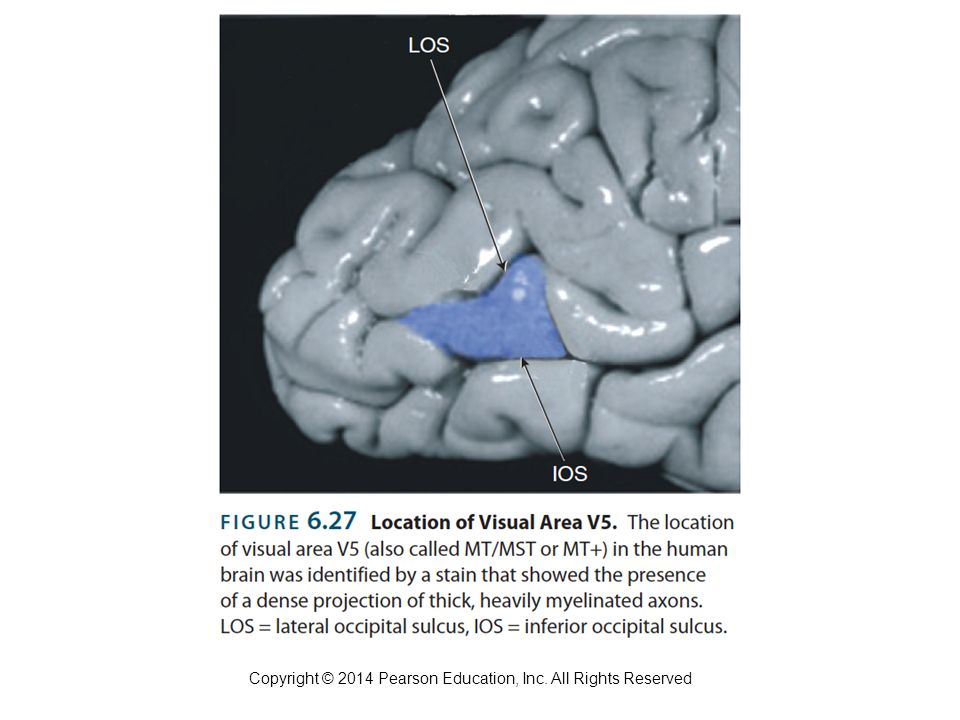
Analysis of Visual Information: Role of the Visual Association Cortex Perception of Movement Studies with Humans Functional imaging studies suggest that a motion-sensitive area (usually called MT/MST) is found within the inferior temporal sulcus of the human brain (Dukelow et al., 2001). However, a more recent study suggests that this region is located in the lateral occipital cortex, between the lateral and inferior occipital sulci (Annese, Gazzaniga, and Toga, 2004). Copyright © 2014 Pearson Education, Inc. All Rights Reserved
. Copyright © 2014 Pearson Education, Inc. All Rights Reserved.")
85
Analysis of Visual Information: Role of the Visual Association Cortex Perception of Movement Studies with Humans akinetopsia Inability to perceive movement, caused by damage to area V5 (also called MST) of the visual association cortex. Copyright © 2014 Pearson Education, Inc. All Rights Reserved

86
Analysis of Visual Information: Role of the Visual Association Cortex Perception of Movement Studies with Humans intraparietal sulcus (IPS) The end of the dorsal stream of the visual association cortex; involved in perception of location, visual attention, and control of eye and hand movements. Copyright © 2014 Pearson Education, Inc. All Rights Reserved

Similar presentations


 Allyn & Bacon 20041 Chapter 6 Vision This multimedia product and its contents are protected under copyright law. The following are prohibited.>")


