Download presentation
Presentation is loading. Please wait.

1
NON-PROTEIN NITROGENOUS COMPOUNDS Dr O. Morebise

2
In addition to serving as building blocks for proteins, amino acids are precursors of many nitrogen- containing compounds that have important physiologic functions. These molecules include porphyrins, neurotransmitters, hormones, purines, and pyrimidines.

3
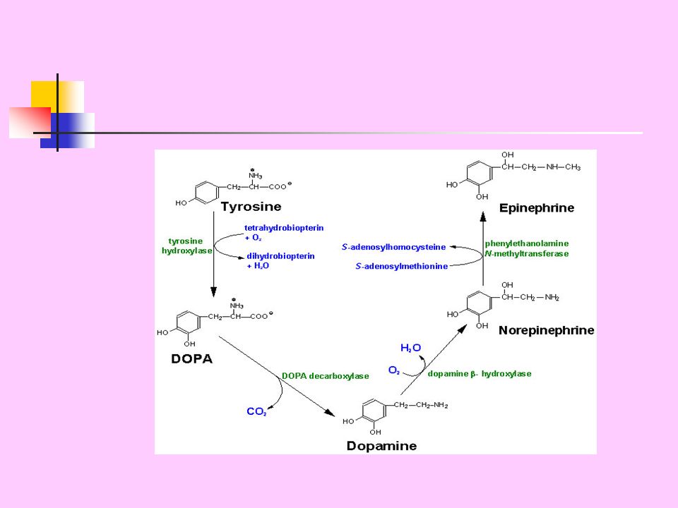
CATECHOLAMINES Dopamine, norepinephrine and epinephrine (adrenalin) are biologically active amines that are collectively termed catecholamines. Dopamine and norepinephrine function as neurotransmitters in the brain and the autonomic nervous system. Norepinephrine and epinephrine are also synthesized in the adrenal medulla.

4
Outside the nervous system, norepinephrine and its methylated derivative, epinephrine act as regulators of carbohydrate and lipid metabolism. Norepinephrine and epinephrine are released from storage vesicles in the adrenal medulla in response to fright, exercise, cold, and low levels of blood glucose. They increase the degradation of glycogen and triacylglycerol, as well as increase blood pressure and the output of the heart. These effects are part of a coordinated response to prepare the individual for emergencies, and are often called the “fight-or-flight” reactions.

6
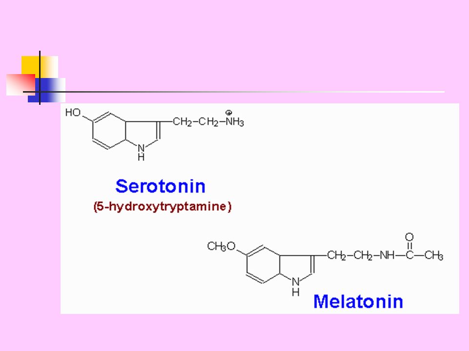
Serotonin Serotonin is synthesized and stored at several sites in the body. Largest amount of serotonin is found in cells of the intestinal mucosa. Smaller amounts occur in platelets and in the central nervous system. Serotonin is synthesized from tryptophan, which is hydroxylated in a reaction analogous to that catalyzed by phenylalanine hydroxylase.

7
Pathway for serotonin and melatonin synthesis from tryptophan. Abbreviations: TPH = tryptophan hydroxylase, DHPR = dihydropteridine reductase, H 2 B = dihydrobiopterin, H 4 B = tetrahyrobiopterin, 5-HT = 5- hydroxytryptophan, AADC = aromatic L-amino acid decarboxylase, SNA = serotonin N-acetylase, HOMT = hydroxyindole-O-methyltransferase.

9
Histamine Histamine is a chemical messenger. It mediates a wide range of cellular responses, including allergic and inflammatory reactions, gastric acid secretion, and neurotransmission in parts of the brain. It is secreted by mast cells as a result of allergic reactions or trauma. It is formed by decarboxylation of histidine in a reaction requiring pyridoxal phosphate.

10
Note: Histamine is a powerful stimulant of gastric secretion, a constrictor of bronchial smooth muscle and a vasodilator (capillaries and arterioles) that causes a fall in blood pressure. Histamine is liberated in the skin as a result of injury.

11
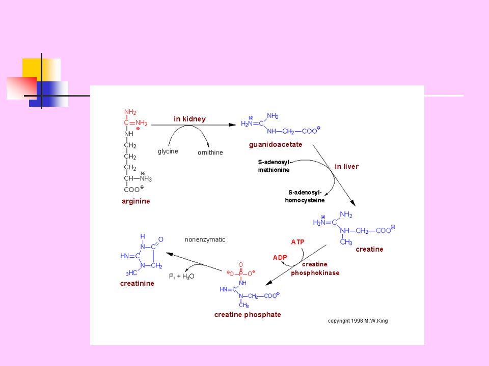
Creatine Creatine phosphate (phosphocreatine) is a high-energy compound that can reversibly donate a phosphate group to ADP to form ATP. Creatine phosphate provides a small but rapidly mobilized reserve of high-energy phosphates that can be used to maintain the intracellular level of ATP during the first few minutes of intense muscular contraction. The amount of creatine phosphate in the body is proportional to the muscle mass.

12
Synthesis of creatine and creatinine Creatine is synthesized from glycine and the guanido group of arginine, plus a methyl group from S-adenosylmethionine. Creatine is reversibly phosphorylated to creatine phosphate by creatine kinase, using ATP as the phosphate donor. The presence of creatine kinase in the plasma is indicative of tissue damage, and is used in the diagnosis of myocardial infarction.

14
Degradation of creatine Creatine and creatine phosphate spontaneously cyclize at a slow, but constant, rate to form creatinine, which is excreted in the urine. The amount of creatinine excreted is proportional to the total creatine phosphate content of the body, and thus can be used to estimate muscle mass. When muscle mass decreases for any reason (e.g., from paralysis or muscular dystrophy), the creatinine content of the urine falls.
, the creatinine content of the urine falls..")
15
In addition, any rise in blood creatinine is a sensitive indicator of kidney malfunction, because creatinine is normally rapidly removed from the blood and excreted. A typical adult male excretes about 15 mmol of creatinine per day. The constancy of this excretion is sometimes used to test the reliability of collected 24-hour urine samples—too little creatinine in the submitted sample may indicate an incomplete sample.

16
Melanin Melanin is a pigment that occurs in several tissues in the body, particularly in the eye, hair, and skin. It is synthesized in the epidermis by pigment-forming cells called melanocytes. Its function is to protect underlying cells from the harmful effects of sunlight. The first step in melanin formation from tyrosine is a hydroxylation to form DOPA, catalyzed by the copper- containing enzyme tyrosine hydroxylase (tyrosinase). Subsequent reactions leading to the formation of brown and black pigments are also thought to be catalyzed by tyrosine hydroxylase or to occur spontaneously.
. Subsequent reactions leading to the formation of brown and black pigments are also thought to be catalyzed by tyrosine hydroxylase or to occur spontaneously..")
17
Synthesis of melanin from tyrosine or DOPA

18
Chemical structure of melanin Melanin is a very complex absorbing material. Melanins from natural sources fall into two general classes: Eumelanin The most abundant type of human melanin. It is a black-to-dark-brown insoluble material found in brown and black skin and hair, and in the retina; cross-linked polymers of 5,6- dihydroxyindoles, usually linked to proteins; levels are decreased in certain types of albinism.

19
Pheomelanin A yellow-to-reddish-brown sulfur-containing alkali-soluble material found in red hair and red feathers. A variety of low molecular weight pheomelanins are called "trichromes". Elevated levels of pheomelanins are found in the rufous type of albinism. Rufous albinism: A pigmentary anomaly of blacks, characterized by red or yellow-red hair color, copper-red skin, and often by dilution of iris pigment. Syn: xanthism

20
The melanins are considered to be polymers of the basic building blocks shown in the Figure. However, the details of the polymerization and the role of protein linkages in the natural melanin complex are not known. Curly red lines indicate sites of attachment to the extended polymer and possibly to proteins.

21
PORPHYRINS Porphyrins: cyclic compounds that readily bind metal ions, usually Fe 2+ or Fe 3+ Most prevalent metalloporphyrin: heme – prosthetic group for Hemoglobin Myoglobin Cytochromes Catalase Tryptophan pyrrolase These heme proteins are constantly being broken down and recycled 6-7 g Hb synthesized daily to replace those lost in catabolism. Formation and degradation of porphyrin component of Hb are quantitatively important for N balance in body.

22
Ring structure: Porphyrins are cyclic molecules Linkage of 4 pyrrole rings through methenyl bridges

23
Distribution of side chains: Side chains can be ordered around tetrapyrrole ring in 4 different ways – designated I through IV Only type III porphyrins are of physiologic importance in humans Type III porphyrins contain an asymmetric substitution on ring D. Type I porphyrins contain a symmetric arrangement of substituents and may be synthesized in higher amounts in congenital erythropoietic porphyria.

24
Congenital erythropoietic porphyria The deficient enzyme is uroporphyrinogen III cosynthase ( Uroporphyrinogen III synthase).
.")
25
PROTOPORPHYRIN IX

26
Heme It is the final product of porphyrin synthetic pathway. Consists of one Fe 2+ ion coordinated in center of tetrapyrrole ring of protoporphyrin IX. Protoporphyrin IX is quantitatively the most important porphyrin in humans.

27
Major sites of heme biosynthesis The major sites of heme biosynthesis are the liver (where the rate of synthesis is highly variable) and the erythrocyte-producing cells of the bone marrow (where the rate is generally constant). Initial reaction and last 3 steps of formation of porphyrins occur in mitochondria Intermediate steps occur in cytosol Mature RBCs lack mitochondria and therefore cannot synthesize heme All the carbon and nitrogen atoms are provided by glycine and succinyl CoA.

28
Porphyrias Caused by inherited or acquired defects in heme synthesis accumulation and ’sed excretion of porphyrins or precursors. Porphyrias classified as erythopoietic or hepatic depending on whether enzyme deficiency occurs in RBC or liver. Congenital erythopoietic porphyria is an autosomal recessive disease; virtually all others are autosomal dominant. Each porphyria accumulation of unique pattern of intermediates due to enzyme deficiency in heme synthetic pathway.

29
Clinical Manifestations: Those with an enzyme defect leading to the accumulation of tetrapyrrole intermediates show photosensitivity— i.e., their skin itches and burns (pruritis) when exposed to visible light. Accumulation of ALA and porphobilinogen e.g. acute intermittent porphyria abdominal pain and neuropsychiatric disturbances.

30
Treatment: Severity of symptoms can be diminished by injection of hemin ’se of ALA synthase Avoidance of sunlight and ingestion of -carotene (free radical scavenger) helpful
 helpful")
Similar presentations


>")


>")

