Download presentation
Presentation is loading. Please wait.

3
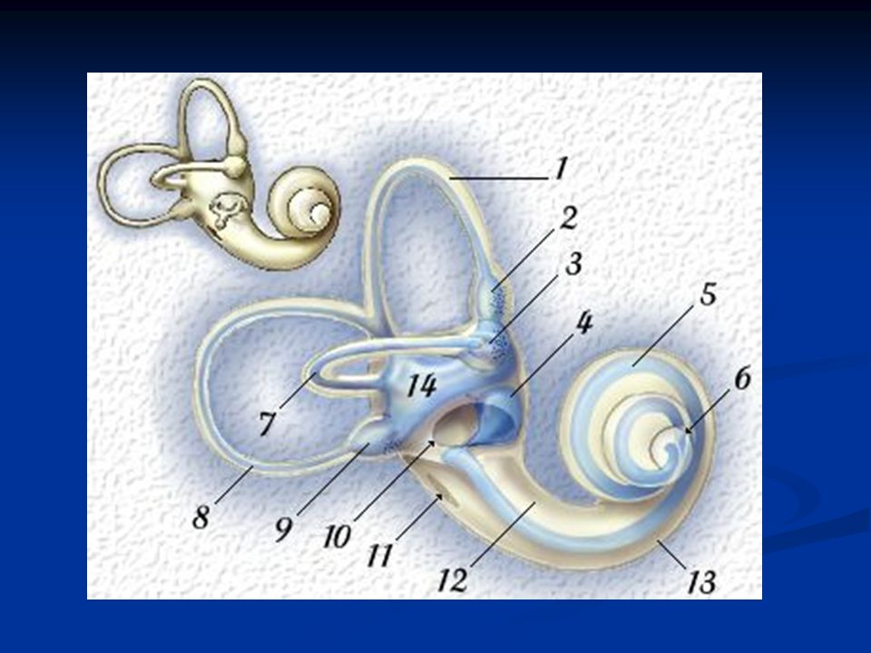
Review of Cochlear Anatomy Bony Capsule Bony Capsule Semicircular Canals Semicircular Canals Vestibule Vestibule Scala Tympani Scala Tympani Scala Vestibuli Scala Vestibuli Cochlear Aqueduct Cochlear Aqueduct Vestibule Aqueduct Vestibule Aqueduct Membranous Labyrinth Semicircular Canals Membranous Ampullae Utricle Saccule Utricular Duct Saccular Duct Endolympatic Duct Endolympatic Sac Cochlear Duct Canal Reunions

4
Winding of the Cochlea

6
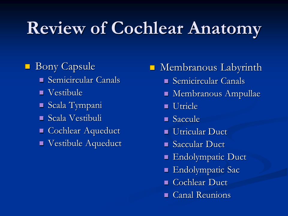
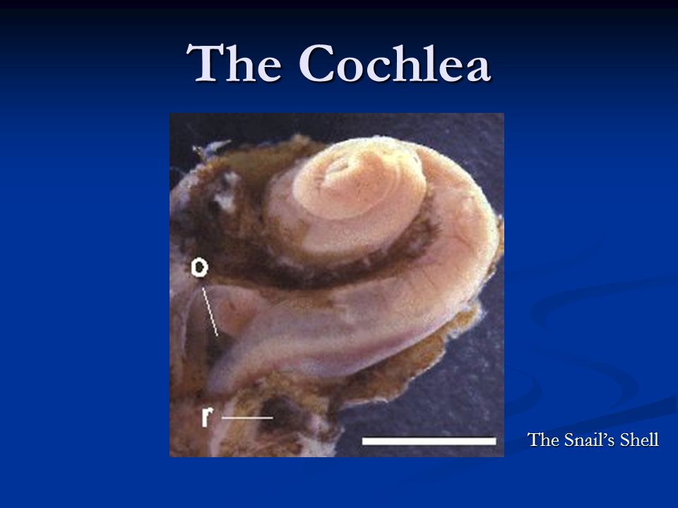
The Cochlea The Snail’s Shell

7
The Cochlea This figure shows the location of the cochlea with respect to the external ear canal. The cochlea is shown here as the blue spiral structure, which resembles a snail. (The word cochlea is derived from the Latin word for a snail shell) The cochlea is responsible for converting sounds which enter the ear canal, from mechanical vibrations into electrical signals. This process, known as transduction, is performed by specialized sensory cells within the cochlea. The electrical signals, which code the sound's characteristics, are carried to the brain by the auditory nerve.
 The cochlea is responsible for converting sounds which enter the ear canal, from mechanical vibrations into electrical signals. This process, known as transduction, is performed by specialized sensory cells within the cochlea. The electrical signals, which code the sound s characteristics, are carried to the brain by the auditory nerve..")
8
The Cochlea The cochlea is partially divided into an upper duct called the scala vestibuli and a lower duct, the scala tympani, by a thin bony shelf called the osseous spiral lamina. It is a narrow shelf of bone arising from the modiolar side of the cochlea. This division is completed by the membranous cochlear duct or scala media. Its floor is formed by the basilar membrane. The vestibular membrane, or Reissner’s membrane, forms the roof of the scala media.

9
Stria Vascularis The stria vascularis is a very important structure. It secretes endolymph.

10
The Organ of Corti 1. Inner Hair Cell 2. Outer Hair Cells 3. Inner tunnel of Corti 4. Basilar membrane 5. Reticular Membrane 6. Tectorial membrane 7. Deiters cells (outer phalangeal cells) 8.Space of Nuel 9.Cells of Hensen 10.Inner Sulcus
 8.Space of Nuel 9.Cells of Hensen 10.Inner Sulcus.")
11
Sensory Receptor Organ Sensory Receptor Cells Sensory Receptor Cells Special neurons that are capable of interacting with the environment Special neurons that are capable of interacting with the environment (i.e. responsive to specific factors in the environment such as light, vibration, heat, etc.) (i.e. responsive to specific factors in the environment such as light, vibration, heat, etc.) Accessory Structures Accessory Structures Framework for the sensory cells. They act as a FUNNEL for the stimulus to the receptor cells. Framework for the sensory cells. They act as a FUNNEL for the stimulus to the receptor cells. (i.e. for the eye: lens & cornea funnel to rods. (i.e. for the eye: lens & cornea funnel to rods. for the ear: ossicular chain & TM funnel to hair cells.) for the ear: ossicular chain & TM funnel to hair cells.)
 (i.e. responsive to specific factors in the environment such as light, vibration, heat, etc.) Accessory Structures Accessory Structures Framework for the sensory cells. They act as a FUNNEL for the stimulus to the receptor cells. Framework for the sensory cells. They act as a FUNNEL for the stimulus to the receptor cells. (i.e. for the eye: lens & cornea funnel to rods. (i.e. for the eye: lens & cornea funnel to rods. for the ear: ossicular chain & TM funnel to hair cells.) for the ear: ossicular chain & TM funnel to hair cells.).")
12
The Typical Neuron Three parts Three parts Receptive pole or dendritic zone Receptive pole or dendritic zone Transmission apparatus or axon Transmission apparatus or axon Distribution apparatus or presynaptic zone Distribution apparatus or presynaptic zone

13
Hair Cells Inner Hair Cells Outer Hair Cells

14
Inner Hair Cells (IHC) There is one row of approx. 3,500 inner hair cells. These cells receive about 95% of the innervation from the afferent nerve fibers from the acoustic portion of the VIII nerve. FYI--The cuticle capped apex of an IHC is slightly concave and bears about 48 stereocilia, oriented parallel to the longitudinal axis of the cochlea. The cell body height of the IHC is fairly constant over the extent of the entire cochlea. Stereocilia vary in length and diameter within an individual hair cell and also among the individual turns of the cochlea. The number of cells also varies along the length of the basilar membrane. Bredberg (1968) found about 80 inner hair cells per mm at the basal end and 155 cells per millimeter at the apical end.
 found about 80 inner hair cells per mm at the basal end and 155 cells per millimeter at the apical end..")
15
Outer Hair Cells (OHC) There are three rows of approx. 13,500 outer hair cells. Although they are much greater in number than the inner hair cells, they receive only about 5% of the innervation of the afferent nerve fibers from the acoustic portion of the VIII nerve. These cells contain muscle-like filaments that contract upon stimulation and fine tune the response of the basilar membrane to the movement of the traveling wave. The OHC cilia are in a “W” shape.

16
VIII Nerve The VIII nerve has two branches, the cochlear nerve and the vestibular nerve. These nerves form a common trunk when exiting the internal auditory meatus. The cochlear nerve arises from the spiral ganglion of the cochlea. The peripheral fibers pass to the hair cells of the cochlea through the canal of the modiolus and continue into the internal auditory meatus. The vestibular nerve conveys the impression of equilibrium and orientation in three dimensional space. It arises from cells in the vestibular ganglion. Three peripheral branches supply the utricle, ampullae and the saccule.

17
The End. Hope that you enjoyed this show. More to come.

Similar presentations


 3.vestibule (vestibular.>")


 INNER EAR.>")


