Download presentation
Presentation is loading. Please wait.

1
CHAPTER 9- Auditory System
ANATOMY OF HEARING CHAPTER 9- Auditory System

2
STRUCTURES OF HEARING Audition- the process associated with hearing
Essential element of verbal communication Energy Transducer- converts acoustic energy into electrochemical energy

3
The Ear The three parts of the ear- Outer, Middle and Inner

4
OUTER EAR THREE BASIC COMPONENTS Pinna or Auricle- It serves as a collector of sound to be processed at deeper levels. It is primarily made of a cartilagenous framework External Auditory Meatus- ear canal. It is approximately 7 mm in diameter and 2.5 cm long. Tympanic Membrane- most medial portion of the EAM

5
Look at the person next to you and examine- find all of these parts
Helix forms the curled margin of the pinna- most distal border (outer lobe) Antihelix- immediately anterior to the helix (inside lobe) Bifucates superiorly which is the crura anthelix Triangular fossa- space between helix and crus of helix Scaphoid fossa- posterior space between the helix and antihelix Concha Cava- entrance to the ear canal/External Auditory Meatus Tragus- a flap of epithelium covered cartilage Antitragus- posterior and inferior to tragus Lobule- Below the antitragus
 Antihelix- immediately anterior to the helix (inside lobe) Bifucates superiorly which is the crura anthelix. Triangular fossa- space between helix and crus of helix. Scaphoid fossa- posterior space between the helix and antihelix. Concha Cava- entrance to the ear canal/External Auditory Meatus. Tragus- a flap of epithelium covered cartilage. Antitragus- posterior and inferior to tragus. Lobule- Below the antitragus.")
6
PARTS OF EXTERNAL AUDITORY MEATUS
Outer 1/3 is lined with hairs and has cerumen- traps insects and dirt which protects the medial most point of the outer ear, the Tympanic Membrane Osseus Meatus- medial 2/3 of the canal surrounded by the temporal bone Isthmus- junction of the cartilaginous and bony framework of the ear canal .

7
The lateral third of the canal is comprised of cartilage and the medial two-thirds is the bony meatus of the temporal bone. At the end of the EAM is the Tympanic Membrane which is the structure separating the outer and middle ear.

8
Look at the shape of the Outer ear- external auditory meatus

9
Tympanic Membrane Tympanic Membrane- thin 3 layer sheet of tissue
Fibrocartilaginous ring that fits into the tympanic sulcus, a groove in the temporal bone. The epithelial cover of the pinna continues into the EAM and is the outer layer of the TM Middle layer is fibrous tissue that provides structure Inner layer is the lining of the middle ear TM is slightly concave oval structure with Umbo the most depressed portion of this concavity

10
Outer to Middle Ear Pay attention to the shape and how the middle ear ossicles are attached

11
Tympanic Membrane

12
MIDDLE EAR Occupied by three of the smallest bones of the body
Ossicles- The bones of the ear Malleus Incus Stapes Tympanic Muscles Stapedius Tensor Tympani Landmarks of the Middle Ear Medial Wall Anterior Wall Posterior Wall and Floor

13
Middle ear or tympanic cavity is a box
Superior wall or “roof” is thin plate of bone separating the tympanic cavity from the cranium Inferior wall or “floor” is a thin bone that separates the tympanic cavity from the internal jugular vein. Tympanic nerve, branch of the glossopharyngeal nerve pass through the floor Lateral wall (membranous) is the tympanic membrane Medial wall (laryrinthine) is the oval window, round window, chorda tympani nerve (facial nerve),
 is the tympanic membrane. Medial wall (laryrinthine) is the oval window, round window, chorda tympani nerve (facial nerve),")
14
Middle Ear

15
Ossicles of Middle Ear Let’s talk about the individual bones of the middle ear Malleus Handle or Manubrium is the long process, thin neck and a head Anterior and lateral processes provide points of attachment for the ligaments Stapes Head (Caput) of the stapes articulates with the lenticular process of the incus Neck of the stapes bifurcates to become an arch of the anterior and posterior crura which attach to the footplate The footplate of the stapes rests in the oval window of the temporal bone held in place by the annular ligament Incus The long process of the incus is nearly parallel with the manubrium of the malleus The short process projects posteriorly The end of the long process bends medially forming the lenticular process
 of the stapes articulates with the lenticular process of the incus. Neck of the stapes bifurcates to become an arch of the anterior and posterior crura which attach to the footplate. The footplate of the stapes rests in the oval window of the temporal bone held in place by the annular ligament. Incus. The long process of the incus is nearly parallel with the manubrium of the malleus. The short process projects posteriorly. The end of the long process bends medially forming the lenticular process.")
16
Tympanic Membrane Umbo- most distal point of attachment of the inner TM to the malleus bone in middle ear The handle/manubrium attaches to the TM along its length, terminating with the lateral process. This attachment of the lateral process with the TM forms the anterior and posterior malleolar folds and the pars flaccida

17
Malleus Largest of the 3 bones- attaches to the TM

18
Malleus

19
Incus Shaped like an anvil
Intermediate communicating link of the ossicular chain The body of the incus articulates with the head of the malleus by means of the malleolar facet- saddle joint Malleus and Incus move as a unit upon movement of the TM

21
Stapes Shaped like a stirrup
Incus/Stapes articulation is a ball and socket joint

22
Stapes

23
Ligaments in Middle Ear
Strategically placed ligaments suspend the ossicles from the walls of the middle ear cavity Anterior ligament of the malleus Superior ligament of the malleus Lateral ligament of the malleus Superior ligament of the incus Posterior ligament of the incus Annular ligament

24
Middle Ear Ligaments Superior ligament of the malleus- holds the head of the malleus within the epitympanic recess Anterior ligament of the malleus binds the neck of the malleus to the anterior wall of the middle ear Lateral ligament of the malleus attaches the head of the malleus to the lateral wall Posterior ligament of the incus suspends the incus by means of its short process Superior ligament of the incus binds the incus to the epitympanic recess Annular ligament attaches the footplate of the stapes to the oval window of the cochlea

25
Middle Ear Muscles Stapedial Smallest striated muscle in the body
Originates from the posterior wall Inserts into the posterior surface of the stapes Innervated by the facial nerve Contraction rotates the stapes posteriorly Stiffens the middle ear ossicles Reduces strength of the signal reaching the cochlea- protects from loud sounds

26
Middle Ear Muscles Tensor Tympani
Originates from the anterior wall just superior to the eustachian tube Inserts into the upper manubrium malli Contraction pulls the malleus anteromedially Innervated by trigeminal nerve Stiffens the middle ear ossicles Reduces strength of the signal reaching the cochlea- protects from loud sounds

27
Tympanic Muscles Stapedius muscle
Embedded in the bone of the posterior wall of the middle ear Inserts into the posterior neck of the stapes Upon contraction the stapes is rotated posteriorly Innervation of the stapedius is by means of the stapedial branch of the VII facial nerve Tensor Tympani Housed in bone Originates from the cartilaginous part of the Eustachian tube and greater wing of the sphenoid and inserts into the upper manubrium malli Contraction pulls the malleus anteromedially and stiffens ossicular chain Innervation by the V trigeminal nerve

28
Medial Wall Middle ear or tympanic cavity is a box- Air filled cavity
Lateral wall (membranous) is the tympanic membrane Medial wall (laryrinthine) is the oval window, round window, chorda tympani nerve (facial nerve), Promontory of the cochlea Promontory of the semicircular canal Looking from the tympanic membrane into the inner ear (medial wall) Fenestra vestibuli or fenestra ovalis: Oval window is where the footplate of the stapes is embedded Fenestra cochlea; fenestra rottunda: Round window which is sealed by the secondary tympanic membrane and marks the entrance into the scala tympani of the cochlea Promontory- between the two above- created by the basal turn of the cochlea. The prominence of the lateral semicircular canal can be seen above the oval window The prominence of the facial nerve can be seen in between the oval window and the lateral semicircular canal
 is the tympanic membrane. Medial wall (laryrinthine) is the oval window, round window, chorda tympani nerve (facial nerve), Promontory of the cochlea. Promontory of the semicircular canal. Looking from the tympanic membrane into the inner ear (medial wall) Fenestra vestibuli or fenestra ovalis: Oval window is where the footplate of the stapes is embedded. Fenestra cochlea; fenestra rottunda: Round window which is sealed by the secondary tympanic membrane and marks the entrance into the scala tympani of the cochlea. Promontory- between the two above- created by the basal turn of the cochlea. The prominence of the lateral semicircular canal can be seen above the oval window. The prominence of the facial nerve can be seen in between the oval window and the lateral semicircular canal.")
29
Anterior Wall Separates the tympanic cavity from the carotid artery
Entrance to the Eustachian tube Internal carotid artery courses Canal for the tensor tympani arises from this wall

30
Posterior Wall and Floor
Mastoid Antrum- sinus cavity that opens to the mastoid air cells Prominence of the stapedial pyramid where the tendon of the stapedius arises before attaching to the neck of the stapes Chorda Tympani and VII Facial nerve may be seen to continue to the medial wall Beneath the floor of the middle ear cavity lies the jugular bulb and Mastoid air cells Floor is thin bone that separates the tympanic cavity from the internal jugular vein- Glossopharyngeal nerve passes through the floor of the tympanic cavity

31
Eustachian Tube Extends downward, forward and medially from the tympanic cavity to the nasopharynx Lateral part is bony (osseous) Medial part is cartilage and connective tissue It is normally closed to protect the middle ear It opens during yawning and swallowing Equalizes pressure between the middle ear and external pressure Drains the middle ear cavity
 Medial part is cartilage and connective tissue. It is normally closed to protect the middle ear. It opens during yawning and swallowing. Equalizes pressure between the middle ear and external pressure. Drains the middle ear cavity.")
32
Eustachian Tube

33
Eustachian Tube

34
Eustachian Tube

35
INNER EAR Houses the vestibular system – needed for balance
Houses the cochlea- needed for hearing Osseous-Bony Labyrinth Outer system Membranous Labyrinth Internal system

36
Osseous “Bony” Labyrinth
A series of ducts and cavities Lie within the petrous portion of the Temporal bone Vestibule Semicircular Canals Coiled Cochlea

37
Osseous Vestibule Oval window is within the lateral wall of the vestibule Vestibular Aqueduct is in the medial wall Vestibule is the space between the entrance to the cochlea and the semicircular canals It houses 3 prominent recesses Spherical- vestibular nerve passes through Cochlear- communicates between the vestibule and the basal end of the cochlear duct Elliptical-communicates between the utricle and the ampullae

38
Vestibule

39
Osseous Semicircular Canals
House the sense organs for the movement of the body in space Near the opening to the vestibule in each canal is an enlargement that houses the ampulla A series of three rings attached to a ball ( the vestibule) Anterior (anterior, vertical, superior) Posterior (posterior vertical) Horizontal (lateral)
 Anterior (anterior, vertical, superior) Posterior (posterior vertical) Horizontal (lateral)")
40
Semicircular Canals

41
SEMICIRCULAR CANALS Anterior Vertical Canal Posterior Vertical Canal
Senses movement of your head as it moves toward your shoulder Posterior Vertical Canal Sense movement if you move your head to nod “yes” Lateral horizontal Canal Helps your brain differentiate rotatory movement toward the left vs. right- shaking your head “no”

42
Inner Ear Utricle and Saccule
Mediating the sense of acceleration of your head in space, such as in sudden movement or falling

43
Utricle and Saccule

44
OSSEOUS COCHLEAR LABYRINTH
Looks like a coiled snail shell Modiolus- core of the labyrinth which is a finely perforated bone Fibers of the VIII vestibulocochlear nerve pass through these perforations

46
Cochlear Duct- Scala Media houses the sensory organ for hearing
Divided into two incomplete chambers by the Osseous spiral lamina which is an incomplete bony shelf protruding from the modiolus Scala vestibuli Scala Tympani

48
OSSEOUS COCHLEAR LABYRINTH
Osseous spiral lamina becomes progressively smaller approaching the apex Space between it and the opposite wall of the labyrinth increases The apex where the two chambers formed by the incomplete lamina become hooklike which forms the helicotrema which is the region where the scala tympani and scala vestibuli will communicate

49
Inner Ear 2 ¾ turns from the base to the apex around a central core of a bone called the modiolus Small perforations in the modiolus and the spiral lamina shelf allow passage of the auditory nerve fibers

50
Round Window provides communication between the scala tympani and the middle ear space
Oval window permits communication between the scala vestibuli and the middle ear space Perilymph fluid fills the scala vestibuli and the scala tympani

52
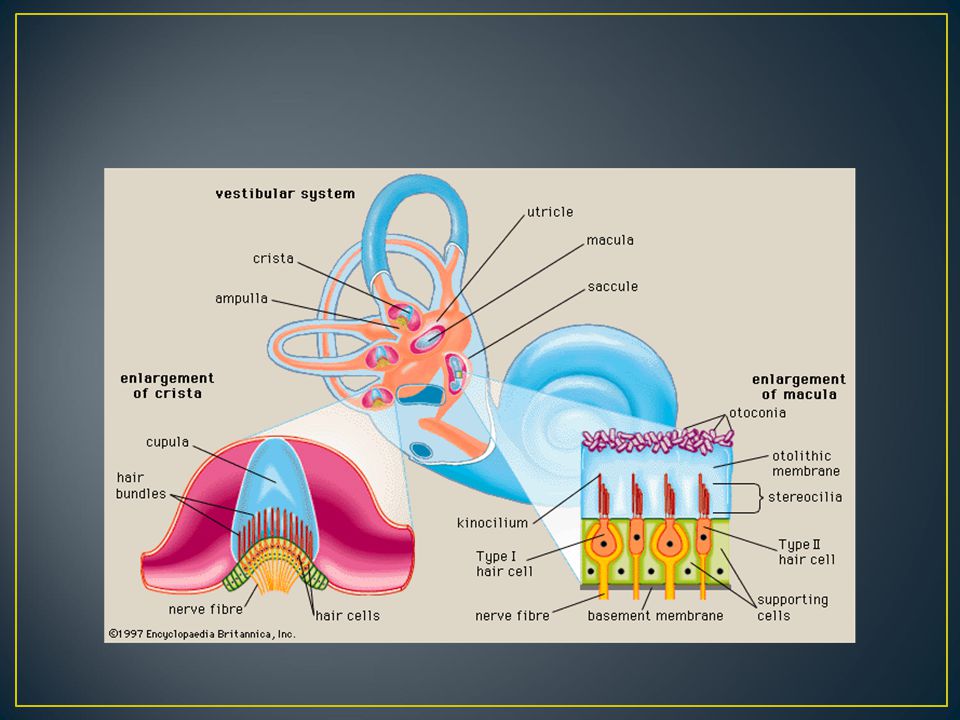
MEMBRANOUS LABYRINTH A fluid-filled sac that rests within the cavity of the osseous labyrinth- filled with endolymph fluid Vestibular system- Houses the vestibular organ Membranous labyrinth is in each ampulla, saccule and utricle Parallels the bony labyrinth

54
Membranous Vestibular System
Each ampulla houses a crista ampularis which is the sense organ for movement Houses the vestibular organ Ampulla- expanded region of the semicircular canals Utricle and Saccule- houses the otolithic organs of the vestibular system Communicate via the endolymphatic duct

56
Membranous Cochlear Duct
Cochlear duct forms a small portion of the membranous labyrinth Fluid in cochlear duct is endolymph Cochlear Duct- membranous labyrinth within the Cochlea Called the Scala Media-Resides between the scala vestibuli and scala tympani Houses the sensory apparatus for hearing

57
Membranes of the Organ of Corti
Reissner’s Membrane Divides the Scala vestibuli from the Scala Media Ceiling of the Organ of Corti Basilar Membrane Divides the Scala Media from the Scala tmpani Organ of Corti sits on it Floor of the Organ of Corti Tectorial Membrane Semitransparent and gelatinous-like Overlays hair cells- outer hair cells are embedded

59
MEMBRANOUS LABYRINTH Cochlear Duct
Organ of Corti has four rows of hair cells resting on a bed of Deiters’ cells for support Tunnel of Corti- separates 3 rows of outer hair cells from the single row of inner hair cells

60
Hair Cells Outer Hair Cells Inner Hair Cells
Three rows- approximately 12,000 Broadens to four rows at the apical end At the surface of each hair cell, forms a “W” or “V” pattern Shaped like a test tube Inner Hair Cells Modiolar side of the tunnel of Corti 3500 Hair cells Form a single row stretching from base to apex Upper surface of each hair cell forms a slight “U” pattern Shaped like a test tube or gourd

61
Hair Cell Innervation Each inner hair cell is connected to as many as 10 VIII vestibulocochlear nerve fibers referred to as “many-to-one” innervation Each outer hair cell shares its innervation with 10 other outer hair cells, all being innervated by the same VIII nerve fiber (one to many

62
Cochlea

63
Cochlea

Similar presentations


 Describe structurec and functions in the outer, middle and inner ear.>")


 ceruminous glands B) lacrimal glands C) meibomian glands D) apocrine.>")