Download presentation
Presentation is loading. Please wait.

1
Neural Tube, Somites and the Placenta
Lecture 3

2
Neurulation Neural tube formation
Notochord stimulates the ectoderm to form the neural tube Cells between the outer ectodermal layer and the neural tube will develop into the neural crest Will eventually form parts of the peripheral nervous system, pigment cells (melanocytes), endocrine system and musculoskeletal system
, endocrine system and musculoskeletal system.")
3
In amphibians

9
Neural Tube Defects anencephaly

10
Spina bifida

11
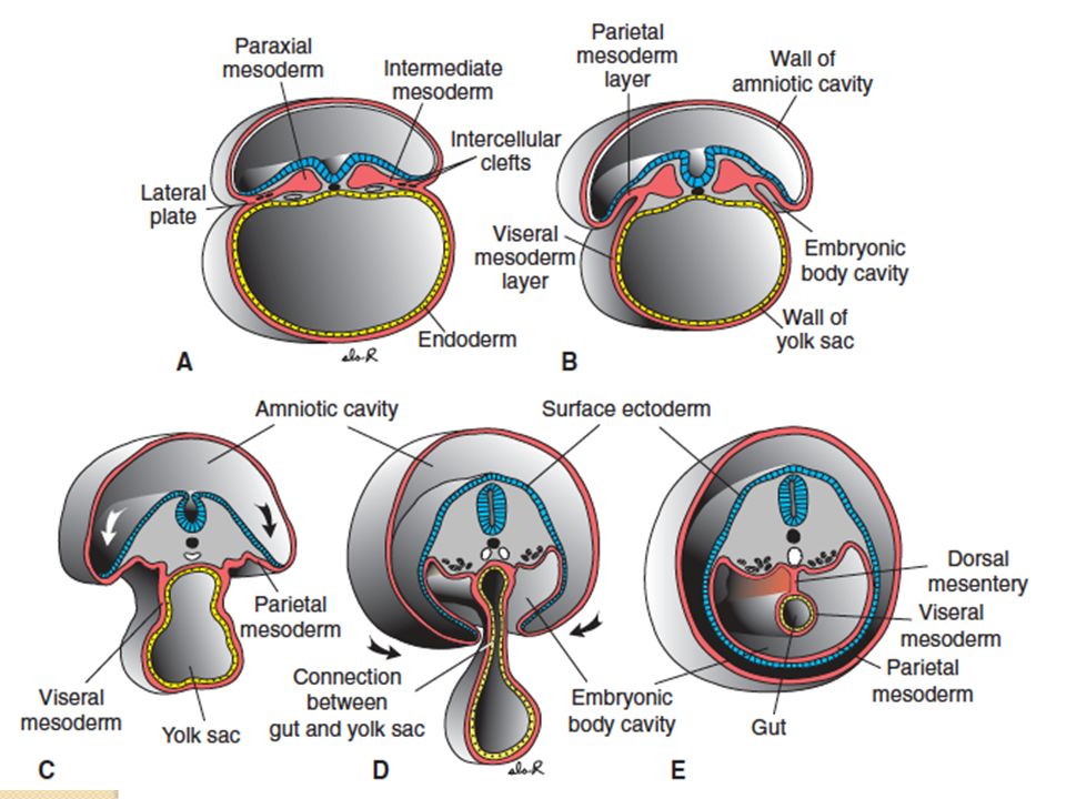
Somite differentiation
During neural tube formation, the mesoderm transforms into special structures called somites Landmark to identify the age of the embryo Develops into the specific organ systems

12
Somite differentiation
Paraxial mesoderm: just lateral to the neural tube = forms the somites Intermediate mesoderm: gives rise to the urogenital system Lateral mesoderm Somatic/Parietal mesoderm and somatopleure Splanchnic/Visceral mesoderm and splanchnopleure Coelom = between somatic and splanchnic mesoderm

13
Somite formation in chick embryo

14
Somite differentiation
Somites are committed to produce structures characteristic of their level along the body axis Somites near the neural tube will develop into sclerotomes eventually form parts of the vertebrae, ribs and sclapula Outer cells of the somite become the dermatomyotome becomes muscle (myotome cells) and dermis (dermatome cells)
 and dermis (dermatome cells)")
15
Somite differentiation
Myotome Medial myotome = axial muscles Lateral myotome = will migrate as muscles of the limbs May help determine age of embryo

16
Somite differentiation
Age of Chick embryo Number of somites 22 hours 1-2 24 hours 3-4 27 hours 8 30 hours 10 33 hours 12 38 hours 17 55 hours 29 72 hours 36

17
Somite differentiation
Age of human embryo (days) Number of somites 20 1-4 21 4-7 22 7-10 23 10-13 24 13-17 25 17-20 26 20-23 27 23-26 28 26-29 29 29-34 30 34-35
 Number of somites")
18
Somite differentiation
Lateral mesoderm separates to 2 layers, with a coelum between them Somatic/Parietal mesoderm Forms limb muscles together with the myotomes Forms lining of the peritoneal, pleural and pericardiac cavities Splanchnic/Visceral mesoderm and splanchnopleure Forms wall of the gut tube Forms outer covering of the internal organs

21
Somite formation in humans

23
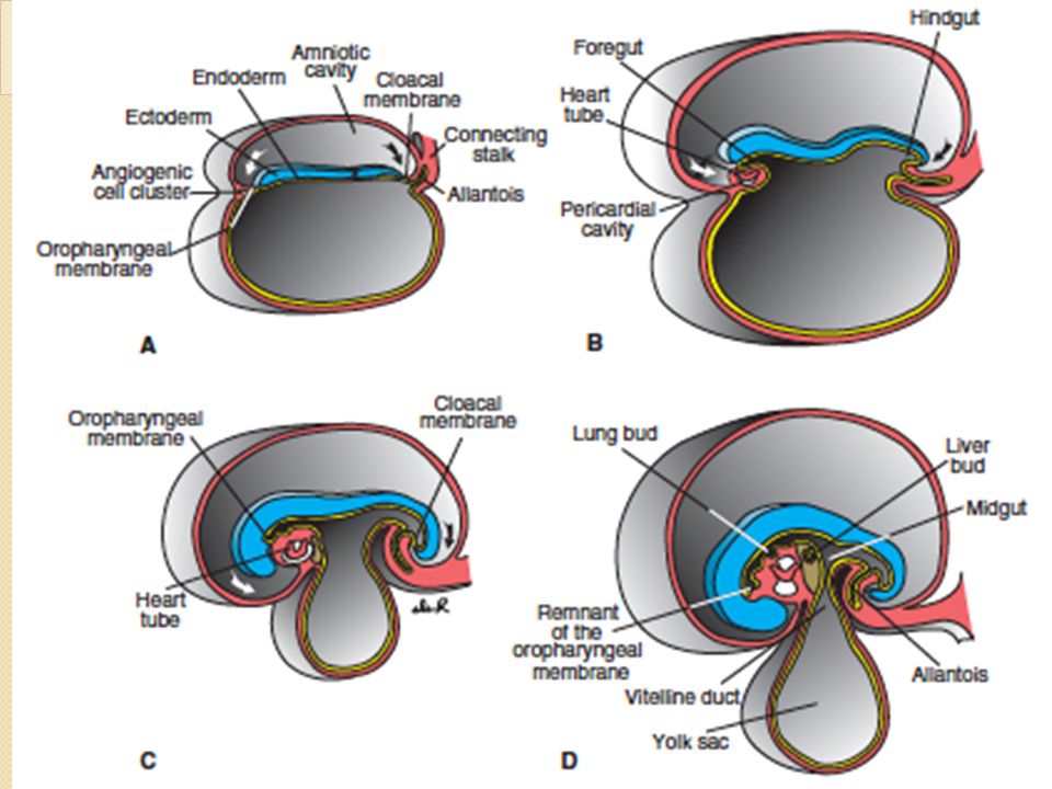
Extraembryonic Membranes
Amnion ectodermally derived Encloses entire embryo in a fluid filled sac Yolk sac Endodermally derived Encloses the yolk, which provides nutrients for the embryo Induces formation of blood and blood vessels in the yolk

24
Extraembryonic Membranes
Allantois Endodermally derived From the hindgut Acts as a reservoir for storing or removing wastes and to mediate gas exchange Chorion or serosa Outermost extraembryonic membrane Site for gas exchange for oviparous animals (egg laying) In mammals, also acts for nutrition, excretion, filtration and synthesis of hormones
 In mammals, also acts for nutrition, excretion, filtration and synthesis of hormones.")
25
Extraembryonic membranes in the Chick
Formation of the yolk sac Splanchnopleure extends into the outer boundary of the yolk, eventually enclosing it Beginning of primitive gut formation Foregut Midgut Hindgut Yolk duct and stalk Formation of the omphalomesenteric arteries and veins brings nutrients from yolk to the embryo Initial site of red blood cell production

26
Extraembryonic membranes in the Chick
Formation of the amnion Derived from the inner layer of the somatopleure Begins at the head fold, followed next at the tail fold and lateral folds Head fold, tail fold and lateral folds meet each other to completely enclose the embryo, as they join together at the amniotic raphe

27
Extraembryonic membranes in the Chick
Formation of the chorion Derived from the outer layer of the somatopleure Outermost covering Extends over the yolk sac Cavity between chorion and amnion = chorioamniotic cavity Functions to transport Calcium from the egg shell into the embryonic circulation

28
Extraembryonic membranes in the Chick
Formation of the allantois Arises from the endoderm of the hindgut Extends and eventually fuses with the chorion 2 Functions As the respiratory system of the chick embryo to become the chorioallantoic membrane transports oxygen from outside of the shell to the embryo and releases CO2 As a reservoir for metabolic wastes of the embryo (uric acid)
")
36
Extraembryonic membranes in humans
Trophoblast cells invade uterus Formation of the amnion A cavity forms within the epiblast layer of the inner cell mass Allantois Is replaced by the placenta Yolk sac (previously the blastocoele) Only for RBC production Nutrients are supplied by the placenta Chorion Forms placenta Fuses with amniotic cavity and uterus
 Only for RBC production. Nutrients are supplied by the placenta. Chorion. Forms placenta. Fuses with amniotic cavity and uterus.")
37
Formation of the placenta
Maternal blood enters the placenta through the intervilous spaces via endovascular invasion by cytotrophoblast cells

38
Functions of the placenta
Exchange of nutrition, metabolic and gaseous products Transmission of maternal antibodies Primary respiratory center of the fetus Production of hormones Progesterone = to develop uterus Estrogen = to develop uterus and to promote milk production by the mother Beta HCG = maintains the corpus luteum Somatomammotropin = gives fetus priority on blood glucose

39
Amniotic Fluid Initially produced by amniotic cells and fluid transported from maternal blood Serves as a protective cushion Fetus drinks amniotic fluid and urinates back the fluid

44
Cells from yolk sac endoderm transform into splanchnic mesoderm and form cavities within the
Cytotrophoblast + amnion mesoderm = extraembryonic somatic mesoderm somatopleure (ectoderm + mesoderm) Yolk sac endoderm + amnion mesoderm = extraembryonic splanchnic mesoderm splanchnopleure (endoderm + mesoderm)
 Yolk sac endoderm + amnion mesoderm = extraembryonic splanchnic mesoderm splanchnopleure (endoderm + mesoderm)")
45
Extraembryonic Somatic mesoderm Extraembryonic Splanchnic mesoderm
Cytotrophobast + syncitiotrophoblast = primary villa (precursor of chorionic villi)
")
47
Chorionic villi

51
Maternal-Fetal barrier
No direct contact between maternal and fetal blood Embryo is considered a foreign organism Prevent invasion by the maternal immune system and protection of embryo from toxic substances ingested by the mother Diffusion of substances from maternal blood to fetal blood in placenta and vice versa

56
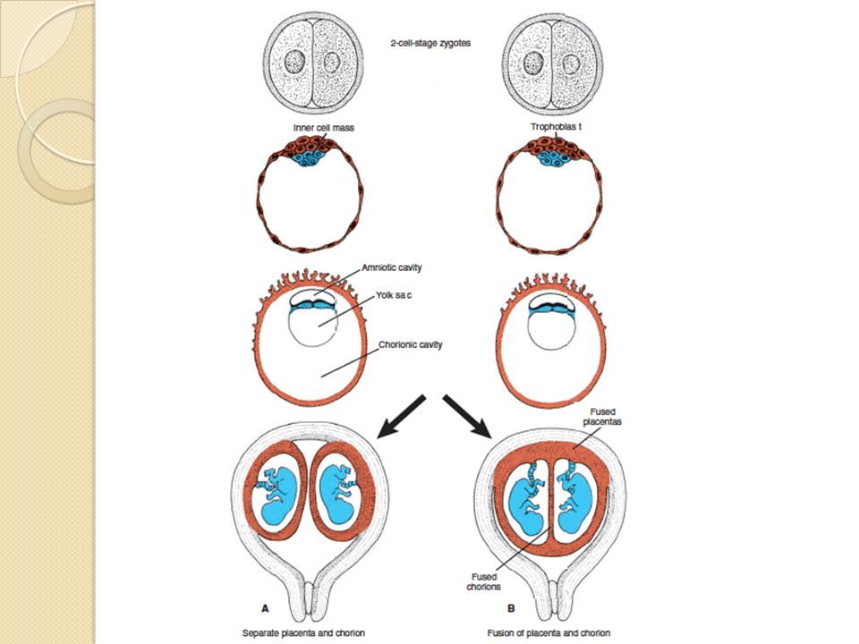
Placenta and twin pregnancy
Dichorionic diamniotic Monochorionic diamniotic Monochorionic monoamniotic

59
Twin-Twin Problems

Similar presentations


>")


>")

>")


