Download presentation
Presentation is loading. Please wait.

2
GROSS IMAGE OF A NORMAL AND A CIRRHOTIC LIVER
Cirrhosis Irregular surface Slide 6 GROSS IMAGE OF A NORMAL AND A CIRRHOTIC LIVER Gross images of two livers. On the left a normal liver with a smooth surface and homogeneous appearance. On the right a cirrhotic liver with an irregular surface and nodules that give it a heterogeneous appearance. Nodules

3
Causes of Liver Disease: Cirrhosis
Viral Hepatitis: B and C Genetic Liver Disease: Hemochromatosis, Wilson’s Disease Alcohol NASH (non alcoholic steatohepatitis) Autoimmune Liver Disease Cryptogenic
 Autoimmune Liver Disease. Cryptogenic.")
4
Synthetic Function:measured by Bilirubin, albumin and INR
Two aspects of Liver Disease Synthetic Function:measured by Bilirubin, albumin and INR Portal Hypertension

7
NORMAL VASCULAR ANATOMY OF THE LIVER
Hepatic vein Coronary vein Sinusoid Liver Slide 43 NORMAL VASCULAR ANATOMY OF THE LIVER The liver has a dual blood supply: portal vein and hepatic artery. The portal vein is formed by the union of the superior mesenteric vein (that collects blood from the bowel) with the splenic vein (that collects blood from the spleen). Normally, blood from the coronary vein drains into the portal system. After progressive ramifications, blood from the portal vein and hepatic artery join at the hepatic sinusoids, a specialized capillary system that is extensively interconnected. Hepatic sinusoidal blood drains into hepatic venules and then into collecting veins that unite to form the hepatic veins through which blood leaves the liver, draining into the vena cava and the right heart. Portal vein Splenic vein Hepatic artery Inferior vena cava Superior mesenteric vein Inferior mesenteric vein
 with the splenic vein (that collects blood from the spleen). Normally, blood from the coronary vein drains into the portal system. After progressive ramifications, blood from the portal vein and hepatic artery join at the hepatic sinusoids, a specialized capillary system that is extensively interconnected. Hepatic sinusoidal blood drains into hepatic venules and then into collecting veins that unite to form the hepatic veins through which blood leaves the liver, draining into the vena cava and the right heart. Portal vein. Splenic vein. Hepatic artery. Inferior. vena cava. Superior. mesenteric vein. Inferior. mesenteric vein.")
8
Portal systemic collaterals Cirrhotic Liver
ARCHITECTURAL LIVER DISRUPTION IS THE MAIN MECHANISM THAT LEADS TO AN INCREASED INTRAHEPATIC RESISTANCE Portal systemic collaterals Cirrhotic Liver Distorted sinusoidal architecture leads to increased resistance Slide 48 ARCHITECTURAL LIVER DISRUPTION IS THE MAIN MECHANISM THAT LEADS TO AN INCREASED INTRAHEPATIC RESISTANCE The deposition of fibrous tissue and the formation of nodules, disrupts the architecture of the liver, leading to an increased resistance to flow and to portal hypertension. Vessels that normally drain into the portal system, such as the coronary vein, reverse their flow and become porto-systemic collaterals. Additionally, with portal hypertension, the spleen increases in size and sequesters platelets and other formed blood cells leading to hypersplenism. Portal vein Splenomegaly

9
Complications of Cirrhosis:
Esophageal and gastric Varices Ascites Hepatic Encephalopathy Hepatorenal Syndrome

10
DIAGNOSIS OF CIRRHOSIS – CAT SCAN
CAT Scan in Cirrhosis Slide 23 DIAGNOSIS OF CIRRHOSIS – CAT SCAN This slide shows typical computed tomography findings in compensated cirrhosis. The contour of the liver is irregular, there is obvious splenomegaly and the presence of collaterals indicates portal hypertension and secures the diagnosis of cirrhosis. Liver with an irregular surface Collaterals Splenomegaly

11
VARICES INCREASE IN DIAMETER PROGRESSIVELY
Slide 82 VARICES INCREASE IN DIAMETER PROGRESSIVELY Both development of varices and growth of small varices occurs at a rate of 7-8% per year. Although there are no identified clinical predictors for the development of varices, factors associated with variceal growth are Child B/C cirrhosis, alcoholic etiology and presence of red wale marks on initial endoscopy. Merli et al., J Hepatol 2003; 38: 266 No varices Small varices Large varices 7-8%/year 7-8%/year Merli et al. J Hepatol 2003;38:266

12
PROGNOSTIC INDICATORS OF FIRST VARICEAL HEMORRHAGE
Slide 97 PROGNOSTIC INDICATORS OF FIRST VARICEAL HEMORRHAGE In a prospective study, the presence of the following clinical features was associated with a high probability of developing variceal hemorrhage: large variceal size, Child B/C and the presence of red wale markings on varices. North Italian Endoscopic Club. N Engl J Med 1988; 319: 983 Variceal hemorrhage Varix with red signs Predictors of hemorrhage: Variceal size Red signs Child B/C NIEC. N Engl J Med 1988; 319:983

13
Acute Variceal Bleeding Initial Management
Assessment of severity/resuscitation airway protection for massive bleed or if agitated Blood and fluid replacement Pharmacology therapy Octreotide Endoscopy for diagnosis and therapy Variceal ligation Sclerotherapy Balloon tamponade

14
Acute Variceal Bleeding Endoscopic Ligation
Bleeding controlled in 90% Rebleeding rate reduced to 30% Band to “obliteration”

15
Acute Variceal Bleeding Sclerotherapy
Bleeding controlled in 80-95% Rarely used now- massive bleeds?

16
ENDOSCOPIC IMAGES OF GASTRIC VARICES
Slide 189 ENDOSCOPIC IMAGES OF GASTRIC VARICES The arrow on the panel on the left shows an actively bleeding gastric varix. The panel on the right shows the varix after treatment with cyanoacrylate. Pretreatment cyanoacrylate Post-treatment cyanoacrylate

17
Treatment of Varices / Variceal Hemorrhage
MANAGEMENT OF PATIENTS WITH MEDIUM/LARGE VARICES WITHOUT PRIOR HEMORRHAGE Treatment of Varices / Variceal Hemorrhage No varices Small varices No hemorrhage 1) -blockers (propranolol, nadolol) indefinitely 2) Endoscopic variceal ligation in patients intolerant to -blockers Medium/ large varices No hemorrhage Slide 121 MANAGEMENT OF PATIENTS WITH MEDIUM/LARGE VARICES WITHOUT PRIOR HEMORRHAGE In patients with medium/large varices who have never bled, non-selective beta-blockers (propranolol, nadolol) should be considered first line therapy in the prevention of first variceal hemorrhage. Endoscopic band ligation can be considered in patients intolerant to beta-blockers or who have contraindications to beta-blockers. Variceal hemorrhage Recurrent hemorrhage
 -blockers (propranolol, nadolol) indefinitely. 2) Endoscopic variceal ligation in patients intolerant to -blockers. Medium/ large varices. No hemorrhage. Slide 121. MANAGEMENT OF PATIENTS WITH MEDIUM/LARGE VARICES WITHOUT PRIOR HEMORRHAGE. In patients with medium/large varices who have never bled, non-selective beta-blockers (propranolol, nadolol) should be considered first line therapy in the prevention of first variceal hemorrhage. Endoscopic band ligation can be considered in patients intolerant to beta-blockers or who have contraindications to beta-blockers. Variceal. hemorrhage. Recurrent. hemorrhage.")
18
Variceal Bleeding Bleeding Ectopic Varices

19
Tips Transjugular Intrahepatic Portosystemic Shunt

20
ENDOSCOPIC IMAGES OF MILD AND SEVERE PORTAL HYPERTENSIVE GASTROPATHY
Slide 192 ENDOSCOPIC IMAGES OF MILD AND SEVERE PORTAL HYPERTENSIVE GASTROPATHY Mild portal hypertensive gastropathy (left panel) is characterized by a mosaic pattern. Severe gastropathy consists of a mosaic pattern with superimposed red signs. Mosaic pattern Mosaic pattern + red spots Carpinelli et al. Ital J Gastroenterol Hepatol 1997; 29:533
 is characterized by a mosaic pattern. Severe gastropathy consists of a mosaic pattern with superimposed red signs. Mosaic pattern. Mosaic pattern + red spots. Carpinelli et al. Ital J Gastroenterol Hepatol 1997; 29:533.")
23
Ascites Malignancy Chronic Liver Disease Heart Failure Turberculosis
Miscellaneous Nephrogenic Pancreatic Fulminant Hepatic Failure Biliary Leak

24
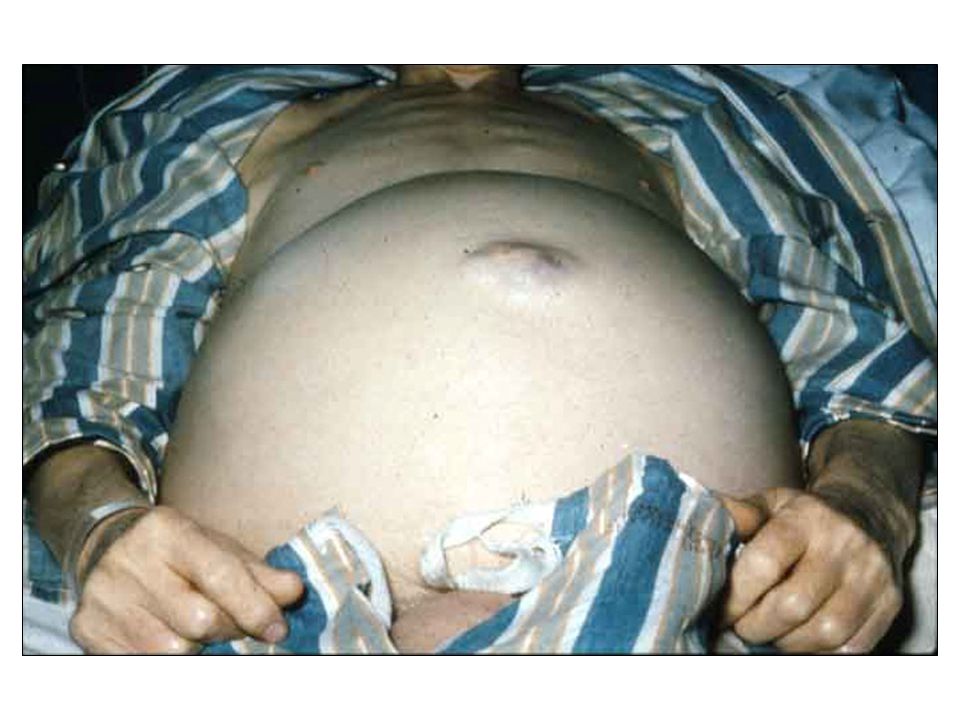
Ascites

26
Ascites Pathogenesis: Hypotheses
Underfill Overflow Vasodilation Cirrhosis Portal Hypertension Primary renal sodium retention (? Stimulus) Peripheral arterial vasodilation Ascites Effective Intravascular volume Effective Intravascular volume Blood volume Renal sodium retention Renal sodium retention Ascites
 Peripheral arterial vasodilation. Ascites. Effective Intravascular volume. Effective Intravascular volume. Blood volume. Renal sodium retention. Renal sodium retention. Ascites.")
28
Ascites Diagnosis Paracentesis
Indications New-onset ascites Admission to hospital Clinical deterioration Fever Contraindications None

29
Ascites Fluid Analysis
Routine Cell count Culture Albumin Protein Optional Glucose LDH Amylase Gram stain TB smear and culture Cytology Triglyceride

30
Ascites Serum-Ascites Albumin Gradient
[Albumin] – [Albumin] Serum Ascites High (> 11 g/L) Cirrhosis; alcoholic hepatitis Cardiac disease Massive liver metastases Fulminant hepatic failure Hepatic outflow block Portal vein thrombosis Low (< 11 g/L) Peritoneal carcinomatosis Tuberculous peritonitis Pancreatic duct leak Biliary leak Nephrotic syndrome Serositis
 Cirrhosis; alcoholic hepatitis. Cardiac disease. Massive liver metastases. Fulminant hepatic failure. Hepatic outflow block. Portal vein thrombosis. Low (< 11 g/L) Peritoneal carcinomatosis. Tuberculous peritonitis. Pancreatic duct leak. Biliary leak. Nephrotic syndrome. Serositis.")
31
Ascites

32
Ascites

33
Ascites Initial Therapy
Sodium restriction Diuretics Spironolactone +/- furosemide Stepwise increase as needed to maximal doses Large volume paracentesis (for tense ascites)
")
34
Ascites Therapy of Refractory Ascites

35
Ascites Complications

36
Spontaneous Bacterial Peritonitis (SBP)
")
37
Spontaneous Bacterial Peritonitis:SBP
Usually caused by gram-negative bacteria and usually only one organism: only 50% of cultures positive If neutrophil count high in ascites: ie > 250/cc-treat with IV Cefotaxime, even if culture negative Prophylaxis for SBP or recurrent SBP controversial: high resistance rates develop

38
SURVIVAL AFTER DEVELOPMENT OF SPONTANEOUS BACTERIAL PERITONITIS (SBP)
1.0 .8 .4 .2 .6 3 6 12 24 36 Probability of survival (%) Slide 297 SURVIVAL AFTER DEVELOPMENT OF SPONTANEOUS BACTERIAL PERITONITIS (SBP) The development of SBP in a cirrhotic patient with ascites carries a poor prognosis. In a cohort of 75 patients with SBP the one-year survival was 38%, with a median survival of only 9 months. Tito L, et al., Hepatology 1988; 8(1): 27-31 Months Tito et al., Hepatology 1988; 8:27
 Slide 297. SURVIVAL AFTER DEVELOPMENT OF SPONTANEOUS BACTERIAL PERITONITIS (SBP) The development of SBP in a cirrhotic patient with ascites carries a poor prognosis. In a cohort of 75 patients with SBP the one-year survival was 38%, with a median survival of only 9 months. Tito L, et al., Hepatology 1988; 8(1): Months. Tito et al., Hepatology 1988; 8:27.")
39
Hepatic Encephalopathy
Reversible neuropsychiatric abnormalities Asterixis and abnormal EEG Hepatic failure and/or portosystemic shunting

41
Hepatic Encephalopathy

43
BLOOD AMMONIA LEVELS ONLY LEAD TO CONFUSION
“Blood ammonia levels cause as much confusion in those requesting the measurement as in the patients in whom they are being measured” Slide 356 BLOOD AMMONIA LEVELS ONLY LEAD TO CONFUSION Adrian Reuben Hepatology 2002;35:983

44
Hepatic Encephalophathy

45
Hepatic Encephalophathy Precipitants

46
Hepatic Encephalophathy Actions of Lactulose

47
Hepatorenal Syndrome Identify other causes
Establish circulatory volume Avoid nephrotoxic agents Consider hemodialysis Evaluate for liver transplatation

48
Hepatorenal Syndrome Evidence for a Functional Disorder

53
Hematologic Manifestations of Cirrhosis
Mild Anemia and Macrocytosis Neutropenia secondary to splenic sequestration Thrombocytopenia secondary to splenic sequestration Prolonged INR secondary to decreased production of clotting factors by liver

54
Hepatocellular Carcinoma
Other names are Hepatoma, Primary Liver Cell Cancer Develop in Cirrhotic livers especially Hepatitis B and C May produce a protein tumour marker: alphafetoprotein Ultrasound screening every 6 months for early detection

58
Complications of Cirrhosis

59
Child-Pugh Criteria 1 point 2 points 3 points Bilirubin (mg/dL)
Albumin (g/L) PT (sec prolonged) Ascites Encephalopathy <2 >35 1-3 none 2-3 28-35 4-6 Slight 1-2 >3 <28 >6 Moderate 3-4 Grades: A = 5-6 points; B = 7-9 points; C = 10-15
 PT (sec prolonged) Ascites. Encephalopathy. <2. > none Slight >3. <28. >6. Moderate Grades: A = 5-6 points; B = 7-9 points; C =")
60
Causes of death in Cirrhosis
Infection Variceal bleeding Hepatic encephalopathy Hepatocellular carcinoma

61
# Hospitalized cirrhotic patients
SPONTANEOUS BACTERIAL PERITONITIS (SBP) IS THE MOST COMMON INFECTION IN CIRRHOTIC PATIENTS Spontaneous Bacterial Peritonitis (SBP) is the Most Common Infection in Cirrhotic Patients 150 125 100 # Hospitalized cirrhotic patients 75 50 Slide 264 SPONTANEOUS BACTERIAL PERITONITIS (SBP) IS THE MOST COMMON INFECTION IN CIRRHOTIC PATIENTS In a study of 1,567 admissions for decompensated cirrhosis, bacterial infections developed at admission or during hospitalization in 507 (32%) patients (572 infectious episodes). SBP was the most common type of bacterial infection, present in 138 (24%) of the cases, followed by urinary tract infections (UTI) and pneumonia. Fernández J, et al., Hepatology 2002; 35: 140 25 SBP UTI Pneumonia Procedure- related Spontaneous Bacteremia Fernández et al., Hepatology 2002; 35:140
 IS THE MOST COMMON INFECTION IN CIRRHOTIC PATIENTS. Spontaneous Bacterial Peritonitis (SBP) is the Most Common Infection in Cirrhotic Patients # Hospitalized cirrhotic patients Slide 264. SPONTANEOUS BACTERIAL PERITONITIS (SBP) IS THE MOST COMMON INFECTION IN CIRRHOTIC PATIENTS. In a study of 1,567 admissions for decompensated cirrhosis, bacterial infections developed at admission or during hospitalization in 507 (32%) patients (572 infectious episodes). SBP was the most common type of bacterial infection, present in 138 (24%) of the cases, followed by urinary tract infections (UTI) and pneumonia. Fernández J, et al., Hepatology 2002; 35: SBP. UTI. Pneumonia. Procedure- related. Spontaneous. Bacteremia. Fernández et al., Hepatology 2002; 35:140.")
62
SURVIVAL TIMES IN CIRRHOSIS
Decompensation Shortens Survival 100 80 Median survival ~ 9 years All patients with cirrhosis 60 Probability of survival 40 Slide 16 SURVIVAL TIMES IN CIRRHOSIS In a prospective cohort study of 257 patients with compensated cirrhosis of different etiologies, median survival for all patients (including those who developed decompensation) was approximately 9 years, while it was significantly lower in patients who developed a decompensating event (ascites, jaundice, encephalopathy or hemorrhage), in whom the median survival was only 1.6 years. Gines et al. Hepatology 1987; 7:122 20 Decompensated cirrhosis Median survival ~ 1.6 years 20 40 60 80 100 120 140 160 180 Months Gines et. al., Hepatology 1987;7:122
 was approximately 9 years, while it was significantly lower in patients who developed a decompensating event (ascites, jaundice, encephalopathy or hemorrhage), in whom the median survival was only 1.6 years. Gines et al. Hepatology 1987; 7: Decompensated cirrhosis. Median survival. ~ 1.6 years Months. Gines et. al., Hepatology 1987;7:122.")
63
Liver Transplantation
Only proven treatment for advanced cirrhosis Long waiting list: insufficient donors Allocation by MELD score (Bilirubin, creatinine, INR) Patients with recent alcohol or drug abuse, extrahepatic malignancies or other major co-morbidities excluded
 Patients with recent alcohol or drug abuse, extrahepatic malignancies or other major co-morbidities excluded.")
64
Treatment of Fulmimant Liver Failure
Supportive measures Transfer to transplant centre if encephalopathic ?Mars Improve with time or need liver transplant 1st on Waiting list for Transplant

65
GROSS IMAGE OF A NORMAL AND A CIRRHOTIC LIVER
Cirrhosis Irregular surface Slide 6 GROSS IMAGE OF A NORMAL AND A CIRRHOTIC LIVER Gross images of two livers. On the left a normal liver with a smooth surface and homogeneous appearance. On the right a cirrhotic liver with an irregular surface and nodules that give it a heterogeneous appearance. Nodules

66
Causes of Liver Disease: Cirrhosis
Viral Hepatitis: B and C Genetic Liver Disease: Hemochromatosis, Wilson’s Disease Alcohol NASH (non alcoholic steatohepatitis) Autoimmune Liver Disease Cryptogenic
 Autoimmune Liver Disease. Cryptogenic.")
Similar presentations


 that progresses to cirrhosis Replacement of liver tissue.>")
 Cause ( Etiology) Complication Complication.>")


