Download presentation
Presentation is loading. Please wait.

1
Chapter 3 – Cell Structure Cells: Microscopic, Characteristics and Size

2
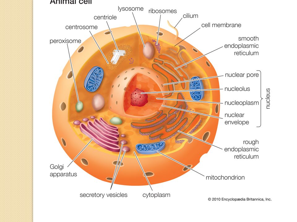
Cell Membrane Consistency of a bubble not a hard shell Protection is like a gated community – only certain objects can pass through Phospholipid – a phosphate group and two fatty acids Lipid Bilayer – nonpolar tails make up the interior and polar heads appear on interior and exterior

3
Nucleus The “brain” of the cell – controlling all the activities Surrounded by a double layer – the nuclear envelope to separate it from cytoplasm Stores DNA (genetic information) Nucleolus – partially assembly of ribosomes location Nuclear Pores – locations where ribosomes are transferred to the nucleolus
 Nucleolus – partially assembly of ribosomes location Nuclear Pores – locations where ribosomes are transferred to the nucleolus")
4
DNA & Cell Specialization DNA hold instructions for every expression a cell gives off Each cell has specialized instruction for their “duty” Instructions can code for a specific cell Example ◦ A cell can be a skin or muscle cell just by reading the information coded in the DNA ◦ A skin cell cannot just become a muscle cell and then a skin cell again

5
Production of Proteins Endoplasmic Reticulum – a system of internal membranes that move proteins and other substances through the cell Rough ER – location where ribosomes are attached and transports the proteins made Vesicle – a small sac that pinches of the ER to transport substances like proteins Smooth ER – no ribosomes – creates lipids and breaks down toxic substances

7
Section 1 - Looking at Cells Cells Under the Microscope Human body cells smaller than a grain of sand Hooke & Cork Cells ◦ Thin slice of cork has tiny boxes that were named cells Anton van Leeuwenhoek ◦ Discovered single-celled organisms based off Hooke's observations

8
Measuring Cell Structures Metric or International Systems (SI) Based on powers of 10 = micro (1/millionth of a meter) Table 1
 Based on powers of 10 = micro (1/millionth of a meter) Table 1")
9
Characteristics of Microscopes Light Microscopes ◦ light passes through one or more lenses to enlarge an image Electron Microscope ◦ beam of electrons forms an image Magnification ◦ quality of enlarging an image ◦ example: 200x = 200 times larger

10
Characteristics Continued. Resolution ◦ measure of clarity of the image Electron microscope has better magnification and resolution

11
Section 2 - Cell Features The Cell Theory - Observed by Schleiden, Schwann & Virchow All living things are made of one or more cells Cells are the basic unit of structure and function in organisms All cells arise from existing cells

12
Cell Size Small cells: ◦ more efficient than large cells ◦ able to diffuse through membranes ◦ have higher surface area to volume ratio Table 2

13
Common Features of Cells Cell Membrane: controls which particles enter and leave the cell Cytoplasm: jelly-like substance that holds all the internal materials in place Ribosome: a cellular structure that produces proteins DNA: genetic code that provides instructions for all cellular processes

14
Prokaryotes smallest and simplest cells lacks a nucleus and other structures example: bacteria

15
Characteristics of Prokaryotes Exist within a large range of environments ◦ some need oxygen and other don’t, some make their own food Cytoplasm split into compartments since there are no internal structures DNA is circular and located near the center of the cell (where the nucleus would be)
")
16
Characteristics continued Cell wall: gives structure and support outside of the cell membrane ◦ only in plants and fungi ◦ made of polysaccharide strands connected by amino acids (building blocks of proteins) Flagella: long, thread-like structure used for movement
 Flagella: long, thread-like structure used for movement")
17
Eukaryotes Eukaryote: ◦ an organism whose cells have a nucleus Nucleus: ◦ internal compartment containing DNA Cilia: ◦ small hair-like structures used for movement

18
Cytoskeleton Interior framework of an animal cell that contains protein fibers Supports the shape of the cell Three types of fibers ◦ Actin fibers ◦ Microtubules ◦ Intermediate fibers

19
Types of Fibers Actin fibers Microtubules Intermediate fibers

20
Actin Fibers Just beneath the cell surface Contraction and expansion plays a major role in providing the cell’s shape

21
Microtubules Acts as a “highway” system Transports information from the nucleus to different parts of the cell Motor proteins move RNA proteins along these microtubules to transport information required for cell activities

22
Intermediate Fibers Frame of the cytoskeleton Ribosomes and enzymes reside on these fibers in certain location around the cell to provide efficient metabolic activities

23
Packing of Proteins Golgi apparatus – flattened, membrane sacs that packaging and distributing center ◦ Modifies and adjusts proteins from the ER Lysosomes – spherical organelles that contain digestive enzymes

24
Processing of Proteins Rough ER ribosomes produce proteins Vesicles transport proteins from rough ER to Golgi Golgi processes proteins and packages them into new vesicles Vesicles leave to move outside of the cell membrane Vesicles can also remain within the cytoplasm like lysosomes that break down proteins, nucleic acids, lipids & carbs

25
Mitochondria An organelle that obtains energy from organic compounds to produce ATP The amount of mitochondrion that reside in the cell depends on requirement amount of energy that cell has ◦ ex. muscle cell – high or low? Have their own DNA and ribosomes ◦ Most of their protein is made by free ribosomes

26
Unique Structures of Plant Cells Cell Wall – a wall that is made up of proteins, carbohydrates and polysaccharide cellulose that protects the cell from damage Chloroplasts – uses light energy to produce carbohydrates from water and carbon dioxide Central Vacuole – large membrane- bound sac that stores water, ions, nutrients and wastes

Similar presentations


>")


