Download presentation
Presentation is loading. Please wait.

1
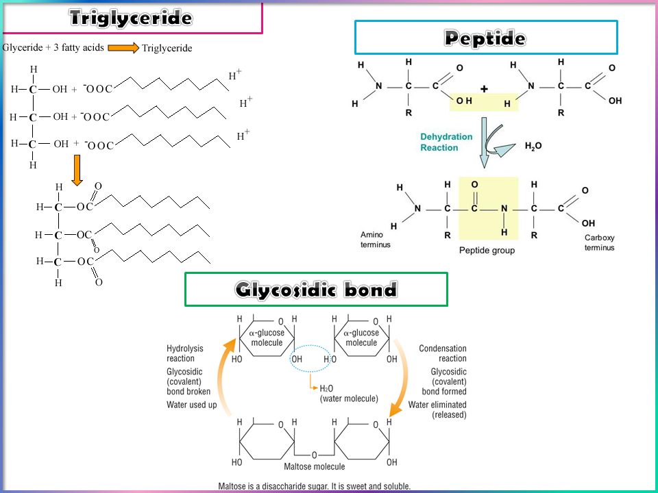
1.Biochemistry 1.1.1What are we made of? 1.List the main elements found in living organisms. 2.State that some elements are needed in trace amounts (details not required). 3. List the key elements which are present as inorganic ions: Mg 2+, Fe 2+, Ca 2+, PO 4 3– 4.The importance of water in terms of its polarity, ability to form hydrogen bonds, surface tension, as a solvent, thermal properties, as a metabolite. 1.1.2 Carbohydrates 1.State the structural difference between α (alpha) and β (beta) glucose. 2.Describe the formation and breakage of glycosidic bonds in the synthesis and hydrolysis of a disaccharide. 3.Describe the molecular structure of alpha-glucose as an example of a monosaccharide carbohydrate. 4.Explain how the structure of glucose relates to its function in living organisms. 1.1.3 Useful carbohydrates 1.Describe the formation and breakage of glycosidic bonds in the synthesis and hydrolysis of a polysaccharide. 2.Compare and contrast the structure and functions of starch (amylose) and cellulose. 3.Describe the structure of glycogen. 4.Explain how the structures of starch and glycogen relate to their functions in living organisms. 5.Compare and contrast the structure and functions of amylose and cellulose. 6.Explain how the structure of cellulose relates to its function in living organisms. 1.1.4 Amino acids and proteins 1.Describe the structure of an amino acid. 2.Describe the formation and breakage of peptide bonds in the synthesis and hydrolysis of dipeptides and polypeptides. 3.Explain the term primary structure 4.Explain the term secondary structure with reference to hydrogen bonding. 5.Explain the term tertiary structure with reference to hydrophobic and hydrophilic interactions, disulfide bonds and ionic interactions. 1.1.5 Proteins in action 1.Explain the term quaternary structure with reference to the structure of haemoglobin. 2.Describe the structure of a collagen molecule. 3.Compare the structure and function of haemoglobin and collagen. 1.1.6 Lipids 1.State that lipids (fats and oils) are a range of biological molecules including triglycerides. 2.Compare the structure of triglycerides and phospholipids. 3.Explain how the structure of triglyceride and phospholipid and cholesterol molecules relates to their functions in living organisms. 1.1.7 Review Biochemistry review Focus on: Numeracy and kinetics (continue in enzymes)
. 3. List the key elements which are present as inorganic ions: Mg 2+, Fe 2+, Ca 2+, PO 4 3– 4.The importance of water in terms of its polarity, ability to form hydrogen bonds, surface tension, as a solvent, thermal properties, as a metabolite Carbohydrates 1.State the structural difference between α (alpha) and β (beta) glucose. 2.Describe the formation and breakage of glycosidic bonds in the synthesis and hydrolysis of a disaccharide. 3.Describe the molecular structure of alpha-glucose as an example of a monosaccharide carbohydrate. 4.Explain how the structure of glucose relates to its function in living organisms Useful carbohydrates 1.Describe the formation and breakage of glycosidic bonds in the synthesis and hydrolysis of a polysaccharide. 2.Compare and contrast the structure and functions of starch (amylose) and cellulose. 3.Describe the structure of glycogen. 4.Explain how the structures of starch and glycogen relate to their functions in living organisms. 5.Compare and contrast the structure and functions of amylose and cellulose. 6.Explain how the structure of cellulose relates to its function in living organisms Amino acids and proteins 1.Describe the structure of an amino acid. 2.Describe the formation and breakage of peptide bonds in the synthesis and hydrolysis of dipeptides and polypeptides. 3.Explain the term primary structure 4.Explain the term secondary structure with reference to hydrogen bonding. 5.Explain the term tertiary structure with reference to hydrophobic and hydrophilic interactions, disulfide bonds and ionic interactions Proteins in action 1.Explain the term quaternary structure with reference to the structure of haemoglobin. 2.Describe the structure of a collagen molecule. 3.Compare the structure and function of haemoglobin and collagen Lipids 1.State that lipids (fats and oils) are a range of biological molecules including triglycerides. 2.Compare the structure of triglycerides and phospholipids. 3.Explain how the structure of triglyceride and phospholipid and cholesterol molecules relates to their functions in living organisms Review Biochemistry review Focus on: Numeracy and kinetics (continue in enzymes).")
2
Biomolecule Name of testReagentsInstructions Colour change for positive result Colour change for negative result Protein Reducing sugars Non- reducing sugars Starch Lipid

4
The test for Protein The biuret test is a chemical test used for detecting the presence of peptide bonds. In the presence of peptides, a copper(II) ion forms a violet-coloured complex in an alkaline solution 1.Using a syringe, add 2 cm 3 of protein suspension to a test tube. 2.Using a clean syringe, add 2 cm 3 of sodium hydroxide and shake to mix. 3.Add a small amount of copper sulfate drop by drop, shaking between each addition. 4.Check for any colour change. Do not get confused with the blue colour of the copper sulfate. 5.Repeat the test using a clean test tube and syringes and substituting distilled water for the protein suspension. 6.Check for any colour change.
 ion forms a violet-coloured complex in an alkaline solution 1.Using a syringe, add 2 cm 3 of protein suspension to a test tube. 2.Using a clean syringe, add 2 cm 3 of sodium hydroxide and shake to mix. 3.Add a small amount of copper sulfate drop by drop, shaking between each addition. 4.Check for any colour change. Do not get confused with the blue colour of the copper sulfate. 5.Repeat the test using a clean test tube and syringes and substituting distilled water for the protein suspension. 6.Check for any colour change..")
5
The test for Lipids Lipids do not dissolve in water, but do dissolve in ethanol. This characteristic is used in the emulsion test. Do not start by dissolving the sample in water, but instead shake some of the test sample with about 4 cm³ of ethanol. Decant the liquid into a test tube of water, leaving any undissolved substances behind. If there are lipids dissolved in the ethanol, they forming a will precipitate in the water, forming a cloudy white emulsion cloudy white emulsion.

6
The test for Starch The Iodine test is used to test for the presence of starch. Iodine solution — iodine dissolved in an aqueous solution of potassium iodide — reacts with the starch (starch–iodine complex forming as the iodine fits into the starch helix) A positive result will produce a purple-black colour.
 A positive result will produce a purple-black colour..")
7
Carbohydrates can be classed as either : 1. Reducing sugars e.g. all Monosaccharides and some disaccharides. 2. Non reducing sugars e.g. Sucrose and Polysaccharides. Reducing sugars contain a chemical group which can donate electrons to reduce another chemical. Reducing form of glucose Aldehyde group donates electrons e-e- Cu 2+ Cu 1+

8
The test for Reducing sugars Add a few drops of Benedicts solution to the sample. Heat the mixture in a water bath for 10 + minutes. Observation The solution will gradually change from : Blue Green Brown Orange Red Explanation Benedict's solution contains CuSO 4 – this contains Blue Cu 2+ ions Electrons donated by the Aldehyde group from a reducing sugar slowly reduce the blue Cu 2+ into red Cu 1+ Aldehyde group donates electrons (e-) to reduce the blue Cu 2+ in the solution to red Cu 1+ Some reducing sugars do not have an Aldehyde group, but have a Ketone group which donates electrons in a similar way.
 to reduce the blue Cu 2+ in the solution to red Cu 1+ Some reducing sugars do not have an Aldehyde group, but have a Ketone group which donates electrons in a similar way..")
10
The test for Non reducing sugars The Benedicts test gives a negative result with Sucrose and Polysaccharides. To detect non reducing sugars the following test should be done. 1. Heat the sugar with dilute hydrochloric acid. This hydrolyses sugar into smaller units which have Aldehyde groups. 2.The large non reducing is broken into smaller reducing sugars. 3. Neutralise the acid with Sodium Hydroxide 4. Carry out the Benedicts test as normal. Result: Blue Cu 2+ Red Cu 1+

11
Non

12
Answers 1.Most disaccharides are reducing sugars but sucrose is not. This is because of the way that fructose and glucose are joined, within the molecule, making it impossible to reduce the copper(II) ion in Benedict’s reagent. 2.Sodium hydrogencarbonate is added to the tube before testing with Benedict’s solution to neutralise the acid hydrochloric acid. Benedict’s test will not work in an acid environment. 3.The test on sucrose without any pre-treatment (the control) demonstrates that sucrose will not reduce Benedict’s solution unless it is first hydrolysed into its monosaccharide components.
 ion in Benedict’s reagent. 2.Sodium hydrogencarbonate is added to the tube before testing with Benedict’s solution to neutralise the acid hydrochloric acid. Benedict’s test will not work in an acid environment. 3.The test on sucrose without any pre-treatment (the control) demonstrates that sucrose will not reduce Benedict’s solution unless it is first hydrolysed into its monosaccharide components..")
13
Is it a carbohydrate? Add Benedicts’ reagent Heat Add Benedicts’ reagent Heat Green or Red Colour observed Reducing sugar Monosaccharide or some disaccharides Blue Colour observed Add Benedicts’ reagent Heat Add Benedicts’ reagent Heat Boil in dilute acid Neutralise acid Boil in dilute acid Neutralise acid Green or Red Colour observed Non-Reducing sugar Polysaccharide or some disaccharides Blue Colour observed Not a carbohydrate, possibly a ferret

14
Colorimetry is method of determining the concentration of a substance by measuring the relative absorption of light (usually visible) with respect to a known concentration of the substance. The instrument by which these measurements are made is called a colorimeter Light from a suitable source is passed through a light filter to select the most appropriate wavelength of light, some of which is then absorbed by the solution held in a special glass cuvet (a sort of 'test tube'). The amount of light absorbed is called, and measured as, the absorbance which is a function of the coloured solute concentration. The amount of light that travels through the sample is called, and measured as, the transmission which is a function of the coloured solute concentration.
. The amount of light absorbed is called, and measured as, the absorbance which is a function of the coloured solute concentration. The amount of light that travels through the sample is called, and measured as, the transmission which is a function of the coloured solute concentration..")
16
The semi quantitative basis of the Benedicts test the concentration and relative amounts of reducing and non reducing sugars in a solution can be estimated by the Benedict's test. To look at concentration the quicker the colour change from Blue Cu 2+ to Red Cu 1+ the stronger the sugar concentration – there will be more Aldehyde groups donating electrons to reduce the Cu 2+. To look at relative amounts of reducing/non reducing sugars if you carry out the reducing sugar test and get a slow colour change and then carry out the non reducing test and get a rapid colour change...... what does this suggest about the relative amounts of reducing and non reducing sugars in the sample?

17
Describe how the concentration of glucose in a solution may be determined using colorimetry Use known concentrations of glucose (0.2, 0.4, 0.6, 0.8, 1.0 mol dm -3 etc) 1.Heat with Benedicts solution 2.Using the same volumes of glucose solutions each time 3.Using excess Benedicts 4.Benedict’s changes to green / yellow / orange / brown / red colour 5.Remove precipitate by filtering 6.Zero the colorimeter using water 7.Use a filter 8.Read the absorbance Less absorbance of filtrate = more sugar present Plot a calibration curve by plotting absorbance against glucose concentration Use the reading of the unknown glucose solution and read off graph to find concentration
 1.Heat with Benedicts solution 2.Using the same volumes of glucose solutions each time 3.Using excess Benedicts 4.Benedict’s changes to green / yellow / orange / brown / red colour 5.Remove precipitate by filtering 6.Zero the colorimeter using water 7.Use a filter 8.Read the absorbance Less absorbance of filtrate = more sugar present Plot a calibration curve by plotting absorbance against glucose concentration Use the reading of the unknown glucose solution and read off graph to find concentration")
18
Describe how the concentration of glucose in a solution may be determined using colorimetry

Similar presentations


 # Carbon atoms = # Oxygen atoms Fx: 1. ENERGY STORAGE…WHY?? The C-H bonds store.>")

.>")


