Download presentation
Presentation is loading. Please wait.

1
Purulent disease of the lungs and pleura. Diseases of the esophagus.
L. Yu. Ivashchuk

2
ACUTE SUPPURATIVE DISEASES OF LUNGS
Abscessing pneumonia - multiple destructive foci 0,3-0,5 cm in size, within 1-2 segments of lungs. Abscess of lungs - purulent or ichorous destruction of pulmonary tissue with formation of one or several cavities, filled by pus, and detached from adjacent parenchyma by a pyogenic capsule. Gangrene of lungs – a diffuse purulent, ichorous necrosis of the tissue without the tendency to demarcation with prompt spreading of necrotic zone and destruction of the parenchyma.

3
Etiology and pathogenesis
disturbances of bronchial patency with the development of atelectasis; infectious inflammatory process in a pulmonary tissue; regional disturbances of blood supply with a further necrosis of areas of pulmonary parenchyma.

4
Symptomatology and clinical course
Signs of a lobar pneumonia Signs of intoxication (general weakness, headache, malaise, suppressed appetite, moderate chest pain, dyspnea, subfebrile temperature). Troubling cough and dyspnea.
. Troubling cough and dyspnea.")
5
Objective signs By percussion – a blunted sound over the site of the purulent focus and perifocal infiltration (at subpleural location of the abscess). By auscultation – rough respiration with a moist rales in the zone of purulent focus. After abscess discharge could be revealed bandbox sound by percussion, by auscultation - moist rales on the background of amphoric respiration.
. By auscultation – rough respiration with a moist rales in the zone of purulent focus. After abscess discharge could be revealed bandbox sound by percussion, by auscultation - moist rales on the background of amphoric respiration.")
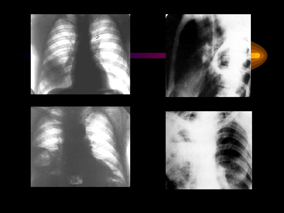
7
Differential diagnostic of lung abscess with central lung carcinoma

8
Differential diagnostic of lung abscess with peripheral lung carcinoma

9
Differential diagnostic of lung abscess with tuberculosis

10
Differential diagnostic of lung abscess with tuberculoma

11
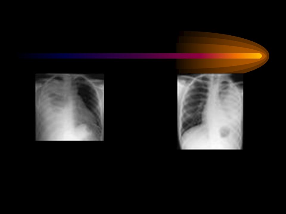
Complications: pulmonary bleeding; pyopneumothorax; pleural empyema;
sepsis; bronchogenic dissemination.

12
PULMONARY BLEEDING TREATMENT I degree – hemorrhage less 300 ml.
ІІІ degree - hemorrhage exceeds 700 ml. TREATMENT 1. Stop the cough 2. Decrease the pulmonary blood pressure 3. Hemostatic therapy

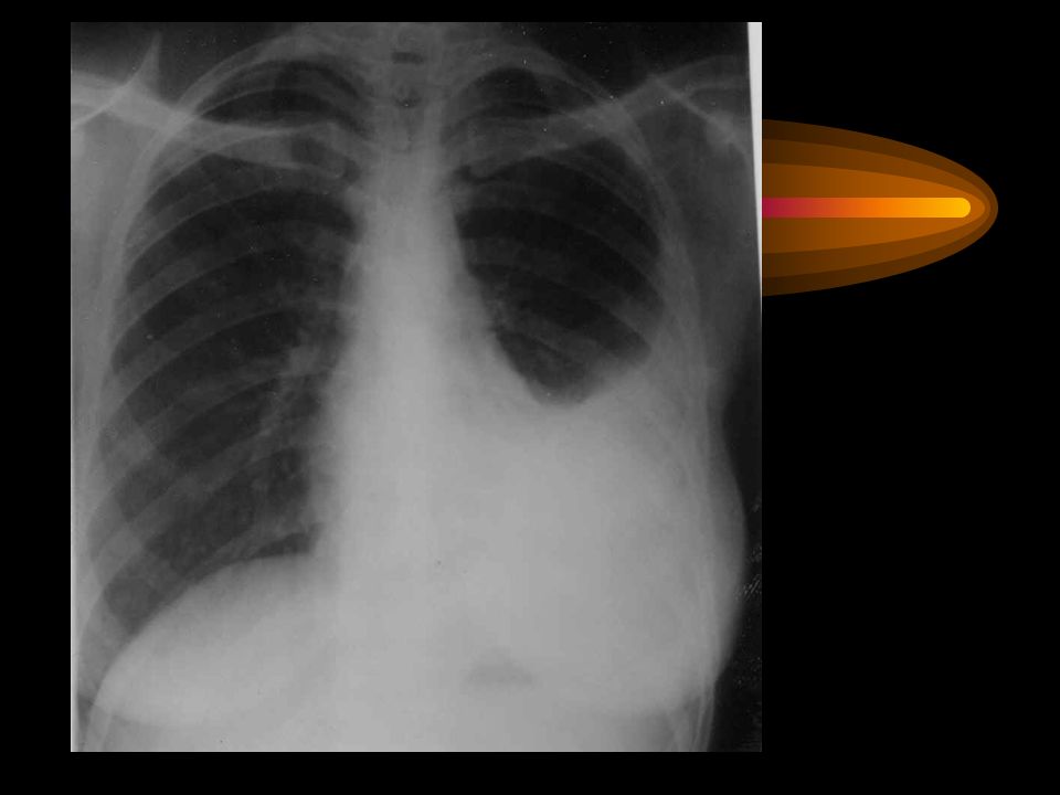
13
PYOPNEUMOTHORAX

14
Tactics and choice of treatment
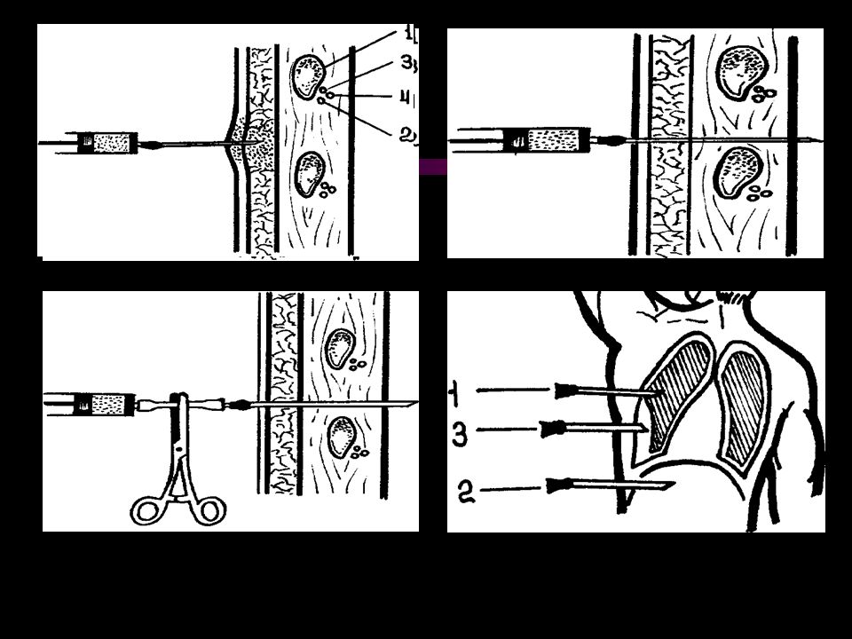
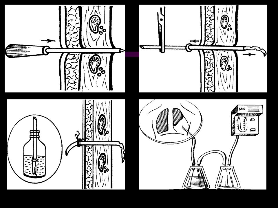
1. The adequate antibacterial, antiinflammatory therapy 2. Evacuation of purulent content of the cavities: active sanation of tracheobronchial tree; repeated punctures or external draining of peripheral cavities. 3. Detoxycation therapy

15
Tactics and choice of treatment

16
4.Immune correction: 5. Desensitizing, antiinflammatory therapy, regulation of activity of proteases (antihistamine, nonsteroid antiinflammatory agents, inhibitors of proteases, antioxidants). 6. Correction of dysfunction of the vital organs and systems, prevention of complications, symptomatic therapy.
. 6. Correction of dysfunction of the vital organs and systems, prevention of complications, symptomatic therapy.")
17
Indications for operative management in acute destructive processes of lungs:
Pulmonary bleeding of ІІ- ІІІ degree; Progression of the process on the background of active and appropriate therapy; Tension pyopneumothorax, which is failed to liquidate by the draining of a pleural space; Impossibility to rule out the suspicion on a malignant tumour.

18
Contraindications decompensation of the vital functions in the terminal stage, bilateral purulent destruction of lungs, concomitant incurable malignant tumours.

19
PLEURAL EMPYEMA The pleural empyema is a purulent inflammation of visceral and parietal pleural membranes, which is associated with accumulation of pus in a pleural space.

20
Etiology and pathogenesis
purulent and destructive processes of lungs, abscesses of abdominal cavity (secondary pleural empyema), open and closed damages of chest, operative approaches on thoracic organs (primary pleural empyema). A secondary pleural empyema occurs in 88 % of the patients.
, open and closed damages of chest, operative approaches on thoracic organs (primary pleural empyema). A secondary pleural empyema occurs in 88 % of the patients.")
21
Classification І. According to the pathogenic factor: Primary.
Secondary. ІІ. According to the clinical course: Acute. Chronic. ІII. According to extension of the process: Focal. Wide-spread.

22
CLINICAL MANIFESTATION
Pain Dyspnea Cough Intoxication By palpation – diminished vocal fremitus on the side of lesion. By percussion over the exudate it is possible to reveal short sound. By auscultation – diminished or absent sound.

23
EMPYEMA FLUID CHARACTERISTICS
pH < Glucose < 40 mg/dL LDH (lactatdehydrogenasa) > 1000 IU/dL Positive culture (50%) Specific gravity > WBC (leucocytes) > 500 cells/mm3 Protein > 2.5 g/dL
 > 1000 IU/dL Positive culture (50%) Specific gravity > WBC (leucocytes) > 500 cells/mm3 Protein > 2.5 g/dL.")
24
Tactics and choice of treatment
The presence of pus in a pleural space is the indication for its elimination. In the place of performed diagnostic thoracentesis carried out the draining of empyema's cavity, its sanation by means of antiseptic solutions. In a focal empyema the aspiration of pus is performed by thoracentesis and only in its inefficiency carried out a draining of pleural space. Intensive antibacterial and antiinflammatory therapy.

30
OPERATIVE APPROACH INDICATION
transformation into the chronic form, that is in case of residual empyema's cavity. VOLUME OF THE OPERATION pleurectomy, decortication of lung. In some cases, when a bronchial fistula and great empyema's cavity has been formed, there is the necessity of performance of resection of lung and corrective thoracoplasty.

Similar presentations


>")


