Download presentation
Presentation is loading. Please wait.

1
Down Syndrome Trisomy 21: Understanding the Problem Richard C. Deth, PhD Department of Pharmaceutical Sciences Northeastern University

2
Key Points: DS (trisomy of Chr21) is a result of Chr21 nondisjunction during meiosis (cell division of egg/sperm) Impaired methylation contributes to DS risk Epigenetic regulation of gene expression is the driver of development Genes on Chr21 can affect development via their effect on cellular oxidative state and methylation status Metabolic support of methylation may help optimize the abilities of DS individuals
 is a result of Chr21 nondisjunction during meiosis (cell division of egg/sperm) Impaired methylation contributes to DS risk Epigenetic regulation of gene expression is the driver of development Genes on Chr21 can affect development via their effect on cellular oxidative state and methylation status Metabolic support of methylation may help optimize the abilities of DS individuals")
3
Down Syndrome Trisomy Chr 21: Trisomy 21 Causes Down Syndrome By: Clare O'Connor, Ph.D. (Biology Department, Boston College) © 2008 Nature Education
 © 2008 Nature Education.")
4
Nondisjunction of linked sister chromatids can occur at two different stages of egg (or sperm) development: Meiosis I and Meiosis II
 development: Meiosis I and Meiosis II")
5
Trisomy begins with maternal gametogenesis, which occurs within the maternal grandmother’s body, during her pregnancy.

6
Faulty Gamete Production (mainly ova) Ovulation and Conception Prenatal Development Postnatal Development Mother and Placenta Breastfeeding and Nutrition Methylation plays an important role in all phases of conception and development Meiosis I Meiosis II Oocytes form during maternal fetal development Mother or Father Maternal Grandmother Years Prophase
 Ovulation and Conception Prenatal Development Postnatal Development Mother and Placenta Breastfeeding and Nutrition Methylation plays an important role in all phases of conception and development Meiosis I Meiosis II Oocytes form during maternal fetal development Mother or Father Maternal Grandmother Years Prophase")
9
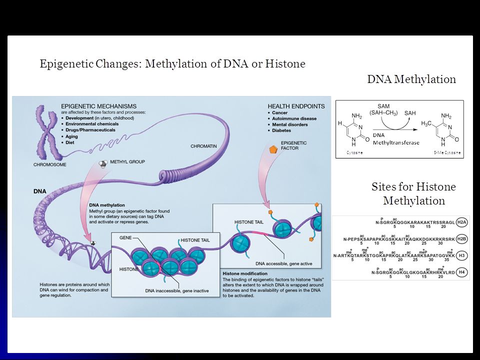
Regulation of gene expression during development X Gene sequence Start site for mRNA synthesis TF Growth Factors NeurotransmittersHormones RNA polymerase mRNA Protein (e.g. enzyme) DNA Transcription Translation DNA DNA + Histone = Heterochromatin Genes are silenced and transcription is blocked Me MBDP (e.g. MeCP2) Histone proteins HMT Me DNMT SAM CpG Transcription Factor Regulation: Epigenetic Regulation: TF binding region CpG No mRNA X
 DNA Transcription Translation DNA DNA + Histone = Heterochromatin Genes are silenced and transcription is blocked Me MBDP (e.g. MeCP2) Histone proteins HMT Me DNMT SAM CpG Transcription Factor Regulation: Epigenetic Regulation: TF binding region CpG No mRNA X.")
11
METHIONINE CYCLE HCY MET SAH SAM ~1,000 Methylation Reactions via 209 MTases ( - ) ATPPP + P i DIETARY PROTEIN THF MethylTHF Methionine Synthase Adenosine Vitamin B12 (Cobalamin) A reversible reaction! ( + ) Global methylation = SAM SAH ( )
 Global methylation = SAM SAH ( ).")
12
Glutathione (GSH) Redox Status γ-Glutamylcysteine Cysteine Cystathionine HCY Methionine Cycle SAH Adenosine > 1,000 Methylation Reactions SAM ( - ) ATP PP + P i THF Methyl-THF Methionine Synthase MET Adenosine D4- SAH ATP PP + P i Dopamine (Attention) Phospholipid Methylation D4- HCY D4- MET D4- SAM Methyl-THF THF D4 Dopamine Receptor PLM Cycle CBS GSH GSSG = Transsulfuration
 Redox Status γ-Glutamylcysteine Cysteine Cystathionine HCY Methionine Cycle SAH Adenosine > 1,000 Methylation Reactions SAM ( - ) ATP PP + P i THF Methyl-THF Methionine Synthase MET Adenosine D4- SAH ATP PP + P i Dopamine (Attention) Phospholipid Methylation D4- HCY D4- MET D4- SAM Methyl-THF THF D4 Dopamine Receptor PLM Cycle CBS GSH GSSG = Transsulfuration")
13
24% decrease in HCY in DS 47% decrease in MET in DS 350% increase in cystathionine 15% decrease in cysteine in DS Consistent with a decrease in methionine synthase activity and an increase in CBS activity

14
25% decrease in combined SAM and SAH in DS 33% increase in adenosine in DS Consistent with a decrease in methionine synthase activity and an increase in CBS activity

15
Glutathione (GSH) Redox Status γ-Glutamylcysteine Cysteine Cystathionine HCY ↓ Methionine Cycle SAH Adenosine > 1,000 Methylation Reactions SAM ( - ) ATP PP + P i THF Methyl-THF Methionine Synthase MET Adenosine D4- SAH ATP PP + P i Dopamine (Attention) Phospholipid Methylation D4- HCY D4- MET D4- SAM Methyl-THF THF D4 Dopamine Receptor PLM Cycle CBS GSH GSSG = ↑ Transsulfuration In DS: Increased transsulfuration Decreased methylation
 Redox Status γ-Glutamylcysteine Cysteine Cystathionine HCY ↓ Methionine Cycle SAH Adenosine > 1,000 Methylation Reactions SAM ( - ) ATP PP + P i THF Methyl-THF Methionine Synthase MET Adenosine D4- SAH ATP PP + P i Dopamine (Attention) Phospholipid Methylation D4- HCY D4- MET D4- SAM Methyl-THF THF D4 Dopamine Receptor PLM Cycle CBS GSH GSSG = ↑ Transsulfuration In DS: Increased transsulfuration Decreased methylation")
16
Methionine synthase has five domains + cobalamin (Vitamin B12) Domains alternate interacting with cobalamin during turnover SAM Domain Cobalamin Domain Cap Domain Cobalamin (vitamin B12) SAM Domain Cobalamin Domain Cap Domain 5-Methyl THF Domain HCY Domain Cobalamin (vitamin B12) 1 2 3 B12 serves as a sensor of redox status
 Domains alternate interacting with cobalamin during turnover SAM Domain Cobalamin Domain Cap Domain Cobalamin (vitamin B12) SAM Domain Cobalamin Domain Cap Domain 5-Methyl THF Domain HCY Domain Cobalamin (vitamin B12) B12 serves as a sensor of redox status")
17
REDOX STATUS: GSH GSSH Methylation Status: SAM SAH ~ 1,000 Methylation Reactions Nitric Oxide Synthesis Phospholipid Methylation DNA/Histone Methylation Gene Expression Arginine Methylation Membrane Properties Creatine Synthesis Cognitive Status Energy Status Catecholamine Methylation Serotonin Methylation Melatonin Sleep

18
Dr. Christina Muratore - 2010 Research Fellow in Neurology Harvard Medical School Domain-specific PCR analysis of MS mRNA in postmortem frontal cortex

19
Age-dependent decrease of Cob and Cap domain mRNA in postmortem frontal cortex in 49 subjects Cobalamin-binding DomainCap Domain

20
Methionine synthase mRNA is lower but protein levels are not different in cortex of autistic subjects

21
Methionine synthase provides redox-sensitive global coordination of metabolism: HOMEOSTASIS

22
RISK OF DS In a number of studies (but not all) impaired methylation status and/or elevated homocysteine in the mother was shown to influence the risk of Down Syndrome (i.e. poor methylation may increase the risk of nondisjunction) But, what about grandma?
 But, what about grandma .")
23
After 10 years (1999–2009) of active research in the field the question of whether or not polymorphisms in folate/Hcy metabolizing genes are associated with increased DS risk is still largely debated in literature, and none of the studied polymorphisms can be firmly considered as an independent DS risk factor[15–40]. Even if MTHFR 677C > T, MTHFR 1298A > C and MTRR 66A > G gene polymorphisms gave positive results in several independent studies, results are still conflicting and inconclusive
![After 10 years (1999–2009) of active research in the field the question of whether or not polymorphisms in folate/Hcy metabolizing genes are associated with increased DS risk is still largely debated in literature, and none of the studied polymorphisms can be firmly considered as an independent DS risk factor[15–40].](http://images.slideplayer.com/25/8196190/slides/slide_23.jpg "Even if MTHFR 677C > T, MTHFR 1298A > C and MTRR 66A > G gene polymorphisms gave positive results in several independent studies, results are still conflicting and inconclusive.")
24
Increased HCY in mothers of DS individuals was found in a number of studies, but was not found in other studies (e.g. in France). MTHFR 677C > T correlates with increased HCY.
. MTHFR 677C > T correlates with increased HCY..")
25
CBS (833 ins 68) Genetic variants in methylation pathway associated with DS
 Genetic variants in methylation pathway associated with DS")
26
Combinations of methylation-related SNPs increase the maternal risk of having a DS baby in some studies (e.g. up to a 7-fold increase).
..")
27
Higher HCY levels are associated with lower IQ.

28
Zampieri et al. (2012) found significant risk associations for maternal age, MTHFR 677 C>T, and Transcobalamin 766 C>G, but decreased risk for BHMT 742 G>A.
 found significant risk associations for maternal age, MTHFR 677 C>T, and Transcobalamin 766 C>G, but decreased risk for BHMT 742 G>A..")
29
DS children have a lower SAM/SAH, indicative of impaired methylation capacity

30
Role of oxidative stress and methylation during development

31
Eggs are rich In cysteine Sperm are rich in selenium GSH ∆ DNA and Histone Methylation ∆ Gene Expression Does Redox Control Development Via Epigenetic Effects? juanv.wordpress.com/

34
Which chromosome 21 genes contribute to DS? Answer: All of them contribute something. Which genes are more important for causing Down Syndrome? Answer: Methylation-related genes Which genes are more important for cognitive development? Answer: Methylation-related genes Chromosome 21 Genes and Down Syndrome

35
Chromosome 21 has about 400 genes. Increased gene dosage from each of them probably contributes to Down Syndrome characteristics. Certain genes deserve special attention for their relationship to oxidative stress and methylation, which are the foundation of development, especially brain development. These genes is located in the region of chromosome 21 (21q21-22) that has been implicated as being most important for DS. Increased activity of the proteins produced by these genes is likely to contribute to DS. Amyloid precursor protein (APP) Cystathionine-beta-synthase (CBS) DNA methyltransferase 3L (DNMT3L) Formiminotransferase cyclodeaminase (FTCD) Superoxide dismutase 1 (SOD1)
 that has been implicated as being most important for DS. Increased activity of the proteins produced by these genes is likely to contribute to DS. Amyloid precursor protein (APP) Cystathionine-beta-synthase (CBS) DNA methyltransferase 3L (DNMT3L) Formiminotransferase cyclodeaminase (FTCD) Superoxide dismutase 1 (SOD1).")
36
Formiminotransferase cyclodeaminase (FTCD): Removes an imino group from forminoglutamate (FIGLU), attaches it to THF, and converts it to methenylTHF. This provides a back-up source of methylTHF to support methionine synthase and methylation.

37
Formiminotransferase cyclodeaminase FIGLU In trisomy, a higher than normal activity of FTCD might provide extra methyl groups to compensate for decreased MTHFR activity. MTHFR

38
AMYLOID PRECURSOR PROTEIN (APP): A cleavage product of amyloid precursor protein (APP), known as Aβ, is though to be the primary cause of Alzheimer’s disease (AD). Amyloid plaques rich in Aβ are found at autopsy in AD brain, but the neurodegeneration is thought to be caused by small Aβ oligomers, starting decades before the onset of AD symptoms. AD is much more common in DS, presumably because the extra APP gene leads to increased Aβ.

39
Early onset familial AD ▫APP ▫Presenilin 1 and 2 Late onset AD ▫APOE4 ▫LRP1 ▫A2M ▫AD5-8 ▫HLA-A ▫NOS3 ▫PAXIP1 ▫MS ▫MTHFR Current Genetics of Alzheimer’s Disease OMIM.org

40
Amyloid Processing http://www.bath.ac.uk/bio-sci/research/profiles/brown-d.html

41
Neurons have impaired transsulfuration and low GSH levels that depend upon growth factor-stimulated cysteine uptake Methionine Synthase HCY MET SAH SAM >1,000 Methylation Reactions ATPPP+P i Adenosine MethylTHF THF Cystathionine Cysteine GSH γ-Glutamylcysteine D4 HCY D4 SAM D4 SAH D4 MET ATP PP+P i MethylTHF THF Phospholipid Methylation Adenosine Dopamine Cysteine ( - ) PI3-kinase ( + ) PARTIALLY BLOCKED IN NEURONAL CELLS EAAT3 Astrocytes Cysteinylglycine GSH GSSG Neurotrophic Growth Factors GSCbl MeCbl SAM OHCbl GSH NEURON Cystine EAAT3
 PI3-kinase ( + ) PARTIALLY BLOCKED IN NEURONAL CELLS EAAT3 Astrocytes Cysteinylglycine GSH GSSG Neurotrophic Growth Factors GSCbl MeCbl SAM OHCbl GSH NEURON Cystine EAAT3")
42
Soluble Aβ oligomers inhibit cysteine uptake, increase oxidative stress and decrease methylation capacity in neuroblastoma cells

43
A-β oligomers decrease DNA methylation and alter expression of redox/methylation pathway genes in neuroblastoma cells

44
Our Amyloid Hypothesis Cysteine EAAT3 SAH SAM HCY MET Cystathionine Cysteine GSH γ- Glutamylcysteine GSSG Cystine Homocystine MethylTHF THF ATPPP+P Methionine Synthase Adenosine DNA Methyl-DNA GSHGSCbl SAM OHCbl MeCbl Cysteinylglycine GSH Healthy Glial Cells (Astrocytes) ( - ) PI3-kinase ( + ) Abeta Oligomers Epigenetic Changes Nate Hodgson PhD Aug 2012
 ( - ) PI3-kinase ( + ) Abeta Oligomers Epigenetic Changes Nate Hodgson PhD Aug 2012")
45
A-β inhibits cysteine uptake, decreases DNA methylation and alters gene expression. This is likely to be a natural role for A-β, promoting oxidative stress in neurons. The extra APP and A-β production occurring in trisomy 21 may produce excessive oxidative stress, with adverse epigenetic consequences.

46
Superoxide dismutase 1 (SOD1) Superoxide anion is a reactive oxygen species (ROS) produced by mitochondria as a by-product during ATP synthesis SOD converts superoxide to hydrogen peroxide
 Superoxide anion is a reactive oxygen species (ROS) produced by mitochondria as a by-product during ATP synthesis SOD converts superoxide to hydrogen peroxide")
47
Data from Waly et al. (In Prep) Interestingly, methionine synthase has B12-dependent SOD activity, which is essential for GSH-dependent reactivation of enzyme activity after B12 oxidation
 Interestingly, methionine synthase has B12-dependent SOD activity, which is essential for GSH-dependent reactivation of enzyme activity after B12 oxidation.")
48
Cystathionine beta synthase (CBS): CBS converts HCY to cystathionine in a vitamin B6- dependent reaction CBS converts HCY to cystathionine in a vitamin B6- dependent reaction CBS activity is increased by SAM but increased by oxidative stress and TNF-alpha CBS activity is increased by SAM but increased by oxidative stress and TNF-alpha Vitamin D was recently shown to increase the level of CBS Vitamin D was recently shown to increase the level of CBS Testosterone decreases CBS activity Testosterone decreases CBS activity CBS also converts cysteine to hydrogen sulfide CBS also converts cysteine to hydrogen sulfide
: CBS converts HCY to cystathionine in a vitamin B6- dependent reaction CBS converts HCY to cystathionine in a vitamin B6- dependent reaction CBS activity is increased by SAM but increased by oxidative stress and TNF-alpha CBS activity is increased by SAM but increased by oxidative stress and TNF-alpha Vitamin D was recently shown to increase the level of CBS Vitamin D was recently shown to increase the level of CBS Testosterone decreases CBS activity Testosterone decreases CBS activity CBS also converts cysteine to hydrogen sulfide CBS also converts cysteine to hydrogen sulfide")
49
Transsulfuration Pathway Glutathione (GSH) Redox Status γ-Glutamylcysteine Cysteine Cystathionine HCY SAH Adenosine > 1,000 Methylation Reactions SAM ( - ) ATP PP + P i THF Methyl-THF Methionine Synthase MET THF GSH GSSG = ~ CBS Cysteine Hydrogen Sulfide SAM Vitamin D Oxidative stress THF-alpha (+) Testosterone (-)
 Redox Status γ-Glutamylcysteine Cysteine Cystathionine HCY SAH Adenosine > 1,000 Methylation Reactions SAM ( - ) ATP PP + P i THF Methyl-THF Methionine Synthase MET THF GSH GSSG = ~ CBS Cysteine Hydrogen Sulfide SAM Vitamin D Oxidative stress THF-alpha (+) Testosterone (-)")
50
Table 2 | Intellectual disability by gender (n=121) MalesFemalesTotal count %count % count % mild912.91427.52319.0 moderate2332.91325.53629.8 severe2130.01937.34033.1 profound1724.359.82218.2 Males are affected more severely than females
 MalesFemalesTotal count %count % count % mild moderate severe profound Males are affected more severely than females")
51
Excessive CBS activity in DS limits methylation by removing HCY from the methionine cycle. The critical balance between methylation and transsulfuration is therefore altered.

53
DNA methyltransferase 3L (DNMT3L): Does not directly attach methyl groups to DNA, but forms a complex with other methyltransferases and alters their activity. Important for gender-specific DNA methylation

54
In the absence of Dnmt3L, neither methylation of most oocyte-methylated gDMRs nor intragenic methylation was observed. There was also genome- wide hypomethylation, and partial methylation at particular retrotransposons, while maintaining global gene expression, in oocytes. Along with the identification of the many Dnmt3L-dependent gDMRs at intragenic regions, the present results suggest that oocyte methylation can be divided into 2 types: Dnmt3L-dependent methylation, which is required for maternal methylation imprinting, and Dnmt3L-independent methylation, which might be essential for endogenous retroviral DNA silencing. The present data provide entirely new perspectives on the evaluation of epigenetic markers in germline cells. In other words, DNMT3L is critical for oocyte methylation.

55
SUMMARY > 400 genes on Chr 21 contribute to Down syndrome. The relatively high survival rates for Chr 21 trisomy indicates that the higher expression of genes creates an acceptable alternative pathway for development. Impaired methylation, importantly involving altered DNA methylation, is a primary factor in causing DS. Altered patterns of methylation continue to be important throughout the lifespan. Metabolic interventions which address oxidative stress and improve methylation capacity may be beneficial. Controlled clinical trials of these interventions are needed.

58
Brain Samples: Autism Tissue Program Harvard Brain Tissue Resource Center Tissue Resource Center (Australia) Stanley Medical Research Foundation and donor families. Collaborators: Antonio Persico Suzanne De la Monte Hamid Abdolmaleky Mostafa Waly Yahya Al-Farsi Grant Support: Autism Research Institute SafeMinds National Autism Association Autism Speaks ACKNOWLEDGEMENTS

Similar presentations


 DNA (gene) mRNA Protein Transcription RNA processing (splicing etc) Translation Folding Post translational modifications Peptides/amino.>")


