Download presentation
Presentation is loading. Please wait.

1
Anatomy & Physiology Unit 5: The Skeletal System 5A: Skeletal Tissues & Basics

2
The Skeletal System Parts of the skeletal system – Bones (skeleton) – Joints – Cartilages – Ligaments Two subdivisions of the skeleton – Axial skeleton – Appendicular skeleton
 – Joints – Cartilages – Ligaments Two subdivisions of the skeleton – Axial skeleton – Appendicular skeleton")
3
The Axial Skeleton Forms the longitudinal axis of the body Divided into three parts – Skull – Vertebral column – Bony thorax

4
The Appendicular Skeleton Appendages that hang from axial Girdles from which the appendages hang Shoulder girdles— – clavicle (collar bone) – scapula (shoulder blade) Arms hang from shoulder girdles Pelvic girdle—coxal bones – Coxal or pelvic bones are 3 fused bones Legs hang from pelvic girdle
 – scapula (shoulder blade) Arms hang from shoulder girdles Pelvic girdle—coxal bones – Coxal or pelvic bones are 3 fused bones Legs hang from pelvic girdle")
6
Functions of Bones Support the body Protect soft organs – Skull and vertebrae for brain and spinal cord – Rib cage for thoracic cavity organs Allow movement due to attached skeletal muscles Store minerals and fats – Calcium and phosphorus – Fat in the internal marrow cavity Blood cell formation (hematopoiesis)
")
7
Bones of the Human Body The adult skeleton has 206 bones Two basic types of bone tissue – Compact bone Homogeneous – Spongy bone Small needle-like pieces of bone Many open spaces

8
Figure 5.1 Spongy bone Compact bone

9
Classification of Bones on the Basis of Shape Bones are classified as: – Long – Short – Flat – Irregular

10
Figure 5.2

11
Classification of Bones Long bones – Typically longer than they are wide – Shaft with heads situated at both ends – Contain mostly compact bone – All of the bones of the limbs (except wrist, ankle, and kneecap bones) – Example: Femur Humerus
 – Example: Femur Humerus")
12
Figure 5.2a

13
Classification of Bones Short bones – Generally cube-shaped – Contain mostly spongy bone – Includes bones of the wrist and ankle – Sesamoid bones are a type of short bone which form within tendons (patella) – Example: Carpals Tarsals
 – Example: Carpals Tarsals")
14
Figure 5.2d

15
Classification of Bones Flat bones – Thin, flattened, and usually curved – Two thin layers of compact bone surround a layer of spongy bone – Example: Skull Ribs Sternum

16
Figure 5.1 Spongy bone Compact bone

17
Figure 5.2c

18
Classification of Bones Irregular bones – Irregular shape – Do not fit into other bone classification categories – Example: Vertebrae Hip bones

19
Figure 5.2b

21
Figure 5.3a Distal epiphysis Diaphysis Proximal epiphysis Articular cartilage Spongy bone Epiphyseal line Periosteum Compact bone Medullary cavity (lined by endosteum) (a)
 (a)")
22
Anatomy of a Long Bone Diaphysis – Shaft – Composed of compact bone Epiphysis – Ends of the bone – Composed mostly of spongy bone

23
Anatomy of a Long Bone Periosteum – Outside covering of the diaphysis – Fibrous connective tissue membrane Perforating (Sharpey’s) fibers – Secure periosteum to underlying bone Arteries – Supply bone cells with nutrients
 fibers – Secure periosteum to underlying bone Arteries – Supply bone cells with nutrients")
24
Figure 5.3c Yellow bone marrow Compact bone Perforating (Sharpey’s) fibers Nutrient arteries Periosteum Endosteum (c)
 fibers Nutrient arteries Periosteum Endosteum (c)")
25
Anatomy of a Long Bone Articular cartilage – Covers the external surface of the epiphyses – Made of hyaline cartilage – Decreases friction at joint surfaces

26
Figure 5.3b Compact bone Spongy bone Articular cartilage (b)
")
27
Anatomy of a Long Bone Epiphyseal plate – Flat plate of hyaline cartilage seen in young, growing bone Epiphyseal line – Remnant of the epiphyseal plate – Seen in adult bones

28
Figure 5.3a Distal epiphysis Diaphysis Proximal epiphysis Articular cartilage Spongy bone Epiphyseal line Periosteum Compact bone Medullary cavity (lined by endosteum) (a)
 (a)")
29
Anatomy of a Long Bone Marrow (medullary) cavity – Cavity inside of the shaft – Contains yellow marrow (mostly fat) in adults – Contains red marrow for blood cell formation in infants In adults, red marrow is situated in cavities of spongy bone and epiphyses of some long bones
 cavity – Cavity inside of the shaft – Contains yellow marrow (mostly fat) in adults – Contains red marrow for blood cell formation in infants In adults, red marrow is situated in cavities of spongy bone and epiphyses of some long bones")
30
Figure 5.3a Distal epiphysis Diaphysis Proximal epiphysis Articular cartilage Spongy bone Epiphyseal line Periosteum Compact bone Medullary cavity (lined by endosteum) (a)
 (a)")
31
Describe everything that is in this photograph.

32
Microscopic Anatomy of Compact Bone Osteon (Haversian system) – A unit of bone containing central canal and matrix rings Central (Haversian) canal – Opening in the center of an osteon – Carries blood vessels and nerves Perforating (Volkmann’s) canal – Canal perpendicular to the central canal – Carries blood vessels and nerves
 – A unit of bone containing central canal and matrix rings Central (Haversian) canal – Opening in the center of an osteon – Carries blood vessels and nerves Perforating (Volkmann’s) canal – Canal perpendicular to the central canal – Carries blood vessels and nerves")
33
Figure 5.4a Compact bone Periosteal blood vessel Periosteum Perforating fibers Central (Haversian) canal Perforating (Volkmann’s) canal Blood vessel Spongy bone Blood vessel continues into medullary cavity containing marrow Lamellae (a) Osteon (Haversian system)
 canal Perforating (Volkmann’s) canal Blood vessel Spongy bone Blood vessel continues into medullary cavity containing marrow Lamellae (a) Osteon (Haversian system)")
34
Microscopic Anatomy of Bone Lacunae – Cavities containing bone cells (osteocytes) – Arranged in concentric rings called lamellae Lamellae – Rings around the central canal – Sites of lacunae
 – Arranged in concentric rings called lamellae Lamellae – Rings around the central canal – Sites of lacunae")
35
Figure 5.4b Lamella Canaliculus Lacuna Central (Haversian) canal (b) Osteocyte
 canal (b) Osteocyte")
36
Figure 5.4c Osteon Lacuna Central canal Interstitial lamellae (c)
")
37
Microscopic Anatomy of Bone Canaliculi – Tiny canals – Radiate from the central canal to lacunae – Form a transport system connecting all bone cells to a nutrient supply

38
Figure 5.4b Lamella Canaliculus Lacuna Central (Haversian) canal (b) Osteocyte
 canal (b) Osteocyte")
39
Describe everything that is in this photograph.

40
Bone Matrix (obj 5): 2 components— Organic Collagen fibers Reinforces bones Gives bones some degree of flexibility & resilience w/o collagen—brittle bone disease Inorganic Inorganic salts – Especially calcium salts but other minerals as well Makes bones hard & supportive w/o hard, supportive bones—rickets (soft bones)
: 2 components— Organic Collagen fibers Reinforces bones Gives bones some degree of flexibility & resilience w/o collagen—brittle bone disease Inorganic Inorganic salts – Especially calcium salts but other minerals as well Makes bones hard & supportive w/o hard, supportive bones—rickets (soft bones)")
41
Types of Bone Cells Slide 5.15 Copyright © 2003 Pearson Education, Inc. publishing as Benjamin Cummings Osteocytes Mature bone cells Osteoblasts Bone-forming cells Osteoclasts Bone-destroying cells Break down bone matrix for remodeling and release of calcium Bone remodeling is a process by both osteoblasts and osteoclasts

42
Formation of the Human Skeleton In embryos, the skeleton is primarily hyaline cartilage During development, much of this cartilage is replaced by bone—Endochondral Ossification However, between flat bones of the skull arise from layers of dense connective tissue— Intramembranous Ossification Cartilage remains in isolated areas – Bridge of the nose – Parts of ribs – Joints

43
Bone Growth (Ossification) Epiphyseal plates allow for lengthwise growth of long bones during childhood – New cartilage is continuously formed – Older cartilage becomes ossified Cartilage is broken down Enclosed cartilage is digested away, opening up a medullary cavity Bone replaces cartilage through the action of osteoblasts
 Epiphyseal plates allow for lengthwise growth of long bones during childhood – New cartilage is continuously formed – Older cartilage becomes ossified Cartilage is broken down Enclosed cartilage is digested away, opening up a medullary cavity Bone replaces cartilage through the action of osteoblasts")
44
Bone Growth (Ossification) Bones are remodeled and lengthened until growth stops – Bones are remodeled in response to two factors Blood calcium levels Pull of gravity and muscles on the skeleton – Bones grow in width (called appositional growth)
 Bones are remodeled and lengthened until growth stops – Bones are remodeled in response to two factors Blood calcium levels Pull of gravity and muscles on the skeleton – Bones grow in width (called appositional growth)")
45
Figure 5.5 In a fetus In an embryo Bone collar Hyaline cartilage model Bone starting to replace cartilage In a child Medullary cavity New center of bone growth Hyaline cartilage Epiphyseal plate cartilage Growth in bone length New bone forming Invading blood vessels Epiphyseal plate cartilage Articular cartilage Spongy bone New bone forming Growth in bone width

46
Figure 5.5, step 1 In an embryo Bone collar Hyaline cartilage model Bone starting to replace cartilage

47
Figure 5.5, step 2 In a fetus Medullary cavity New center of bone growth Hyaline cartilage Growth in bone length Invading blood vessels

48
Figure 5.5, step 3 In a child Epiphyseal plate cartilage New bone forming Invading blood vessels Epiphyseal plate cartilage Articular cartilage Spongy bone New bone forming Growth in bone width

49
Figure 5.6 Bone growth Bone grows in length because: Bone remodeling Growing shaft is remodeled as: Cartilage grows here. Cartilage is replaced by bone here. Cartilage grows here. Cartilage is replaced by bone here. 1 2 3 4 1 2 3 Bone is resorbed here. Epiphyseal plate Articular cartilage Bone is resorbed here. Bone is added by appositional growth here.

50
Types of Bone Cells Osteocytes—mature bone cells Osteoblasts—bone-forming cells Osteoclasts—giant bone-destroying cells – Break down bone matrix for remodeling and release of calcium in response to parathyroid hormone Bone remodeling is performed by both osteoblasts and osteoclasts

51
Skeletal Changes Throughout Life Osteoporosis – Bone-thinning disease afflicting 50 percent of women over age 65 20 percent of men over age 70 – Disease makes bones fragile and bones can easily fracture – Vertebral collapse results in kyphosis (also known as dowager’s hump) – Estrogen aids in health and normal density of a female skeleton
 – Estrogen aids in health and normal density of a female skeleton")
53
Osteoporosis

54
Figure 5.36

55
Bone Fractures Fracture—break in a bone Types of bone fractures – Closed (simple) fracture—break that does not penetrate the skin – Open (compound) fracture—broken bone penetrates through the skin Bone fractures are treated by reduction and immobilization
 fracture—break that does not penetrate the skin – Open (compound) fracture—broken bone penetrates through the skin Bone fractures are treated by reduction and immobilization")
56
Common Types of Fractures Comminuted—bone breaks into many fragments Compression—bone is crushed Depressed—broken bone portion is pressed inward Impacted—broken bone ends are forced into each other Spiral—ragged break occurs when excessive twisting forces are applied to a bone Greenstick—bone breaks incompletely

57
Repair of Bone Fractures Hematoma (blood-filled swelling) is formed Break is splinted by fibrocartilage to form a callus Fibrocartilage callus is replaced by a bony callus Bony callus is remodeled to form a permanent patch
 is formed Break is splinted by fibrocartilage to form a callus Fibrocartilage callus is replaced by a bony callus Bony callus is remodeled to form a permanent patch")
58
Figure 5.7 Internal callus (fibrous tissue and cartilage) Hematoma forms. Fibrocartilage callus forms. Bony callus forms. Bone remodeling occurs. 1 23 4 Hematoma Bony callus of spongy bone Spongy bone trabecula New blood vessels External callus Healed fracture

59
Figure 5.7, step 1 Hematoma forms. Hematoma 1

60
Figure 5.7, step 2 Internal callus (fibrous tissue and cartilage) Hematoma forms. Fibrocartilage callus forms. Hematoma Spongy bone trabecula New blood vessels External callus 1 2

61
Figure 5.7, step 3 Internal callus (fibrous tissue and cartilage) Hematoma forms. Fibrocartilage callus forms. Bony callus forms. Hematoma Bony callus of spongy bone Spongy bone trabecula New blood vessels External callus 1 2 3

62
Figure 5.7, step 4 Internal callus (fibrous tissue and cartilage) Hematoma forms. Fibrocartilage callus forms. Bony callus forms. Bone remodeling occurs. Hematoma Bony callus of spongy bone Spongy bone trabecula New blood vessels External callus Healed fracture 1 2 3 4

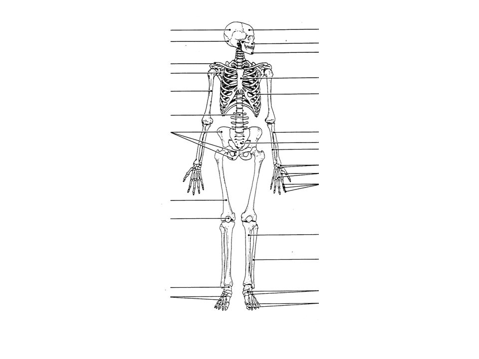
64
Figure 5.8a (a) Anterior view Phalanges Metatarsals Tarsals Fibula Tibia Patella Femur Metacarpals Phalanges Carpals Ulna Radius Vertebra Humerus Rib Sternum Scapula Clavicle Facial bones Cranium Skull Thoracic cage (ribs and sternum) Vertebral column Sacrum
 Anterior view Phalanges Metatarsals Tarsals Fibula Tibia Patella Femur Metacarpals Phalanges Carpals Ulna Radius Vertebra Humerus Rib Sternum Scapula Clavicle Facial bones Cranium Skull Thoracic cage (ribs and sternum) Vertebral column Sacrum")
65
Figure 5.8b (b) Posterior view Fibula Tibia Femur Metacarpals Phalanges Carpals Radius Ulna Vertebra Humerus Rib Scapula Clavicle Cranium Bones of pectoral girdle Upper limb Bones of pelvic girdle Lower limb
 Posterior view Fibula Tibia Femur Metacarpals Phalanges Carpals Radius Ulna Vertebra Humerus Rib Scapula Clavicle Cranium Bones of pectoral girdle Upper limb Bones of pelvic girdle Lower limb")
Similar presentations


 Joints ► Cartilages Ligaments ► Divided.>")


 Joints Cartilages Ligaments Divided into two divisions Axial skeleton –>")

>")

