Download presentation
Presentation is loading. Please wait.

1
Flagellates Species Sites parasitizedDiseases involvedEpidemic regions Leishmania donovani macrophagevisceral leishmaniasis Africa, Asia, Mediterranean region Giardia lambliasmall intestinediarrheacosmopolitan Trichomonas vaginalis vagina, urethra, epididymis, prostate gland vaginitis, urethritiscosmopolitan Trypanosoma brucei blood and lymphoid tissues African trypanosomiasis (sleeping sickness) Africa Trypanosoma cruzi blood and connective tissue Chagas diseaseLatin America
 Africa Trypanosoma cruzi blood and connective tissue Chagas diseaseLatin America")
2
Leishmania Leishmania

3
Introduction Introduction Leishmania is a kinetoplastid flagellate Inhabits macrophages Leishmaniasis (Kala-azar, black fever) is a zoonosis transmitted by the bite of sandflies Leishmaniasis affects 12 million people in 88 countries, 4 million new cases per year
 is a zoonosis transmitted by the bite of sandflies Leishmaniasis affects 12 million people in 88 countries, 4 million new cases per year")
4
Three main forms of leishmaniasis Visceral : involving liver, spleen, and bone marrow – Leishmania donovani Mucocutaneous : involving mucous membranes of the mouth and nose – Leishmania braziliensis Cutaneous : involving the skin at the site of a sandfly bite – Leishmania tropica, Leishmania mexicana, Leishmania major IntroductionIntroduction

5
Mucocutaneous Leishmaniasis nasal mucosa and septum are involved Leishmania braziliensis

6
Cutaneous Leishmaniasis Oriental sore Eastern Mediterranean Self-cured disease, sterilizing immunity Leishmania tropica, Leishmania mexicana, Leishmania major

7
Endemic Areas for Leishmaniasis BMJ 2003;326:378

8
Cutaneous Leishmaniasis Visceral Leishmaniasis

9
Morphology Morphology Minute elliptical body, 2- 5 µ m Very short flagellum barely beyond the cell surface Single prominent nucleus – located at one side Cytoplasm- containing kinetoblast; basal body 1.Amastigote (Leishman-Donovan body, LD body ) Leishmania donovani
 Leishmania donovani")
10
Amastigote Bone marrow smear -- Giemsa stain Morphology

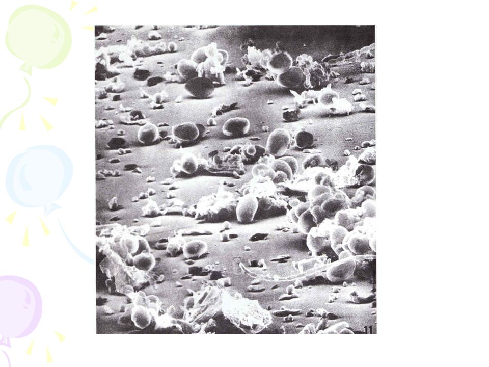
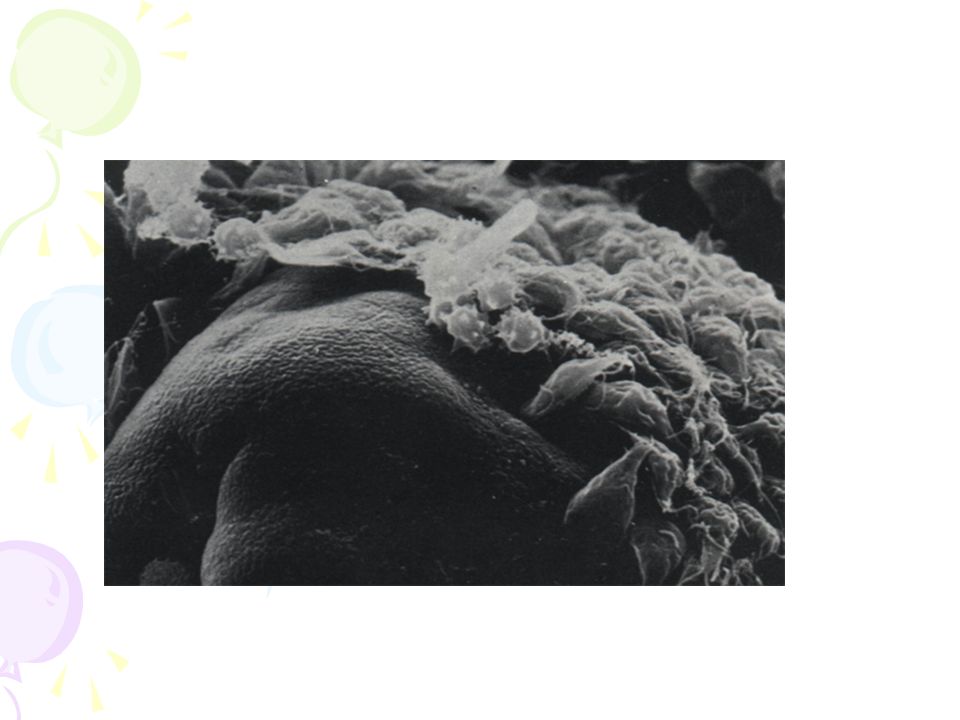
11
Large numbers of amastigotes in a M

12
M broken with the release of amastigote which may invade other M , start asexual multiplication again Morphology Amastigote

13
2. Promastigote Fusiform -shaped with 1 free flagellum Usually gather together in the medium as a mum Morphology

14
Life cycle Life cycle

15
2-host pattern, mammal insect vector Asexual multiplication occurred either in macrophages or in insect Blood feeding of female sandfly, bring amastigote to vector host gut Amastigote Promastigote(3d) binary fission to fill the mouth part (7d) Life cycle
 binary fission to fill the mouth part (7d) Life cycle")
16
Macrophages bring proliferated amastigotes all over the body Dogs and rodents may serve as a reservoir hosts Amastigotes may also be transferred by blood transfusion Life cycle

17
Phlebotomus , sandfly Small , about 1/3 of a mosquito, 3mm, yellow-gray Life cycle

18
Pathogenesis Pathogenesis Mechanism of parasites entering macrophages Mechanism of hypersplenism Anemia, leukopenia, and thrombocytopenia

19
Clinical features Clinical features Infections range from asymptomatic to progressive, fully developed kala-azar. Incubation period is usually 2 – 4 months. Symptoms – Begin with low-grade fever and malaise, followed by progressive wasting, anemia, and protrusion of the abdomen from enlarged liver, spleen and lymph nodes. Nephrosis: IC deposition –type III hypersensitivity

20
Clinical features Clinical features Fatal after 2 – 3 years if not treated. Immediate cause of death is usually an invasion of a secondary pathogen that the body is unable to combat. Gain acquired immunity after effectively cured In acute cases with chills, fevers up to 40 ℃ and vomiting; death may occur within 6–12 months.

21
Two children with visceral leishmaniasis Splenomegaly

22
Leishmaniasis Patients

23
Special clinical features for Kala-azar cases Post-kala-azar dermal leishmaniasis (PKDL) :
 :")
24
Etiological examination –Puncture smear: bone marrow – safe, first choice –Skin biopsy –Tissue or aspirate cultivation –Animal inoculation Diagnosis

25
Animal Xenodiagnosis hamster infected with L.donovani

26
Immuno-diagnosis –Antibody detection –Circulation antigen detection Molecular biology methods –DNA probe test –PCR Diagnosis

27
Injection of Antimony Gluconate( 葡萄糖酸锑钠 ) cure rate would be 97.4 %。 Pentamidine or stilbamidine for antimony-resistant patients Spleen excision Treatment
 cure rate would be 97.4 %。 Pentamidine or stilbamidine for antimony-resistant patients Spleen excision Treatment")
28
Prevention Treat the patients Suppress the reservoir: dogs, rats, gerbils, other small mammals and rodents Suppress the vector: Sandfly Prevent sandfly bites: Personal Protective Measures Most important at night Sleeves down Insect repellent Permethrin soaked uniforms Permethrin soaked bed nets

29
Giardia lamblia Giardia lamblia

30
Introduction Introduction Flagellated protozoan parasite Colonizes and reproduces in the small intestine Cause giardiasis (travelers diarrhea) Person to person transmission Common in many developing countries
 Person to person transmission Common in many developing countries")
31
trophozoite Nucleus Adhensive disc Median body flagellum axostyles Morphology Morphology

32
" smiley face " symbol

33
cysts

34
Life cycle Life cycle

35
Life cycle Trophozoites in duodenum, jejunum and upper ileum Rapid division to produce large number Infective stage: mature cyst Infective route: fecal-oral transmission Infected person may produce approximately 9 billion cysts per day, which are passed in feces Infection occurs after oral ingestion of as few as 10 to 25 cyst

37
Pathogenesis Asymptomatic cyst passer (5 to 15%) acute self-limited diarrhea (25 to 50%) chronic syndrome of diarrhea malabsorption and weight loss Symptomatic giadiasis is characterized by acute onset of diarrhea abdominal cramps, bloating and flatulence feelings of malaise, nausea vomiting, fever and tenesmus occur less commonly stools may be profuse and watery, but later they are commonly greasy, and foul-smelling Vitamin B 12 deficiency
 acute self-limited diarrhea (25 to 50%) chronic syndrome of diarrhea malabsorption and weight loss Symptomatic giadiasis is characterized by acute onset of diarrhea abdominal cramps, bloating and flatulence feelings of malaise, nausea vomiting, fever and tenesmus occur less commonly stools may be profuse and watery, but later they are commonly greasy, and foul-smelling Vitamin B 12 deficiency")
39
Diagnosis Wet, saline mounts: trophozoite (Symptomatic giadiasis ) Iodine dye techniques: cyst (Asymptomatic cyst passer ) Biopsy tissue/duodenal aspirate stained by Giemsa stain Enzyme immunoassay and fluorescent- antibody monoclonal antigen detection system
 Iodine dye techniques: cyst (Asymptomatic cyst passer ) Biopsy tissue/duodenal aspirate stained by Giemsa stain Enzyme immunoassay and fluorescent- antibody monoclonal antigen detection system")
40
Epidemiology Presumed to be zoonotic, but new evidence indicates that strains may be species specific Host can be humans, primates, cat, dog, beaver, rabbits, etc. World wide distribution Highest incidence in children, young adults and travelers in late summer

41
Prevention and treatment Treat the patients and carrier Proper handling and treatment of water Good personal hygiene on an individual basis Metronidazole, Quinacrine, Furazolidone, Paromomycin and Tinidazole

42
Trichomonas vaginalis Trichomonas vaginalis

43
Introduction Introduction A sexually transmitted infection Only trophozoite stage in life cycle; no cyst form Resides in urogenital tract Trichomoniasis: vaginitis in women, while urethritis in men Wet, saline mounts: trophozoite 160 million cases of infection are acquired annually worldwide (WHO, 2011) Cured with metronidazole or tinidazole
 Cured with metronidazole or tinidazole")
Similar presentations


>")
>")


 Leishmania form Rounded shape, absence of free flagellum,>")
 (VL) Leishmania tropica (CL) Leishmania major (CL) Leishmania aethiopica (CL) Leishmania mexicana (Complex)>")

 Giardia lamblia ( 蓝氏贾第鞭毛虫 ) Intestinal flagellate Giardia lambilia lives in small intestine Giardiasis Diarrhea “traveler’s diarrhea”>")

. caused by intracellular protozoan parasites of the genus Leishmania transmitted by phlebotomine sandflies disease involving.>")
