Download presentation
Presentation is loading. Please wait.

1
Glaucoma

2
Glaucoma Glaucomas are group of diseases causing damage to the optic nerve by the effect of raised ocular pressure on the optic nerve head The intraocular pressure depends on the balance btw production and removal of aqueous humour

3
Pathophysiology Aqueous is produced from ciliary processes in the posterior chamber (by active transport and ultrafiltration) Aqueous leaves the eye through tubercular meshwork (iridocorneal angle)*, Schlemm’s canal and then episceral veins “the conventional pathway” 4% of the aqueous is drained into the supra-choroidal space and via the venous circulation across the sclera “uveoscleral pathway”
*, Schlemm’s canal and then episceral veins the conventional pathway 4% of the aqueous is drained into the supra-choroidal space and via the venous circulation across the sclera uveoscleral pathway")
4
Pathophysiology

5
4%

6
the mechanism by which an elevated intraocular pressure damages nerve fibers:
Raised IO pressure causes “mechanical” damage to the optic nerve axon Raised IO pressure causes “ischemia” of the nerve axon by reducing blood flow to it

7
Classification:

8
Primary glaucoma: This classification depends on: The iris doesn’t
cover the trabecular meshwork (open angle) the iris covers the trabecular meshwork (closed angle) .
 the iris covers. the trabecular. meshwork (closed angle) .")
9
Primary open angle glaucoma
Pathogenesis: The structure of trabecular meshwork appears normal but there is an increased resistance to the outflow, this happened due to: Thinking of the trabecular lamellae which reduces the pore size Reduction in the number of lining trabecular cells Increased extracellular material in the meshwork spaces

10
Chronic open angle glaucoma
Epidemiology: Affects of population over the age of 50 Males equally affected as females May be a family hx, although the exact mode of inheritance is not clear Genetic factors play a rule in developing open angle glucoma: mutation in the myocillin gene (GLC1A) om chromosome 1, optineurin (GLC1E)……..
 om chromosome 1, optineurin (GLC1E)……..")
11
History: -Symptoms depends on the rate of IO pressure rises
History: -Symptoms depends on the rate of IO pressure rises. -Associated with slow rise in pressure and it’s symptomless until pt becomes aware of visual deficit. -Many pts diagnosed via an optometrist.

12
Chronic open angle glaucoma
On examination: The eyes are mainly white and the corneas are clear Perimetry….(for visual field loss) On slit lamp: 1- Measure the ocular pressure using the tonometer (NL pressure is15.5 mmHg); mean(11-21 mmHg) In chronic open angle glaucoma: pressure mmHg In angle closure glaucoma >60 mmHg 2- Exclude other ocular disease that may be 2ry cause for the glaucoma 3- Measure the thinkness of the cornea with a pachymeter*, to adjust the value of IO pressure.
 On slit lamp: 1- Measure the ocular pressure using the tonometer (NL pressure is15.5 mmHg); mean(11-21 mmHg) In chronic open angle glaucoma: pressure mmHg. In angle closure glaucoma >60 mmHg. 2- Exclude other ocular disease that may be 2ry cause for the glaucoma. 3- Measure the thinkness of the cornea with. a pachymeter*, to adjust the value of IO. pressure.")
13
4- Examin the iridocorneal angle by Gonioscopy to confirm that an open angle glucoma is present

14
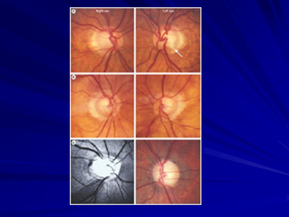
5- Examine the optic disc: Glaucomatous optic disc demonstrating:
1-Increased cupping and central pallor with baring of circumlinear vessel 2-Splinter optic disc hemorrhages 3-Nasalization of the vessels 4-Localized notching of the neural rim between 5-Diffuse thinning of the retinal nerve fiber layer is evident with increased visibility of small vessels and capillaries * In this eye, the disc “neuroretinal rim” is much thinner than in the normal optic disc. The “cup-to-disc ratio” here is about 0.8—much greater than the physiologic limit of 0.4! When the cup-to-disc ratio exceeds 0.4, optic disc cupping is probably pathologic. This patient has glaucoma. The cup-to-disc ratio compares the diameter of the "cup" portion of the optic disc with the total diameter of the optic disc. Notching of the rim,implying focal axonal loss, may also be a sign of glucomatous damage

16
relatively large optic disc with large cupping. However the inferior rim tissue shows a localized notch

20
Treatment Medical treatment Laser treatment Surgical treatment

21
Medical TTT Topical drugs:
Prostaglandin: the 1st line…increasing passage of the aquoeus through uveoscleral pathway B blocker: reduce IO pressure by decreasing aqueous secretion Nonselective: carry the risk of asthma…excecerbate heart block if IO pressure still hight other drugs…laser…surgery (drainage)
")
22
Laser Series of laser burns (50 um)in the meshwork to improve the aqueous outflow Effective initially but the IO pressure may increase slowly

23
Surgery Trabeculectomy: creation of a fistula between the AC & the subconjunctival space

24
Bleb after trabeculectomy

25
Complications of surgery:
1- swallowing of the AC in the immediate postop. Period (cause damage to the lens and cornea) 2- IO infection 3- accelerated cataract development 4- failure to reduce intraocular pressure adequatly 5- an excessivly low pressure (hypotony) which may cause a macular edema
 2- IO infection. 3- accelerated cataract development. 4- failure to reduce intraocular pressure adequatly. 5- an excessivly low pressure (hypotony) which may cause a macular edema.")
26
Normal/low tension glaucoma
when the optic nerve head is susceptible to IO pressure, damage happens even when the IO is NL This type of glucoma is difficult to treat, although lowering the IOP may be beneficial !! Ocular Htn: IO pressure is raised but no optic disc damage

27
Closed angle glaucoma

28
Closed angle glaucoma Occurs in small eyes (hypermetropic) shallow anterior chamber In response to pupil dilation the iris is bunched pressure increased bowing of the iris forward and closing the drainage angle… peripheral iris contact ultimately leads to adheision (peripheral anterior synechiae). Aqueous circulation is reduced no nutrition to the cornea and no O2 delivery to the posterior cornea corneal edema more rise of IO pressure reduced vision . Associated with transient rise of pressure…headache…colored haloes around bright lights during attacks
. Aqueous circulation is reduced no nutrition to the cornea and no O2 delivery to the posterior cornea corneal edema more rise of IO pressure reduced vision . Associated with transient rise of pressure…headache…colored haloes around bright lights during attacks.")
29
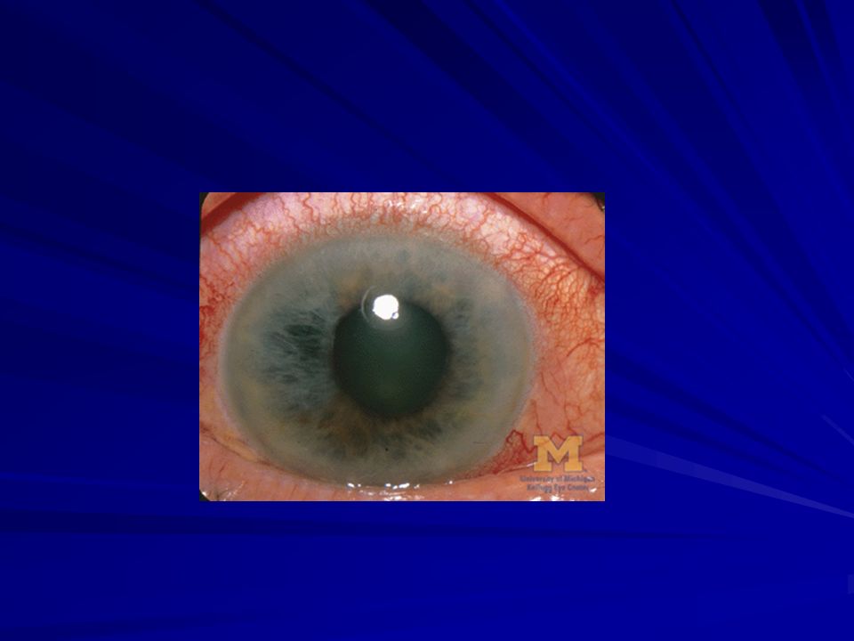
In acute angle closure glucoma, there is an abrupt increase in pressure and the eye becomes photophobic…painful due to ischemia…watering of the eye…loss of vision.. Pt feels unwell…nausea…abdominal pain On exam.: red eye…visual acuity is reduced…cloudy cornea…pupil is oval fixed and dialated

30
Acute angle closure glaucoma of the right eye (intraocular pressure was 42 in the right eye). Note the mid sized pupil on the left that was not reactive to light and conjunctivitis.

32
TTT: If acute must be urgently ttt to prevent permanent damage
Acetazolamide: IV or Oral + topical pilocarpine + B blocker Pilocarpine: constrict the pupil and draw the iris out of the angle B blocker: decrease aqueous secretion

33
Surgery: iridotomy iridectomy

34
2ry glaucoma IO pressure rises due to blockage of the trabecular meshwork… causes: Trauma leads to blood (hyphaema), or damage to the drainage angle (angle recession) Pigment from the iris (pigment dispersion syndrome) Deposition of material produced by the epithelium of the lens, iris and ciliary body (pseudoexfoliative glaucoma) Drugs (steroid-induced glaucoma) (Rubeosis iridis) abnormal iris blood vessels that may obstruct the angle (may accompany proliferative diabetic retinopathy) Choroidal melanoma may push the iris forward causing closure Cataract may swell causing closure Uveitis may cause iris to adhere to the meshwork
, or damage to the drainage angle (angle recession) Pigment from the iris (pigment dispersion syndrome) Deposition of material produced by the epithelium of the lens, iris and ciliary body (pseudoexfoliative glaucoma) Drugs (steroid-induced glaucoma) (Rubeosis iridis) abnormal iris blood vessels that may obstruct the angle (may accompany proliferative diabetic retinopathy) Choroidal melanoma may push the iris forward causing closure. Cataract may swell causing closure. Uveitis may cause iris to adhere to the meshwork.")
35
2ry Glaucome…cont. Much rarer than the primary glaucoma
Sign and symptoms depends on the rate at which the IO pressure rises…mostly are symptomless TTT: same as 1ry + treat the underlying cause In severe cases: laser or cryoprobe are used to ablate the ciliary processes

36
Congenital Glaucoma Uncertain cause
Iridocorneal angle may be developmentally abnormal and covered with a membrane that increase the resistance S &sx: Excessive tearing, photophobia Increased corneal diameter (buphthalmos),,,,, myopia Cloudy cornea (due to edema) Split’s in descemet’s membrane treatment Treatment : surgical by Geniotomy: incision in the trabecular meshwork Trabeculotomy: making a direct passage btw schlemm’s canal and the anterior chamber
,,,,, myopia. Cloudy cornea (due to edema) Split’s in descemet’s membrane treatment. Treatment : surgical by. Geniotomy: incision in the trabecular meshwork. Trabeculotomy: making a direct passage btw schlemm’s canal and the anterior chamber.")
37
Surgery Trabeculotomy

38
Thank you

Similar presentations


 Waxman MD PhD>")


 Giovanni Caboto Club October 3, 2012.>")


. Optic nerve head damage. Corresponding loss.>")


