Download presentation
Presentation is loading. Please wait.

1
Windsor University School of Medicine akolade osanoto
Axillary Artery Windsor University School of Medicine akolade osanoto

2
outline Origin Relations parts and branches Termination

3
Origin and termination
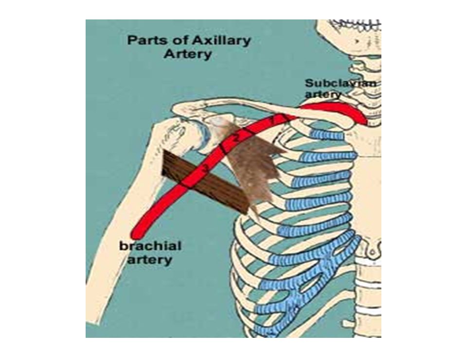
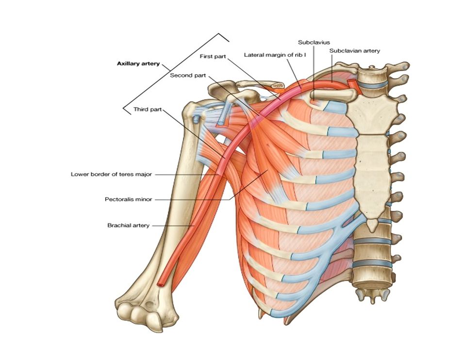
The axillary artery begins at the lateral border of the 1st rib as the continuation of the subclavian artery and ends at the inferior border of the teres major

4
Relation and course It passes posterior to the pectoralis minor into the arm and becomes the brachial artery when it passes the inferior border of the teres major, at which point it usually has reached the humerus

5
PARTS the axillary artery is divided into three parts by the pectoralis minor 1st 2nd 3rd

6
3 parts The first part of the axillary artery is located between the lateral border of the 1st rib and the medial border of the pectoralis minor; it is enclosed in the axillary sheath and has one branche - the superior thoracic artery The second part of the axillary artery lies posterior to pectoralis minor and has two branches the -thoracoacromial and -lateral thoracic arteries which pass medial and lateral to the muscle, respectively.

7
The third part of the axillary artery extends from the lateral border of pectoralis minor to the inferior border of teres major and has three branches. subscapular artery is the largest branch of the axillary artery. the anterior circumflex humeral and posterior circumflex humeral

10
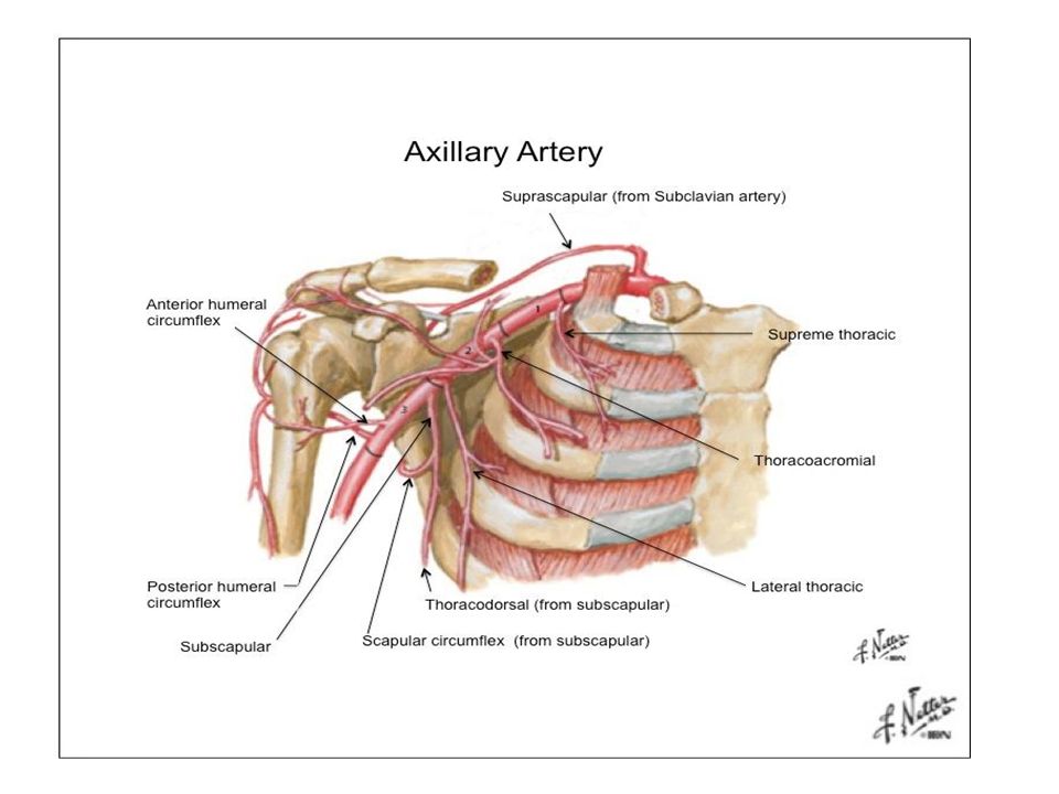
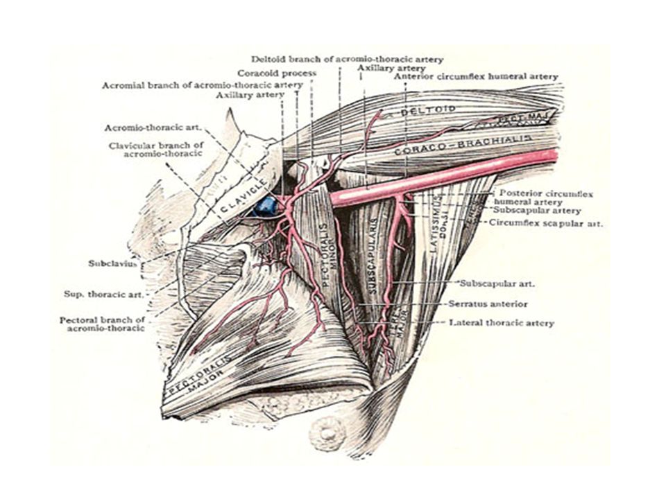
1st part -the superior thoracic artery
arises just inferior to the subclavius Supply: the subclavius, muscles in the 1st and 2nd intercostal spaces, superior slips of the serratus anterior, and overlying pectoral muscles

12
2nd part- The thoracoacromial artery, a short wide trunk, divides into four branches (acromial, deltoid, pectoral, and clavicular) The lateral thoracic artery supplies :the pectoral, serratus anterior, and intercostal muscles, the axillary lymph nodes, and the lateral aspect of the breast.

13
3rd part- The subscapular artery, descends along the lateral border of the subscapularis on the posterior axillary wall. terminates by dividing into the circumflex scapular and thoracodorsal arteries. Supply: muscles on the dorsum of the scapula). It participates in the anastomoses around the scapula
. It participates in the anastomoses around the scapula.")
14
The thoracodorsal artery inferior angle of the scapula ,latissimus dorsi. It also participates in the arterial anastomoses around the scapula. The smaller anterior circumflex humeral artery passes laterally, deep to the coracobrachialis and biceps brachii. It gives off an ascending branch that supplies the shoulder. The larger posterior circumflex humeral artery passes medially through the posterior wall of the axilla via the quadrangular space with the axillary nerve to supply the glenohumeral joint and surrounding muscles

16
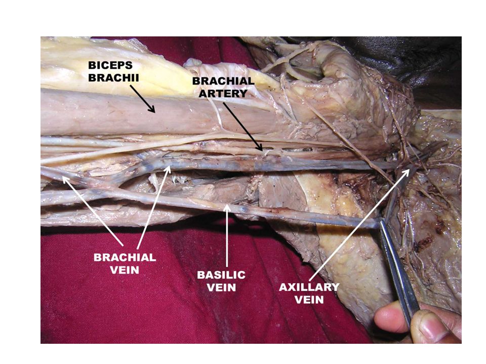
Axillary vein The axillary vein lies initially (distally) on the anteromedial side of the axillary artery, with its terminal part anteroinferior to the artery This large vein is formed by the union of the brachial vein (the accompanying veins of the brachial artery) and the basilic vein at the inferior border of the teres major. Also has 3 parts:the initial, distal end is the third part, whereas the terminal, proximal end is the first part. The axillary vein (first part) ends at the lateral border of the 1st rib, where it becomes the subclavian vein.
 on the anteromedial side of the axillary artery, with its terminal part anteroinferior to the artery. This large vein is formed by the union of the brachial vein (the accompanying veins of the brachial artery) and the basilic vein at the inferior border of the teres major. Also has 3 parts:the initial, distal end is the third part, whereas the terminal, proximal end is the first part. The axillary vein (first part) ends at the lateral border of the 1st rib, where it becomes the subclavian vein.")
Similar presentations