Download presentation
Presentation is loading. Please wait.

1
Copyright Pearson Prentice Hall 1.6 Cell Division

2
Essential Idea Cell division is essential but must be controlled.

3
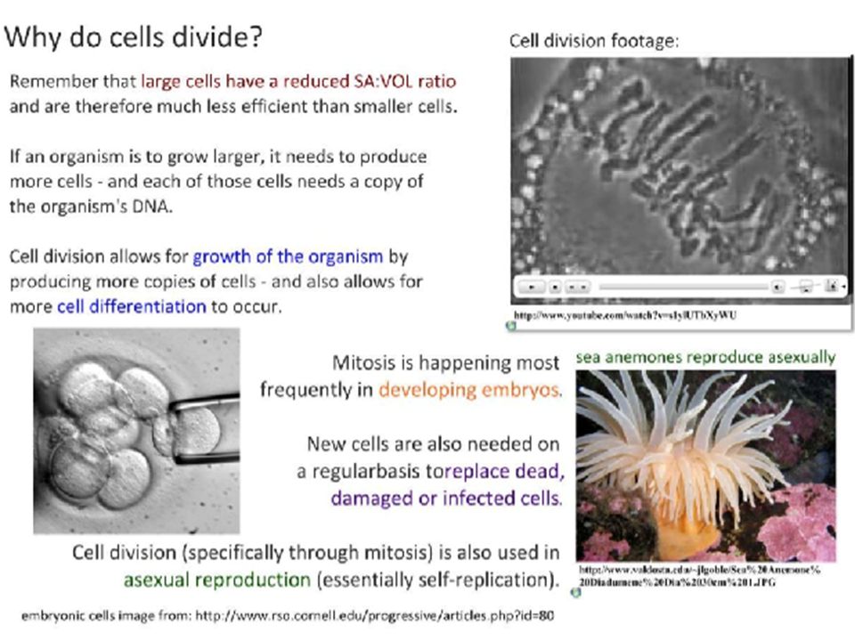
In unicellular organisms, division of one cell reproduces a new organism Multicellular organisms depend on cell division for: Cell size is limited Development from a fertilized cell Growth Repair

4
LE 12-2 Reproduction 100 µm Tissue renewal Growth and development 20 µm200 µm

5
Copyright Pearson Prentice Hall Limits to Cell Growth The larger a cell becomes, the more demands the cell places on its DNA. In addition, a large cell will have trouble moving enough nutrients and wastes across its cell membrane.

6
Copyright Pearson Prentice Hall

7
IB Assessment Statement Outline the stages in the cell cycle, including interphase (G1, S, G2), mitosis and cytokinesis Copyright Pearson Prentice Hall
, mitosis and cytokinesis Copyright Pearson Prentice Hall")
8
LE 12-5 G1G1 G2G2 S (DNA synthesis) INTERPHASE Cytokinesis MITOTIC (M) PHASE Mitosis
 INTERPHASE Cytokinesis MITOTIC (M) PHASE Mitosis")
9
Phases of the Cell Cycle The cell cycle consists of Mitotic (M) phase (mitosis and cytokinesis) Interphase (cell growth and copying of chromosomes in preparation for cell division) Interphase (about 90% of the cell cycle) can be divided into subphases: G 1 phase (“first gap”) S phase (“synthesis”) G 2 phase (“second gap”)
 phase (mitosis and cytokinesis) Interphase (cell growth and copying of chromosomes in preparation for cell division) Interphase (about 90% of the cell cycle) can be divided into subphases: G 1 phase ( first gap ) S phase ( synthesis ) G 2 phase ( second gap )")
10
LE 12-5 G1G1 G2G2 S (DNA synthesis) INTERPHASE Cytokinesis MITOTIC (M) PHASE Mitosis
 INTERPHASE Cytokinesis MITOTIC (M) PHASE Mitosis")
11
Interphase (about 90% of the cell cycle) can be divided into subphases: G 1 phase (“first gap”) First phase of growth New organelles formed Biochemical activity S phase (“synthesis”) chromosomes are copied (DNA replication) Two identical structures are formed called Chromatids Chromatids until mitosis G 2 phase (“second gap”) More growth of cell Preparation for Mitosis
 can be divided into subphases: G 1 phase ( first gap ) First phase of growth New organelles formed Biochemical activity S phase ( synthesis ) chromosomes are copied (DNA replication) Two identical structures are formed called Chromatids Chromatids until mitosis G 2 phase ( second gap ) More growth of cell Preparation for Mitosis")
12
Copyright Pearson Prentice Hall Chromosomes S- Phase of Interphase –Chromosomes are copied –Genetic information (DNA) is passed from one generation to the next on chromosomes. –This process, it is called DNA replication

13
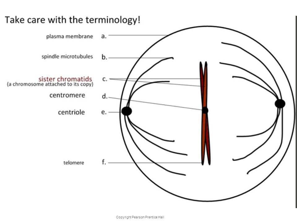
Copyright Pearson Prentice Hall Chromosomes Each chromosome consists of two identical “sister” chromatids. Each pair of chromatids is attached at an area called the centromere. When the cell divides, the chromatids separate. Each new cell gets one chromatid. Sister chromatids Centromere

14
Mitosis is conventionally divided into four phases: Prophase Metaphase Anaphase Telophase Cytokinesis is well underway by late telophase Cell Cycle Animation: http://highered.mcgraw- hill.com/sites/0072495855/student_view0/chapt er2/animation__how_the_cell_cycle_works.html

15
Copyright Pearson Prentice Hall Cell Cycle Events of the Cell Cycle

16
LE 12-6ca G 2 OF INTERPHASE PROPHASEPROMETAPHASE

17
LE 12-6da METAPHASEANAPHASE TELOPHASE AND CYTOKINESIS 10 µm

18
IB Assessment Statement Describe the events that occur in the four phases of mitosis (prophase, metaphase, anaphase and telophase). Copyright Pearson Prentice Hall

19
Mitosis

20
Copyright Pearson Prentice Hall Section 10-2 Prophase Spindle forming Chromosomes (paired chromatids) Centromere Click to Continue Mitosis Prophase
 Centromere Click to Continue Mitosis Prophase")
21
Mitosis Animation: http://highered.mcgraw- hill.com/sites/0072495855/student_view0/c hapter2/animation__mitosis_and_cytokine sis.htmlhttp://highered.mcgraw- hill.com/sites/0072495855/student_view0/c hapter2/animation__mitosis_and_cytokine sis.html Copyright Pearson Prentice Hall

22
Mitosis Prophase Prophase is the first and longest phase of mitosis. The MTOC/ centrosomes separate and take up positions on opposite sides of the nucleus. MTOC – Microtubules organizing center Spindle forming Centromere Chromosomes (paired chromatids)
.")
23
Copyright Pearson Prentice Hall Mitosis Chromatin condenses into chromosomes. The centrioles separate and a spindle begins to form. The nuclear envelope breaks down. Spindle forming Centromere Chromosomes (paired chromatids)
.")
24
Chromatin condenses into chromosomes. Copyright Pearson Prentice Hall

25
MTOC MTOC/ controsomes form the mitotic spindle also called spindle microtubules. The mitotic spindle (spindle microtubules) is an structure that controls chromosome movement during mitosis The MTOC/ centrosomes replicates, forming two MTOC/ centrosomes that move to opposite ends of the cell, as mitotic spindle grows out from them
 is an structure that controls chromosome movement during mitosis The MTOC/ centrosomes replicates, forming two MTOC/ centrosomes that move to opposite ends of the cell, as mitotic spindle grows out from them.")
26
LE 12-7 Spindle Microtubules Chromosomes Sister chromatids MTOC/ Centrosome Metaphase plate spindle microtubules 0.5 µm 1 µm MTOC

27
Copyright Pearson Prentice Hall

29
MTOC Spindle Mitosis Click to Continue Metaphase

30
Copyright Pearson Prentice Hall Mitosis Metaphase The second phase of mitosis is metaphase. The chromosomes line up across the center of the cell. Microtubules connect the centromere of each chromosome to the poles of the spindle. MTOC Spindle

31
Copyright Pearson Prentice Hall Individual chromosomes Anaphase Mitosis Anaphase

32
Copyright Pearson Prentice Hall Mitosis Anaphase Anaphase is the third phase of mitosis. The sister chromatids separate into individual chromosomes. The chromosomes continue to move until they have separated into two groups. Individual chromosomes

33
Copyright Pearson Prentice Hall Nuclear envelope reforming Telophase Mitosis Telophase

34
Copyright Pearson Prentice Hall Mitosis Telophase Telophase is the fourth and final phase of mitosis. Chromosomes gather at opposite ends of the cell and lose their distinct shape.

35
Copyright Pearson Prentice Hall Mitosis A new nuclear envelope forms around each cluster of chromosomes.

36
Copyright Pearson Prentice Hall Cytokinesis

37
Copyright Pearson Prentice Hall Cytokinesis During cytokinesis, the cytoplasm pinches in half. Each daughter cell has an identical set of duplicate chromosomes

38
More animations/ tutorials of mitosis: http://www.johnkyrk.com/mitosis.html http://bcs.whfreeman.com/thelifewire/con tent/chp09/0902001.htmlhttp://bcs.whfreeman.com/thelifewire/con tent/chp09/0902001.html https://www.youtube.com/watch?v=L0k- enzoeOM&feature=player_embeddedhttps://www.youtube.com/watch?v=L0k- enzoeOM&feature=player_embedded Copyright Pearson Prentice Hall

39
Cytokinesis: A Closer Look In animal cells, cytokinesis occurs by a process known as cleavage, forming a cleavage furrow Animation: Cytokinesis Animation: Cytokinesis

40
LE 12-9a Cleavage furrow 100 µm Contractile ring of microfilaments Daughter cells Cleavage of an animal cell (SEM)
")
41
Copyright Pearson Prentice Hall Cytokinesis in Plants In plants, a structure known as the cell plate forms midway between the divided nuclei. Cell wall Cell plate

42
Copyright Pearson Prentice Hall Cytokinesis in Plants The cell plate gradually develops into a separating membrane. A cell wall then begins to appear in the cell plate.

43
LE 12-9b 1 µm Daughter cells Cell plate formation in a plant cell (TEM) New cell wall Cell plate Wall of parent cell Vesicles forming cell plate
 New cell wall Cell plate Wall of parent cell Vesicles forming cell plate")
44
LE 12-10 Nucleus Cell plate Chromosomes Nucleolus Chromatin condensing 10 µm Prophase. The chromatin is condensing. The nucleolus is beginning to disappear. Although not yet visible in the micrograph, the mitotic spindle is starting to form. Prometaphase. We now see discrete chromosomes; each consists of two identical sister chromatids. Later in prometaphase, the nuclear envelope will fragment. Metaphase. The spindle is complete, and the chromosomes, attached to microtubules at their kinetochores, are all at the metaphase plate. Anaphase. The chromatids of each chromosome have separated, and the daughter chromosomes are moving to the ends of the cell as their kinetochore micro- tubules shorten. Telophase. Daughter nuclei are forming. Meanwhile, cytokinesis has started: The cell plate, which will divide the cytoplasm in two, is growing toward the perimeter of the parent cell.

45
Cell Cycle Summary Copyright Pearson Prentice Hall

46
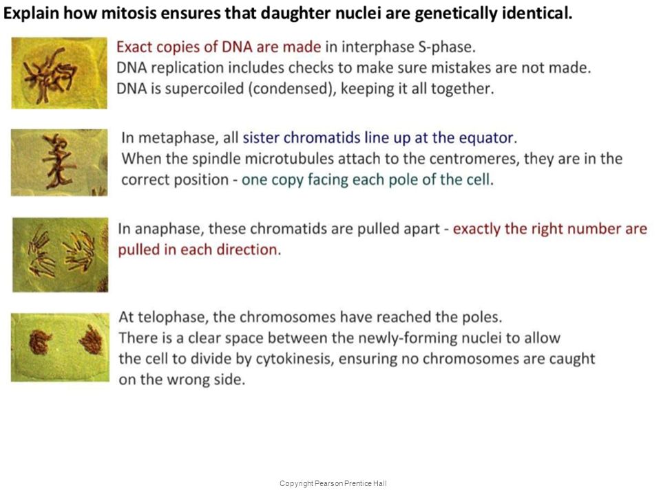
IB ASSESSMENT STATEMENT Explain how mitosis produces two genetically identical nuclei. Copyright Pearson Prentice Hall

48
IB Assessment Statement State that tumours (cancers) are the result of uncontrolled cell division and that these can occur in any organ or Tissue Copyright Pearson Prentice Hall
 are the result of uncontrolled cell division and that these can occur in any organ or Tissue Copyright Pearson Prentice Hall")
49
What is Cancer? Tumours are not foreign invaders. They arise from the same material used by the body to construct its own tissues. Tumours use the same components -human cells- to form the jumbled masses that disrupt biological order and function and, if left unchecked, to bring the whole complex, life sustaining edifice that is the human body crashing down'. –R. Weinberg, R. (1998) One Renegade Cell. London:Phoenix, Science Masters Series. 'Cancer is, in essence a genetic disease' –Volgestein and Kinzler Copyright Pearson Prentice Hall
 One Renegade Cell. London:Phoenix, Science Masters Series. Cancer is, in essence a genetic disease –Volgestein and Kinzler Copyright Pearson Prentice Hall.")
50
Uncontrolled Cell Growth –Cancer is a disorder in which some of the body's own cells lose the ability to control growth. –How are cancer cells different from other cells?

51
Uncontrolled Cell Growth –Cancer cells divide uncontrollably and form masses of cells called tumors that can damage the surrounding tissues. –Cancer cells may break loose from tumors and spread throughout the body, disrupting normal activities and causing serious medical problems or even death.

52
Understandings Cyclins are involved in the control of the cell cycle. Copyright Pearson Prentice Hall

53
Understandings Mutagens, oncogenes and metastasis are involved in the development of primary and secondary tumours. Copyright Pearson Prentice Hall

54
IB Assessment Statement State that growth, embryonic development, tissue repair and asexual reproduction involve mitosis Copyright Pearson Prentice Hall

56
State that growth, embryonic development, tissue repair and asexual reproduction involve mitosis Growth: multicellular organisms increase their size through growth. This growth involves increasing the number of cells through mitosis. These cells will differentiate and specialise their function. Embryonic development is when the fertilised egg cell (zygote) divides to form the multicellular organism. Each cell in the organisms is identical (genetically) to all the other cells. However, each cell will express only a few of its genes to determine its overall specialisms, a process called differentiation. In this way a stem cell may becomes a muscle, or it may become a nerve cell or any one of the many different kinds of cells found in a complex multicellular organism. The best book about this process for the interested reader is Copyright Pearson Prentice Hall
 divides to form the multicellular organism. Each cell in the organisms is identical (genetically) to all the other cells. However, each cell will express only a few of its genes to determine its overall specialisms, a process called differentiation. In this way a stem cell may becomes a muscle, or it may become a nerve cell or any one of the many different kinds of cells found in a complex multicellular organism. The best book about this process for the interested reader is Copyright Pearson Prentice Hall.")
57
State that growth, embryonic development, tissue repair and asexual reproduction involve mitosis Tissue Repair: As tissues are damaged they can recover through replacing damaged or dead cells. This is easily observed in a skin wound. More complex organ regeneration can occur in some species of amphibian. Asexual Reproduction: This the production of offspring from a single parent using mitosis. The offspring are therefore genetically identical to each other and to their “parent”- in other words they are clones. Asexual reproduction is very common in nature, and in addition we humans have developed some new, artificial methods. Bacteria DO NOT asexually reproduce by mitosis but rather by a process called Binary Fission. Copyright Pearson Prentice Hall

Similar presentations