Download presentation
Presentation is loading. Please wait.

1
Malignant Thyroid Disease

2
HISTORY OF PRESENT ILLNESS 10 years PTC slowly growing nodular ant. neck mass 2 years PTC rapid increase in the size of mass 6 months PTC hoarseness & difficulty of swallowing Admission

3
Review of Systems No fever, no weight loss, no tremors No chest pain, no easy fatigability No abdominal pain Past Medical History: unremarkable Family History: unremarkable

4
Physical Exam PR = 100/min RR = 20/min T = 37 o C No exopthalmos 25x20cm multinodular, firm right anterolateral neck mass Palpable cervical adenopathies post. to the sternocleidomastoid

5
SALIENT FEATURES 39 y/o, female Anterior neck mass Hoarseness Difficulty of swallowing

6
Malignant Thyroid Disease 1.What is your clinical impression? What are the differential diagnosis? Thyroid Cancer

7
Thyroid nodules Hx: time of onset, change in size, and associated symptoms such as pain, dysphagia, dyspnea, or choking Pain-raise suspicion of intrathyroidal hemorrhage in a benign nodule, thyroiditis, or malignancy Hoarseness- secondary to malignant involvement of the recurrent laryngeal nerve Increase risk of malignancy: Hx of ionizing radiation and family hx of thyroid cancer

8
PE: Thyroid masses- move with swallowing Hard, gritty, fixed nodules- more likely to be malignant Lymph node involvement- increases the risk of malignancy

9
39 y/0, female from BicolPR=100/min RR=20/min T=37C Growing nodular anterior neck mass(-) exophthalmos Hoarseness and difficulty in swallowing 25x20 multinodular, frim, right anterolat neck mass which moves with deglutition, with a hard nodule (5x3) within the big mass (-) Fever, weight loss, tremorsPalpable cervical adenopathies (-) chest pain, easy fatigability (-)abdominal pain Unremarkable PMH, FH
 exophthalmos Hoarseness and difficulty in swallowing 25x20 multinodular, frim, right anterolat neck mass which moves with deglutition, with a hard nodule (5x3) within the big mass (-) Fever, weight loss, tremorsPalpable cervical adenopathies (-) chest pain, easy fatigability (-)abdominal pain Unremarkable PMH, FH")
10
Specific Tumor Types: Papillary Carcinoma 80 % of all thyroid malignancies in iodine deficient areas and in individuals exposed to external radiation. 2:1 female to male ratio Mean age: 30-40 Euthyroid Slow growing mass with calcification, necrosis, or cystic change apparent grossly Dysphagia, dyspnea, and dysphonia Lymph node metastases

11
Specific Tumor Types: Follicular Carcinoma More common in iodine deficient areas 3:1 female to male ratio Mean age: 50 years Solitary thyroid nodule Cervical lymphadenopathy is uncommon Distant metastasis may be present

12
Hurthle Cell Carcinoma Similar to follicular carcinoma Multifocal Bilateral More likely to metastasize to local nodes and distant sites

13
Medullary Carcinoma Neck mass with palpable cervical lymphadenopathy Dysphagia, dyspnea, dysphonia 1.5:1 female to male ratio Mean age: 50-60, patients with familial disease present at a younger age Unilateral, multicentric

14
Anaplastic Carcinoma Most aggressive Presents in the 7 th or 8 th decade of life Long standing neck mass which rapidly enlarges, may be painful Associated with dysphonia, dysphagia, and dyspnea Palpable lymphnodes

15
1. What is your clinical impression? What are the differential diagnosis? Goiter-may result from iodine deficiency and/or TSH stimulation secondary to inadequate thyroid hormone synthesis - may be diffuse, uninodular, or multinodular -compression due to very large goiters may produce dyspnea and dysphagia

16
Differential Diagnosis: Benign Thyroid Nodule Thyroiditis Lymphadenopathy Metastasis from head and neck cancer

17
3. What work ups are needed, if any? Laboratory Studies Thyroid function –Perform a complete assessment of thyroid function in any patient with thyroid lumps. –Higher-than-normal levels of thyroxine, triiodothyronine and thyroid-stimulating hormone (TSH) may indicate thyroid cancer.
 may indicate thyroid cancer..")
18
TSH suppression test –Cancer is autonomous and does not require TSH for growth, whereas benign lesions do require TSH. –Preoperatively, the test is useful for patients with nontoxic solitary benign nodules and for women with repeated nondiagnostic test results. –Postoperatively, the test is useful for monitoring papillary thyroid cancer cases.

19
Imaging Studies Chest radiography, CT scanning, and MRI – Not usually used in the initial workup of a thyroid nodule, except in patients with clear metastatic disease at presentation.

20
Echography –Performed first in any patient with possible thyroid malignancy. –Noninvasive and inexpensive, and represents the most sensitive procedure for identifying thyroid lesions and for determining the diameters of a nodule. –Useful for localizing lesions when a nodule is difficult to palpate or is deeply seated. –It may be used to help direct a fine-needle aspiration biopsy (FNAB).
..")
21
FNAB FNAB is considered the best first-line diagnostic procedure for a thyroid nodule; FNAB is a safe and minimally invasive procedure. –Sensitivity of the procedure is near 80%, the specificity is near 100%, and errors can be diminished using ultrasonographic guidance. –False-negative and false-positive results occur less than 6% of the time.

22
Histologic Findings Papillary thyroid carcinoma usually appears as a grossly firm mass that is irregular and not encapsulated. Microscopically, it is multifocal, and a net invasion of the lymphatics may be demonstrated. Complete or partial papillary architecture with some follicles is present. The thyrocytes are large and show an abnormal nucleus and cytoplasm with several mitoses. Thyrocytes may have "orphan Annie eyes," that is, large round cells with a dense nucleus and clear cytoplasm. Another typical feature of this cancer is the presence of the psammoma bodies, probably the remnants of dead papillae.

23
Staging The staging of well-differentiated thyroid cancers is related to age for the first and second stages, but it is not related to age for the third and fourth stages. In the staging protocol, T is tumor, N is node, and M is metastasis. Younger than 45 years –Stage I - Any T, any N, M0 (cancer in thyroid only) –Stage II - Any T, any N, M1 (cancer spread to distant organs) Older than 45 years –Stage I - T1, N0, M0 (cancer only in thyroid, may be found in one or both lobes) –Stage II - T2, N0, M0 and T3, N0, M0 (cancer only in thyroid and >1.5 cm) –Stage III - T4, N0, M0 and any T, N1, M0 (cancer spread outside thyroid but not outside of neck) –Stage IV - Any T, any N, M1 (cancer spread to other parts of body)
 –Stage II - Any T, any N, M1 (cancer spread to distant organs) Older than 45 years –Stage I - T1, N0, M0 (cancer only in thyroid, may be found in one or both lobes) –Stage II - T2, N0, M0 and T3, N0, M0 (cancer only in thyroid and >1.5 cm) –Stage III - T4, N0, M0 and any T, N1, M0 (cancer spread outside thyroid but not outside of neck) –Stage IV - Any T, any N, M1 (cancer spread to other parts of body).")
24
3. What are the treatment options? A.TOTAL THYROIDECTOMY B.SUBTOTAL THYROIDECTOMY C.NECK DISSECTION

25
TOTAL THYROIDECTOMY -30%-87.5% of PTC involve the opposite lobe -7%-10% recurrence rate in the contralateral lobe -Lower recurrence rate -For earlier detection and treatment of metastatic CA with radioactive iodine therapy

26
Indications of Total Thyroidectomy: a.) Patients > 40 y/o with papillary or follicular CA b.) Patients with thyroid nodule and history of radiation c.) Patients with bilateral disease
 Patients > 40 y/o with papillary or follicular CA b.) Patients with thyroid nodule and history of radiation c.) Patients with bilateral disease")
27
SUBTOTAL THYROIDECTOMY -Lower incidence of complications A.Hypoparathyroidism B.Recurrent laryngeal nerve injury C.Superior laryngeal nerve injury

28
NECK DISSECTION -For managing lymphadenopathies -For clinically palpable cervical nodes (as in the case) verified by MRI or CT scan
 verified by MRI or CT scan")
29
Management of Patient with Papillary Thyroid CA

31
Management - Surgery 1.Total Thyroidectomy (TTx) –PTA may be multifocal/bilobal –↓ incidence of local recurrence –↓ risk of anaplasia in any residual tissue –↓ incidence of distant recurrence (by facilitating diagnosis of distant metastasis by RAI scan) –↑ sensitivity of blood thyroglobulin (Tg) levels to predict recurrence/persistence
 –PTA may be multifocal/bilobal –↓ incidence of local recurrence –↓ risk of anaplasia in any residual tissue –↓ incidence of distant recurrence (by facilitating diagnosis of distant metastasis by RAI scan) –↑ sensitivity of blood thyroglobulin (Tg) levels to predict recurrence/persistence")
32
Management - Surgery 2.Modified Radical Neck Dissection –Removal of cervical lymph nodes –Spares sternocleidomastoid muscle, internal jugular vein and spinal accessory nerve –All tissue in the anterior triangle of the neck from the hyoid bone to the clavicle is removed –Dissection along the spinal accessory nerve is most important because this is a frequent site of metastatic disease

33
Immediately Post-Op Wound care and analgesia Analgesia Monitor serum thyroglobulin Check for any possible complications Prep patient for RAI scan and treatment

35
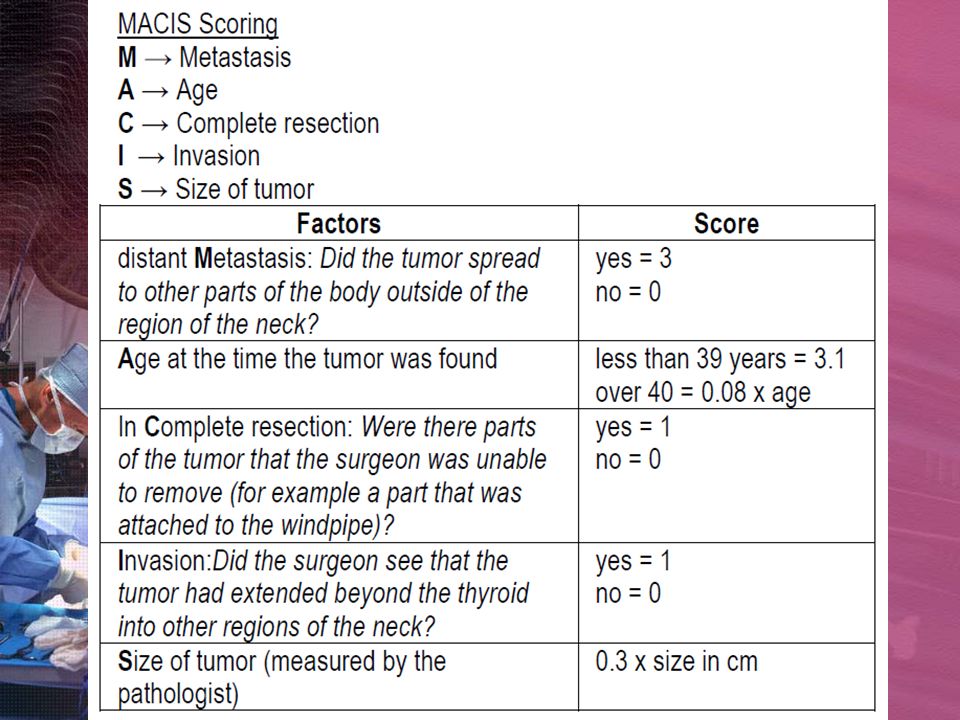
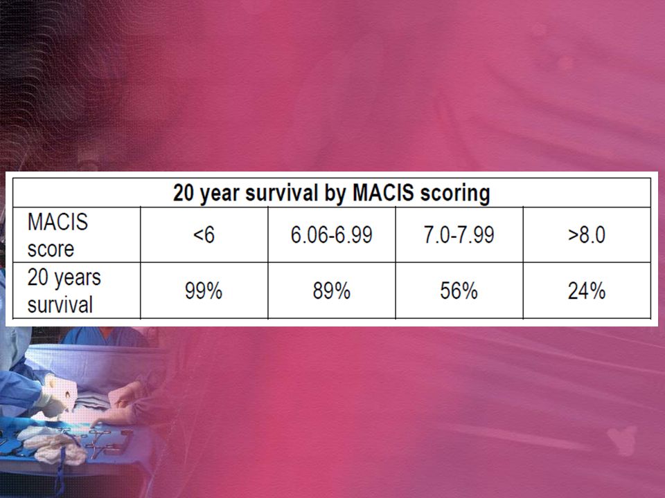
AMES

38
TNM Classification Primary tumor (T) TX: Primary tumor cannot be assessed T0: No evidence of primary tumor T1: Tumor ≤ 1 cm in greatest dimension limited to the thyroid T2: Tumor > 1 cm but ≤ 4 cm in greatest dimension limited to the thyroid T3: Tumor > 4 cm in greatest dimension limited to the thyroid T4: Tumor of any size extending beyond the thyroid capsule
 TX: Primary tumor cannot be assessed T0: No evidence of primary tumor T1: Tumor ≤ 1 cm in greatest dimension limited to the thyroid T2: Tumor > 1 cm but ≤ 4 cm in greatest dimension limited to the thyroid T3: Tumor > 4 cm in greatest dimension limited to the thyroid T4: Tumor of any size extending beyond the thyroid capsule")
39
TNM Classification Regional lymph nodes (N) Cervical and upper mediastinal lymph nodes. NX: Regional lymph nodes cannot be assessed N0: No regional lymph node metastasis N1: Regional lymph node metastasis N1a: Metastasis in ipsilateral cervical lymph node(s) N1b: Metastasis in bilateral, midline, or contralateral cervical or mediastinal lymph node(s)
 N1b: Metastasis in bilateral, midline, or contralateral cervical or mediastinal lymph node(s).")
40
TNM Classification Distant metastases (M) MX: Distant metastasis cannot be assessed M0: No distant metastasis M1: Distant metastasis
 MX: Distant metastasis cannot be assessed M0: No distant metastasis M1: Distant metastasis")
41
AJCC Stage Groupings (Papillary/Follicular) < 45 years old Stage I: Any T, Any N, M0 Stage II: Any T, Any N, M1 ≥ 45 years old Stage I: T1, N0, M0 Stage II: T2 or T3, N0, M0 Stage III: T4, N0, M0 or Any T, N1, M0 Stage IV: Any T, Any N, M1 For undifferentiated (anaplastic) thyroid cancer, stage IV regardless of T, N, M
 < 45 years old Stage I: Any T, Any N, M0 Stage II: Any T, Any N, M1 ≥ 45 years old Stage I: T1, N0, M0 Stage II: T2 or T3, N0, M0 Stage III: T4, N0, M0 or Any T, N1, M0 Stage IV: Any T, Any N, M1 For undifferentiated (anaplastic) thyroid cancer, stage IV regardless of T, N, M")
42
4-6 Weeks Post-Op Serum thyroglobulin determination Radioactive Iodine Scanning and treatment (RRA) –Discontinue L-thyroxine 8 weeks prior to scan First 6 weeks of this: give synthetic T3 Remaining 2 weeks prior to scan: discontinue T3 and recommend low iodine diet –Place patient of L-thyroxine again after procedure –RAI scan looks for persistent/recurrent disease –RAI treatment may destroy microscopic cancer cells –↑ sensitivity of serum Tg improved during follow-up
 –Discontinue L-thyroxine 8 weeks prior to scan First 6 weeks of this: give synthetic T3 Remaining 2 weeks prior to scan: discontinue T3 and recommend low iodine diet –Place patient of L-thyroxine again after procedure –RAI scan looks for persistent/recurrent disease –RAI treatment may destroy microscopic cancer cells –↑ sensitivity of serum Tg improved during follow-up")
43
4-6 Weeks Post-Op TSH Suppression –Via L-thyroxine (which also serves as replacement therapy for TTx) –↓ recurrence by ↓ growth stimulus to any possible residual thyroid cancer cells –circulating TSH levels 0.1 mU/L in low-risk patients < 0.1 mU/mL in high-risk patients
 –↓ recurrence by ↓ growth stimulus to any possible residual thyroid cancer cells –circulating TSH levels 0.1 mU/L in low-risk patients < 0.1 mU/mL in high-risk patients")
44
Post-Op Thyroglobulin Measurement –If patient taking L-thyroxine: < 2 ng/mL –Otherwise: <3 ng/mL If > 3 ng/mL; highly suggestive of metastasis /persistent normal thyroid tissue, especially if TSH also rises (eg. discontinuation of L-thyroxine as prep for AI scan)
.")
45
Long Term PE every 3-6 mo for 2 yrs then annually Serum Tg at 6 and 12 mo then annually 131 I whole body scan (WBS) every year until 2 negative scans Periodic ultrasound and Chest X-ray
 every year until 2 negative scans Periodic ultrasound and Chest X-ray")
46
WBS - Conventional

47
WBS – recombinant thyrotropin Safer, effective means of stimulating 131 I uptake and serum thyroglobulin (Tg) For patients: –Alternative to traditional LT4 withdrawal –Inability to generate endogenous TSH –Unable/unwilling to undergo LT4 withdrawal –Enhance the sensitivity of Tg in thyroid CA follow-up Hypothyroidism is contraindicated
 For patients: –Alternative to traditional LT4 withdrawal –Inability to generate endogenous TSH –Unable/unwilling to undergo LT4 withdrawal –Enhance the sensitivity of Tg in thyroid CA follow-up Hypothyroidism is contraindicated")
48
5. What are the possible complications of your treatment?

49
Bleeding Intraoperative bleeding stains the tissues and obscures important structures. An unrecognized or rapidly expanding hematoma can cause airway compromise and asphyxiation. increases the risk of other anatomic complications Deliberate dissection and fastidious hemostasis are essential to prevent this complication.

50
Injury to the recurrent laryngeal nerve Mechanisms of injury to the RLN include complete or partial transection, traction, contusion, crush, burn, misplaced ligature, and compromised blood supply. The consequence of an RLN injury is true vocal- fold paresis or paralysis. Occurs in <1% of px undergoing thyroidectomy

51
Hypoparathyroidism Parathyroid glands produce parathyroid hormone (PTH), which is intimately involved in the regulation of serum calcium. Direct trauma to the parathyroid glands, devascularization of the glands, or removal of the glands during surgery can cause temporary or permanent shutdown, which results in hypocalcemia. Rate of permanent hypoparathyroidism - <2%. Rate of transient hypoparathyroidism- 50%

52
Injury to the superior laryngeal nerve Trauma to the nerve results in an inability to lengthen a vocal fold and, thus, an inability to create a high-pitched sound. Rate of injury to the external branch of the SLN - 15%

53
Infection Infection was the major cause of death from thyroid surgery during the 1800s. Today, infection occurs in less than 1-2% of all cases. Hypothyroidism Hypothyroidism is an expected sequela of total thyroidectomy. should never be left untreated long enough to elicit signs and symptoms of myxedema (eg, hair loss, large tongue, cardiomegaly). Expect, diagnose, and promptly treat postoperative hypothyroidism.
. Expect, diagnose, and promptly treat postoperative hypothyroidism..")
54
Thank you!

Similar presentations


>")


>")
 M0 - No metastases M1 - Metastases present.>")


. Anatomy Anatomy.>")


