Download presentation
Presentation is loading. Please wait.

1
Male Reproductive System 男性生殖系统 Department of Histology and Embryology Medical college in Three Gorges University

2
The male reproductive organs testis epididymis, ductus deferens seminal vesicle, the prostate, the male urethra, the penis

3
THE TESTIS

6
seminiferous tubules Interstitial tissue

7
seminiferous tubules efferent ductules the duct of the epididymis straight tubules rete testis ductus deferens

8
General Structure of Seminiferous Tubules The wall of each tubule is made up of (1)an outer layer of fibrous tissue which also contains muscle like (myoid) cells. (2) basal lamina (3) Germ cells and sustentacular cells or the cells of sertoli.
 basal lamina (3) Germ cells and sustentacular cells or the cells of sertoli..")
11
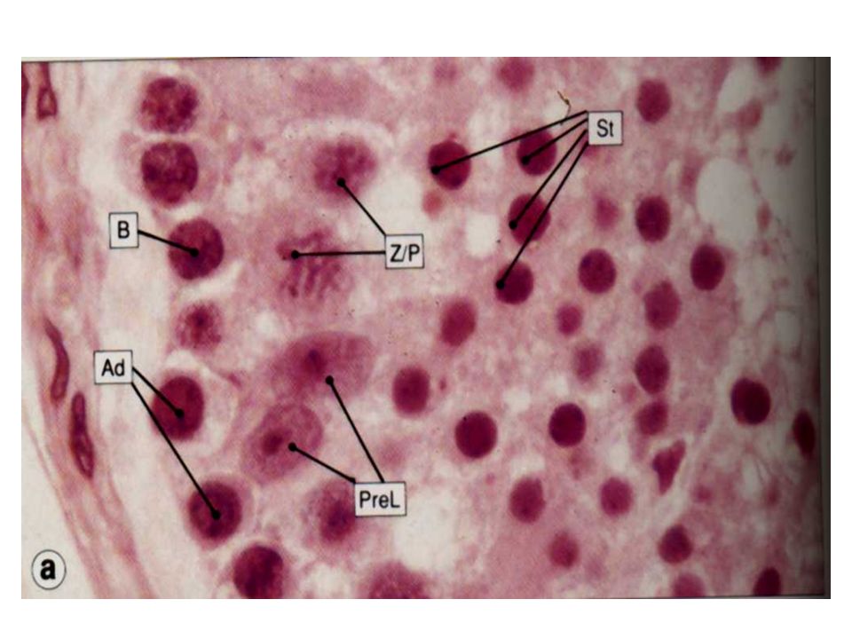
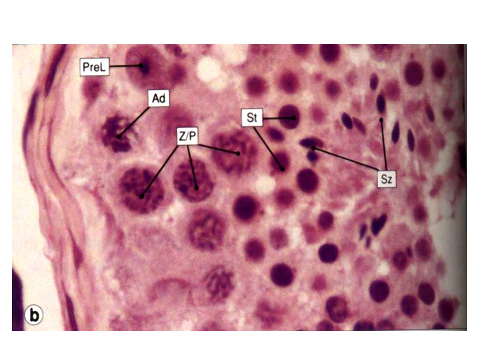
Germ cells: Spermatogonia Primary spermatocytes Secondary spermatocytes Spermatids Spermatozoon

15
(1) Spermatogonia:It is the stem cells of spermatogenesis after puberty. It is about 12um in diameter that are rest on the basement membrane. Type A Type B

16
(2)Primary spermatocytes are formed by mitotic division of spermatogonia. These are large cells with large spherical nuclei. Each primary spermatocyte undergoes meiosis to give rise to two secondary spermatocytes This is the first meiotic division in which the number of chromosomes is reduced to half.

18
(3) Secondary spermatocytes are smaller than primary spermatocytes, and so are their nuclei. We have seen that each secondary spermatocyte has the haploid number of chromosomes. It divides to form two spermatids by the second meiotic division.It rapidly undergoes the second meiotic division and are therefore seldom seen.

19
(4) Spermatid: Each spermatid is a rounded cell with a spherical nucleus. Both cell and nucleus are much smaller than in the case of spermatogonia or spermatocytes. The spermatids undergoes changes in shape, and in the orientation of its organelles, to form a spermatozoon. This process is called spermiogenesis.

21
Structure of a Mature Spermatozoon The spermatozoon has a head, a neck, a middle piece and a principal piece and end piece or tail. The head is covered by a cap called the acrosomic cap, anterior nuclear cap, or galea capitis.

23
Sustentacular Cells or Cells of Sertoli These are tall, slender cells having an irregularly pyramidal or columnar shape. The nucleus lies near the base of the cell. It is light staining and is of irregular shape. There is a prominent nucleolus.

25
The base of each sustentacular cell rests on the basement membrane, spermatogonia being interposed amongst the sustentacular cells. The apex of the sustentacular cell reaches the lumen of the seminiferous tubule. Numerous spermatids, at various stages of differentiation into spermatozoa, appear to be embedded in the apical part of the cytoplasm.

26
Function: Sustentacular cells support developing germ cells and provide them with nutrition. They probably secrete fluid that helps to move spermatozoa along the seminiferous tubules.

27
Sustentacular cells may also act as macrophages. In the adult testis sustentacular cells are less prominent than germ cells. They are more prominent than germ cells before puberty, and in old age.

28
The interstitial tissue: Apart from interstitial cells, the interstitial tissue contains collagen fibres, fibroblasts, macrophages, mast cells, blood vessels and lymphatics.

30
Interstitial Cells The interstitial cells (of Leydig) are large, round or polyhedral cells lying in the connective tissue that intervenes between the coils of seminiferous tubules. Their nuclei are eccentric. The cytoplasm stains lightly and often has a foamy appearance (because of the removal of lipids during processing of tissues).
..")
31
Interstitial cells secrete male sex hormone (testicular androgens).
.")
32
ACCESSORY UROGENITAL ORGANS Structurally, the epididymis consists of two parts. The head is formed by highly convoluted continuations of the efferent ductules. these are lined by ciliated columnar epithelium.

34
The body and tail of the epididymis are made up of the duct of the epididymis, which is greatly coiled on itself. The duct is lined by pseudostratified columnar epithelium in which there are tall columnar cells, and shorter basal cells that do not reach the lumen.

36
The luminal surface of each columnar cell bears non-motile projections that resemble cilia. These stereocilia are seen by EM to be thick microvilli. The basal cells are precursors of the tall cells. The tubules of the epididymis are surrounded by smooth muscle and by a rich network of capillaries.

39
The Ductus Deferens

40
The Prostate

41
concretions

Similar presentations


 produce androgen-testosterone ---gernital ducts: store.>")


 Lecturer of Comparative Anatomy and Embryology Zoology Department, Faculty of Science, MANSOURA UNIVERSITY EGYPT.>")


